
Evaluation of the relationship of CT histogram analysis with survival and local control time in patients with colorectal liver metastases
Prognostic role of CT histogram in crlm
Authors
Abstract
AimColorectal cancer (CRC) is the third most common cause of death, and the presence of liver metastases is the most important factor affecting the survival of patients. In this study, we investigated the relationship between the parameters obtained by CT histogram analysis of liver metastases and the duration of local control and survival.
Methods In our study, the dataset of the study named ‘Colorectal Liver Metastasis’ registered in the ‘Cancer Imaging Archive’ database and CT images of the patients enrolled in this study were used. CT histogram analysis of 197 liver metastatic lesions was performed. The relationship between histogram parameters and survival time and local control time was evaluated by Cox regression analyses.
Results The study included 110 male and 87 female patients (mean age 59.6 ± 8.2 years). The mean follow-up period was 74.2 months, the mean survival period was 66.5 months, and the mean local control period was 53.2 ± 4.1 months. When Cox regression analysis was performed between survival time and tumor histogram parameters, a statistically significant relationship was found between survival time and Skewness value, 1st P value and 99th P value. When Cox regression analysis was performed between local control time and tumor histogram parameters, a statistically significant relationship was found between local control time and the Skewness value 1.P value and 99.P value.
Conclusion Texture analysis from CT, which is frequently used for pre-treatment staging in CRLM, may be useful in early prediction of treatment success and survival and thus in determining treatment strategies.
Keywords
Introduction
Colorectal cancer (CRC) is the third most common cause of death.1 The presence of liver metastases is the most important factor affecting the survival of patients.2 Synchronous colorectal liver metastases (CRLM) are detected in approximately 25 per cent of patients at the time of diagnosis, and more than half of the patients are predicted to develop CRLM during the course of the disease.3 The most common cause of death in patients with CRLM is considered to be liver failure due to the spread of cancer.1 In previous studies, it was reported that the median survival time of untreated patients with CRLM was only 4 months.4 Therefore, it is extremely important to understand the surgical, medical and interventional methods among the treatment options and to determine the most appropriate treatment plan for patients by multidisciplinary teams.
Computed tomography (CT) is a non-invasive imaging technique that is widely used for pre-treatment staging in patients with CRLM, as in other tumor types. Radiomics and texture analysis using CT images are image processing methods that provide a mathematical evaluation of the distribution of grey levels between pixels in an image. This analysis allows the extraction of various quantitative data that may not be discernible through visual inspection.5 Recent studies have shown that radiomic and texture analysis of tumors on CT images can predict survival, local control and metastasis-free survival times in various cancer types.6,7,8,9 Unnecessary surgical procedures can be avoided, and alternative treatment methods can be investigated at an earlier stage by using CT radiomics and tissue analysis to identify patients with low potential for success of surgical resection in relation to tumor heterogeneity. In this study, we examined the correlation between liver metastasis histogram parameters obtained by CT texture analysis and the survival and local control times of patients with CRLM treated with surgical resection. Our aim was to determine whether this approach could provide valuable information and complement existing prognostic factors.
Materials and Methods
We used an anonymized dataset from a study entitled ‘Colorectal Liver Metastases (CRLM)’ registered in ‘The Cancer Imaging Archive (TCIA) database and anonymized CT images of the cases included in this study.10 We obtained pretreatment CT images of 197 cases originally enrolled in the study. Image analysis and statistical evaluation were carried out between January and March 2024. All data acquisition and utilization adhered to TCIA data use guidelines.11 The collected CT images were then reviewed using the ‘Radiant DICOM Viewer 2020.2.3’ software. After evaluation, 197 patients who underwent contrast-enhanced abdominal CT examination with a slice thickness of 1.3 mm were included in our study. In the contrast-enhanced abdominal CT examinations constituting the study group, 120 mL contrast material was administered intravenously at a rate of 3 mL/sec, and the delay time was set as 90 s. For image analysis, a single observer, M.D., with 10 years of experience in radiology, evaluated the CT images of each case using the same window settings (window level 20 and window width 380). The axial image showing the tumor at maximum size was identified and registered. The saved images were then opened using ‘qMaZda v4.6’ software (MaZda for Windows, B11 ver. 4.6, www.eletel.p.lodz.pl/programy/mazda/).12 Tumor boundaries, including necrotic and cystic regions, were manually drawn. Histogram parameters such as mean, variance, skewness, kurtosis, 1st percentile (P), 10th P, 50th P, 90th P and 99th P were calculated over the determined areas and evaluated for each case individually (Figure 1).
Ethical ApprovalThis study was approved by the Ethics Committee of Harran University (Date: 2023-10-10, No: HRU/23.19.26).
Statistical AnalysisStatistical analyses were performed using SPSS version 22.0 (IBM Inc., Armonk, NY, USA). The normal distribution of the numerical data was assessed through a thorough examination of the Kolmogorov-Smirnov and Shapiro- Wilk tests. Descriptive statistics were used to express the results of the study; mean ± standard deviation was applied for numerical data fitting the normal distribution, and median with minimum-maximum values was applied for numerical data not fitting the normal distribution. Categorical data were presented as frequencies and percentages. To compare numerical data of patients with different survival status (surviving or dying) and local control status (successful or unsuccessful), the independent samples t-test was used for data following a normal distribution; the Mann-Whitney U test or Fisher’s exact probability test was used. It was used for data that did not follow a normal distribution. Chi-Square tests were used to evaluate categorical data between these groups. Survival analyses were performed using the Kaplan-Meier method and univariate Cox regression analyses. Hazard ratios (HR) and corresponding 95% confidence intervals (95% CI) were analyzed for various parameters. A significance level of P < 0.05 was accepted to indicate statistical significance.
Reporting GuidelinesThis observational study was reported in accordance with the STROBE guidelines.
Results
The study included 117 male and 80 female patients. The mean age was 59.6 ± 8.2 years. The mean follow-up period was 74.2 months (minimum-maximum: 3.7-131 months), the mean survival period was 66.5 months (minimum-maximum: 3.8-131 months), the mean local control period was 53.2 ± 4.1 months, and the 5-year survival rate was 59%. No significant difference was found when demographic data were evaluated in patients with different survival statuses. The mean age of the surviving patients was 57.6 ± 11.7 years, and the mean age of the lost patients was 63.8 ± 12.7 years (p = 0.26) (Table 1). 70.1 per cent of male patients and 70 per cent of female patients were excised (p = 0.9) (Table 1). No significant difference was found when demographic data were evaluated in patients with different local control statuses. The mean age of patients with successful local control was 57.8 ± 12 years, and the mean age of patients with unsuccessful local control was 60.4 ± 12.4 years (p = 0.155) (Table 1). Progression occurred in 68 per cent of male patients and 72 per cent of female patients. No statistically significant difference was found (p = 0.713) (Table 1). There was no statistically significant difference between the CT histogram parameters of the patients according to survival status (Table 1). According to local control status, there was a significant difference between the mean and 1st percentile values of CT histogram parameters in successful and unsuccessful patients (p values 0.044; 0,024, respectively) (Table 1). When Cox regression analysis was performed between survival time and tumor histogram parameters, a statistically significant relationship was found between survival time and Skewness value (HR: 0.189, 95% CI: 0.064-0.554, p:0.002), 1st P value (HR: 1.088, 95% CI: 1.014-1.168, p:0.019) and 99th P value (HR: 1.077, 95% CI: 1.032-1.124, p:0.001) (Table 2). Cox regression analysis between local control time and tumor histogram parameters showed a statistically significant correlation between local control time and Skewness value (HR: 0.235, 95% CI: 0.098-0.563, p:0. 001), 1st P value (HR: 1.081, 95% CI: 1.013-1.153, p:0.019), and 99th P value (HR: 1.084, 95% CI: 1.047-1.123, p<0.001) (Table 3).
Discussion
The liver is the most common site for distant metastasis in colorectal cancer, with liver metastases occurring in as many as 70% of colorectal cancer patients.13 Liver metastasis is the primary cause of death and holds great significance in the treatment of cancer patients. Currently, hepatic resection is considered the only viable treatment that can ensure long-term survival for those with colorectal liver metastases.14
In our study, the relationship of CT histogram parameters with survival time and local control time in CRLM was investigated. Cox regression analysis showed that the mean value, 50.P, 90.P and 99.P values of histogram parameters were independent predictors of survival time, and the mean value, 1.P and 10.P values of tumor histogram parameters were independent predictors of local control time. It has been shown that radiomics/texture analysis, which enables quantitative evaluation of tumor heterogeneity, can be used to predict survival, metastasis-free survival, early recurrence and local control in various cancer types and that local control and survival times of heterogeneous tumors are lower.15,16,17,18,19 These findings suggest that CT histogram parameters, particularly skewness, can potentially inform clinical decisions. A low skewness value, indicating a more homogeneous tumor texture, may be associated with a more favorable prognosis, supporting curative surgical approaches. In contrast, a high skewness value might reflect structural heterogeneity such as necrosis or irregular vascularization, which are known to be associated with poor treatment response. In such cases, clinicians may consider neoadjuvant therapy before surgery or increased frequency of imaging follow-up. Thus, preoperative CT texture analysis could serve as a non-invasive biomarker to tailor personalized treatment plans.
Since heterogeneity due to differences in vascularity is expected to affect CT texture parameters, CT texture analysis is thought to predict treatment response in CRLM. Kuno et al. evaluated tumor texture properties volumetrically in a texture study performed on pretreatment CT scans of head and neck tumors treated with chemoradiotherapy and found that the geometric and harmonic mean of the tumor showed a statistically significant relationship with local control.20 In our study, texture analysis was performed on a single slice, but it was found that various CT histogram parameters of the tumor showed statistically significant correlation with the duration of local control, as in the study of Kuno et al. In our study, skewness, 1.P and 99.P values showed statistically significant correlation with local control time.
Yuying Li et al. showed that CT histogram parameters may showing the primary tumor in their study with gastric and colon cancer patients with liver metastasis.21 Li Y et al. compared patients with colorectal cancer with and without liver metastases by CT histogram analysis and found significant differences in many radiomics.22 In our study, the primary tumor was not studied, but the differences in the texture analysis of metastatic lesions support the possible difference in the internal structure heterogeneity of primary tumors.
Hu R et al. reported that CT histogram analysis can be a guide in predicting local control in patients with colorectal metastases receiving radiotherapy.23 In our study, we found that histogram analysis may be helpful in predicting both survival and local control success in patients who underwent surgical resection.
Li Z et al. reported that texture analysis may be helpful in differentiating liver metastases from benign and malignant lesions of the primary liver.24 In their study, they stated that primary liver lesions and metastatic lesions are heterogeneous in terms of internal structure, and therefore, differential diagnosis can be made with the help of radiomics. In our study, only metastatic lesions were examined, but the success of the histogram parameters we found in showing survival and local survival supports the usefulness of radiomics in demonstrating internal structure heterogeneity.
Limitations
Our study has some limitations. Since tumor histogram features were measured by a single observer, the inter-observer reproducibility of the measurements could not be evaluated. In future studies, involving multiple radiologists to perform independent measurements will be essential to evaluate interobserver agreement and establish the reproducibility of histogram-based texture analysis. Tumors of different origins were evaluated in the study, and this caused heterogeneity. Evaluation of only tumors with the same origin may increase the reproducibility of the studies. In our study, CT histogram parameters were measured from a single slice because we thought that it could provide practicality in daily routine work intensity. Different results may occur if the analysis is performed to cover the entire volume of the lesions. In this study, only pre- treatment CT images of the patients were analyzed. Evaluation of post-treatment CT scans and investigation of changes in parameters with treatment may provide additional information. Further studies with larger and homogenous patient populations are needed to better evaluate these relationships. Finally, due to the use of an anonymized data set, it was not possible to access some data and correlation with histopathological findings could not be made.
Conclusion
The results of our study showed that CT histogram parameters may provide additional contribution to clinical factors in predicting prognosis in CRLM. Texture analysis from CT, which is frequently used for pre-treatment staging in CRLM, may be useful in early prediction of treatment success and survival and thus in determining treatment strategies.
Declarations
Ethics Declarations
This study was conducted in accordance with internationally accepted ethical standards, including the Declaration of Helsinki. All procedures involving human data were carried out in compliance with institutional and ethical regulations. The authors confirm that all necessary ethical considerations related to study design, data use, and reporting were fulfilled.
Animal and Human Rights Statement
No animal or human subjects were directly involved in this study. The study was conducted using anonymized, publicly available data, and all procedures complied with relevant institutional and ethical guidelines.
Informed Consent
Informed consent was waived due to the retrospective design of the study and the use of anonymized publicly available data.
Data Availability
The data supporting the findings of this article are available from the corresponding author upon reasonable request, due to privacy and ethical restrictions. The corresponding author has committed to share the de-identified data with qualified researchers after confirmation of the necessary ethical or institutional approvals. Requests for data access should be directed to bmp.eqco@gmail.com
Conflict of Interest
The authors declare that there is no conflict of interest.
Funding
None.
Author Contributions (CRediT Taxonomy)
Conceptualization: M.D., S.N.K.
Methodology: M.D., S.N.K.
Formal Analysis: M.D.
Investigation: M.D.
Data Curation: M.D.
Writing – Original Draft Preparation: M.D.
Writing – Review & Editing: S.N.K.
Supervision: S.N.K.
Scientific Responsibility Statement
The authors declare that they are responsible for the article’s scientific content, including study design, data collection, analysis and interpretation, writing, and some of the main line, or all of the preparation and scientific review of the contents, and approval of the final version of the article.
Abbreviations
CI: Confidence Interval
CRC: Colorectal Cancer
CRLM: Colorectal Liver Metastases
CT: Computed Tomography
HR: Hazard Ratio
P: Percentile
SPSS: Statistical Package for the Social Sciences
TCIA: The Cancer Imaging Archive
References
-
Beutler B. Innate immunity: An overview. Mol Immunol. 2004;40(12):845–859. doi: 10.1016/j.molimm.2003.10.005
-
Siperstein AE, Berber E, Ballem N, Parikh RT. Survival After Radiofrequency Ablation of Colorectal Liver Metastases. Ann Surg. 2007;246(4):559–567. doi: 10.1097/SLA.0b013e318155a7b6
-
Jemal A, Siegel R, Ward E, Hao Y, Xu J, Thun MJ. Cancer statistics, 2009. CA Cancer J Clin. 2009;59(4):225–249. doi: 10.3322/caac.20006
-
Lewis AM, Martin RCG. The treatment of hepatic metastases in colorectal carcinoma. Am Surg. 2006;72(6):466–473.
-
Leger S, Zwanenburg A, Leger Ket et al. Comprehensive analysis of tumor sub-volumes for radiomic risk modelling in locally advanced HNSCC. Cancers. 2020;12(10):3047. doi: 10.3390/cancers12103047
-
Leger S, Zwanenburg A, Pilz K, et al. CT imaging during treatment improves radiomic models for patients with locally advanced head and neck cancer. Radiother Oncol. 2019;130:10-17.
-
Rao S, Lambregts DM, Schnerr RS, et al. CT texture analysis in colorectal liver metastases: A better way than size and volume measurements to assess response to chemotherapy? United European Gastroenterol J. 2016;4(2):257–263.
-
Feng M, Zhang M, Liu Y, et al. Texture analysis of MR images to identify the differentiated degree in hepatocellular carcinoma: A retrospective study. BMC Cancer. 2020;20(1):611. doi: 10.1186/s12885-020-07094-8
-
Oh J, Lee JM, Park J, et al. Hepatocellular carcinoma: Texture analysis of preoperative computed tomography images can provide markers of tumor grade and disease-free survival. Korean J Radiol . 2019;20(4):569.
-
Simpson AL, Doussot A, Creasy JM, et al. Computed tomography image texture: A noninvasive prognostic marker of hepatic recurrence after hepatectomy for metastatic colorectal cancer. Ann Surg Oncol. 2017;24(9):2482–2490.
-
Clark K, Vendt B, Smith K, et al. The cancer imaging archive (TCIA): Maintaining and operating a public information repository. J Digit Imaging. 2013;26(6):1045–1057.
-
Szczypiński PM, Klepaczko A. MaZda – A framework for biomedical image texture analysis and data exploration. In: Biomedical Texture Analysis. 2017;1(1):315–347.
-
DeSantis CE, Lin CC, Mariotto AB, et al. Cancer treatment and survivorship statistics, 2014. CA Cancer J Clin. 2014;64(4):252–271.
-
Misiakos EP. Current treatment for colorectal liver metastases. World J Gastroenterol. 2011;17(36):4067.
-
Mahajan A, B G, Wadhwa S, et al. Deep learning based automated epidermal growth factor receptor and anaplastic lymphoma kinase status prediction of brain metastasis in non-small cell lung cancer. Explor Target Antitumor Ther. 2023;4(4):657–668.
-
Huynh E, Coroller TP, Narayan V, et al. CT-based radiomic analysis of stereotactic body radiation therapy patients with lung cancer. Radiotherapy and Oncology. 2016;120(2):258–266.
-
Canahuate G, Wentzel A, Mohamed ASR, et al. Spatially-aware clustering improves AJCC-8 risk stratification performance in oropharyngeal carcinomas. Oral Oncol. 2023;144:106460.
-
Parmar C, Leijenaar RTH, Grossmann P, et al. Radiomic feature clusters and prognostic signatures specific to Lung and Head & Neck cancer. Sci Rep. 2015;5:11044.
-
Lee G, Lee HY, Park H, et al. Radiomics and its emerging role in lung cancer research, imaging biomarkers and clinical management: State of the art. Eur J Radiol. 2017;86:297–307.
-
Kuno H, Qureshi MM, Chapman MN, , et al. CT texture analysis potentially predicts local failure in head and neck squamous cell carcinoma treated with chemoradiotherapy. American Journal of Neuroradiology. 2017;38(12):2334–2340.
-
Li Y, Li J, Meng M, Duan S, Shi H, Hang J. Development and validation of a radiomics nomogram for liver metastases originating from gastric and colorectal cancer. Diagnostics. 2023;13(18):2937.
-
Li Y, Eresen A, Shangguan J, et al. Establishment of a new non-invasive imaging prediction model for liver metastasis in colon cancer. Am J Cancer Res. 2019;9(11):2482–2492.
-
Hu R, Chen I, Peoples J, et al. Radiomics artificial intelligence modelling for prediction of local control for colorectal liver metastases treated with radiotherapy. Phys Imaging Radiat Oncol. 2022;24:36-42.
-
Li Z, Mao Y, Huang W, et al. Texture-based classification of different single liver lesions based on SPAIR T2W MRI images. BMC Med Imaging. 2017;17(1):42.
Figures
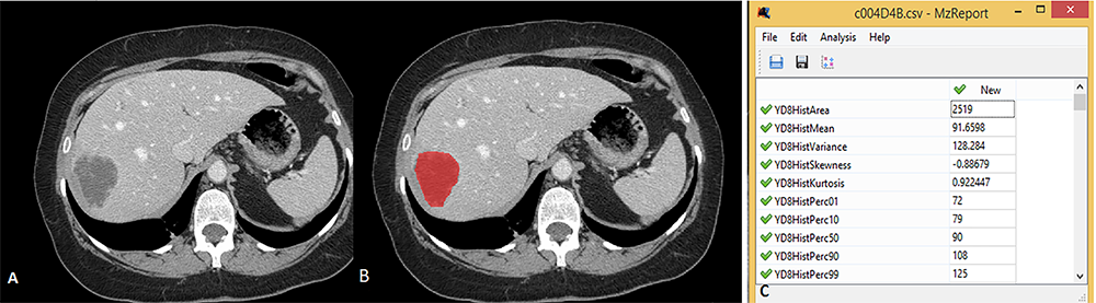
Figure 1. 65-year-old male patient with Colorectal Liver metastasis.. (A) Contrast-enhanced abdominal CT images show a metastatic lesion in the right lobe of the liver. (B) Two-dimensional segmentation of metastasis (red) from the same level using qMazda V.6 software. (C) Quantitative values of the histogram parameters of the area determined in image B
Tables
Table 1. Demographic data and tumor histogram parameters
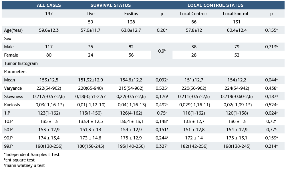
aIndependent Samples t Test bchi-square test cmann whitney u test
Table 2. Investigation of the relationship between tumor histogram parameters and overall survival (Cox regression analysis)
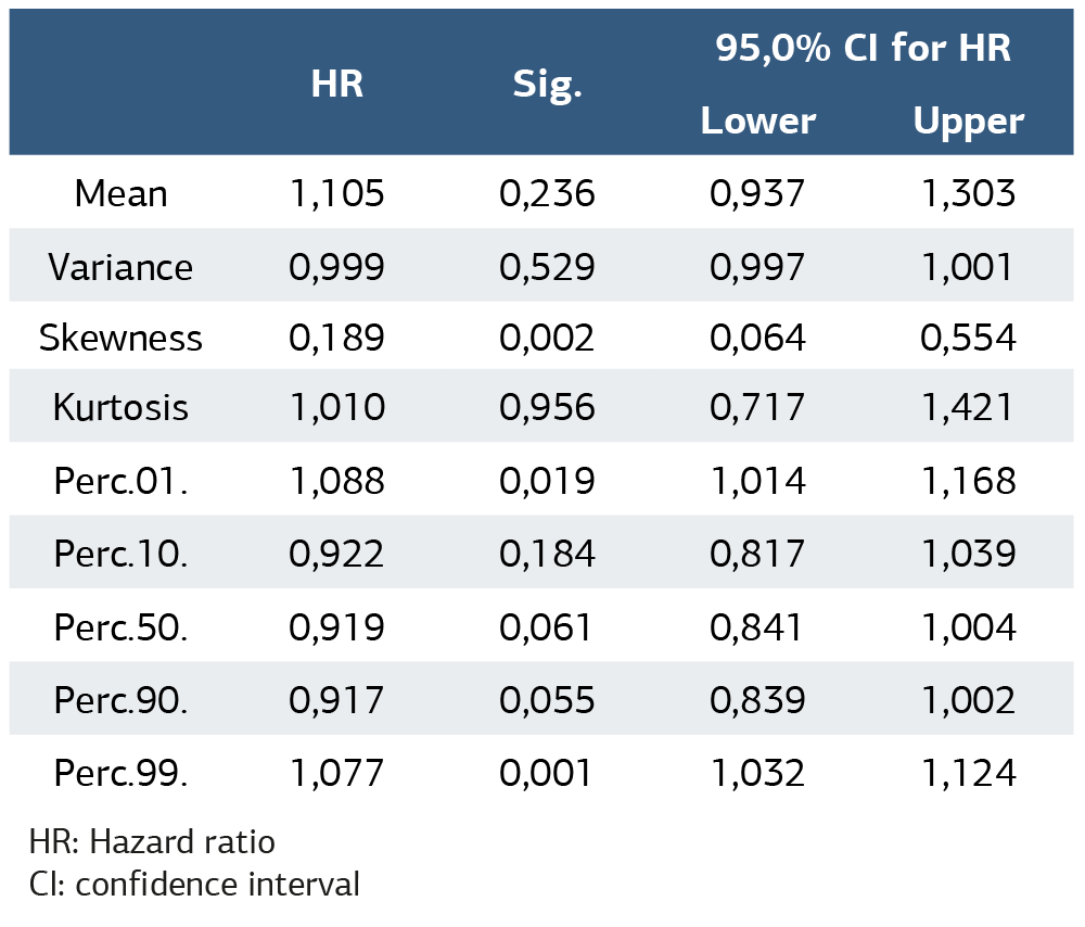
HR: Hazard ratio CI: confidence interval
Table 3. Investigation of the relationship between tumor histogram parameters and local control time (Cox regression analysis)
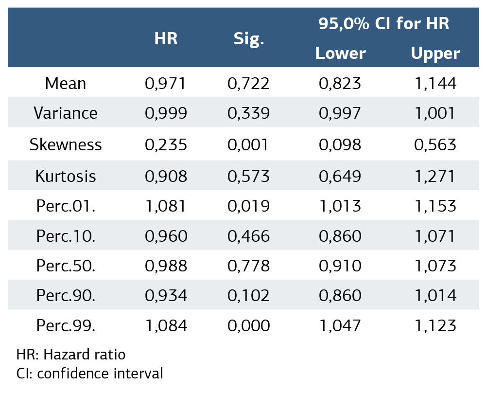
HR: Hazard ratio CI: confidence interval
Additional Information
Publisher’s Note
Bayrakol MP remains neutral with regard to jurisdictional and institutional claims.
Rights and Permissions

This work is licensed under a Creative Commons Attribution-NonCommercial 4.0 International License (CC BY-NC 4.0). To view a copy of the license, visit https://creativecommons.org/licenses/by-nc/4.0/
About This Article
How to Cite This Article
Mehmet Demir, Seda Nida Karaküçük. Evaluation of the relationship of CT histogram analysis with survival and local control time in patients with colorectal liver metastases. Ann Clin Anal Med 2026;17(4):368-372. DOI: 10.4328/ACAM.22808.
- Received:
- July 10, 2025
- Accepted:
- August 11, 2025
- Published Online:
- August 21, 2025
- Printed:
- April 1, 2026