
Abstract
AimOsteoporosis, often resulting from estrogen deficiency, is marked by progressive bone loss, oxidative stress, inflammation, and increased cellular apoptosis. This study evaluated the therapeutic efficacy of Tempol, a superoxide dismutase mimetic, in mitigating osteoporotic changes in an ovariectomized (OVX) rat model.MethodsThirty Female Sprague–Dawley rats were assigned to three groups: Sham, OVX, and OVX treated with Tempol (100 mg/kg/day) for 12 weeks via intraperitoneal injection.ResultsHistological analysis of femoral tissue revealed significant trabecular thinning, increased separation, and reduced osteocyte numbers in the OVX group. These structural impairments were accompanied by decreased serum levels of bone formation markers—alkaline phosphatase (ALP) and osteocalcin—and elevated bone resorption indicators CTX and TRACP-5b. Temporal treatment reversed these changes, improving trabecular architecture and restoring osteocyte density, while normalizing bone turnover markers. Biochemical assays showed that OVX rats had diminished levels of antioxidant enzymes superoxide dismutase (SOD) and catalase (CAT), indicating heightened oxidative stress. Tempol significantly elevated these enzymes, suggesting enhanced antioxidant defense. Furthermore, Tempol suppressed inflammatory cytokines (TNF-α, IL-6, IL-1β) and pro-apoptotic proteins (Bax, caspase-3) and upregulated the anti-apoptotic marker Bcl-2.ConclusionOverall, Tempol demonstrated a protective role against osteoporosis by improving bone structure, enhancing antioxidant capacity, and modulating inflammatory and apoptotic pathways. These findings support Tempol as a promising candidate for the prevention and treatment of postmenopausal osteoporosis.
Keywords
Introduction
Osteoporosis is a systemic metabolic disorder associated with aging and chronic inflammation, characterized by an imbalance in bone remodeling.1 The most prevalent form, postmenopausal osteoporosis, is driven by estrogen deficiency and leads to rapid bone loss following menopause. A major clinical consequence of this bone loss is fracture, a risk for nearly 50% of women aged 50 and older.2,3 Current pharmacological treatments, including anti-resorptive and anabolic agents, are often limited by suboptimal efficacy in certain patients and significant long-term adverse effects.4,5 These challenges highlight the urgent need for novel therapeutic strategies that offer enhanced effectiveness with reduced toxicity.
Research indicates that estrogen deficiency and aging elevate oxidative stress, a key factor in the development of postmenopausal osteoporosis.6,7 This occurs as reactive oxygen species (ROS) trigger apoptosis in bone-forming osteocytes and osteoblasts, thereby suppressing bone formation and encouraging osteoclast activity.8 The resulting apoptotic osteocytes disrupt the balance between bone formation and resorption, impairing bone remodeling and leading to bone loss.9 Furthermore, these dying osteocytes actively signal for osteoclasts to gather at sites of bone resorption, thereby accelerating bone resorption.10
Recent scientific investigations have increasingly focused on synthetic molecules like Tempol for their potent biological properties.9,10 Tempol, a piperidine nitroxide commonly employed as a contrast agent in magnetic resonance spectroscopy, is a versatile synthetic molecule.11 It exists in three readily interconvertible forms: nitroxide, hydroxylamine, and oxoammonium cation, which underpins its reactivity.11 Recognized as a water-soluble, non-toxic cyclic nitroxide, Tempol exhibits powerful antioxidant effects.12 Its unique value lies in its ability to cross cell membranes freely, acting not only as a free radical scavenger but also demonstrating anti-apoptotic and anti-inflammatory activities, thereby enhancing its antioxidant efficacy beyond that of many other compounds.13 Furthermore, Tempol mimics the actions of key antioxidant enzymes, superoxide dismutase (SOD) and catalase, showing potential to prevent organ toxicity by restoring mitochondrial oxidative balance.14 Based on this profile, we posit that Tempol is uniquely positioned to counteract the oxidative stress that drives bone loss.
In the present study, we hypothesized that Tempol would exert anti-osteoporotic effects by scavenging ROS and protecting bone-forming cells, thereby restoring the balance of bone remodeling. To examine this, ovariectomized rats with established bone loss were treated with Tempol for 12 weeks, and its efficacy was evaluated using histological and biomarker analyses.
Materials and Methods
Surgery and Experimental StudySprague–Dawley female rats weighing 200–300 g were housed under 12 h light/dark cycles at 23°C and 60 ± 5% humidity. These animals were placed in a standard rearing environment and adaptively reared for 1 week. Subsequently, rats underwent either bilateral ovariectomy (OVX) or sham surgery, as previously described.15 After three months of standard feeding, a subset of the operated rats was euthanized, and their femurs were extracted to confirm the successful establishment of the osteoporosis model. The remaining animals were randomly assigned to three experimental groups (n = 10 per group): Sham, OVX, and OVX + Tempol. Rats in the OVX + Tempol group received daily intraperitoneal injections of 0.5 mL Tempol (100 mg/kg/day) dissolved in normal saline. TEMPOL powder (4-Hydroxy-TEMPO) was obtained from Sigma-Aldrich (St. Louis, MO, USA), and the saline was purchased from El-Nasr Pharmaceutical Chemicals Company (Egypt). The dosage and route of administration were based on a previous study.16 After 12 weeks of treatment, all rats were sacrificed. Blood and bilateral femurs were immediately harvested for further analysis.Histological ExaminationDistal femur samples were collected and fixed in 4% paraformaldehyde (PFA) in 0.1 M phosphate buffer (pH 7.4) for 48 hours at 4°C. Following fixation, the specimens were rinsed in phosphate-buffered saline (PBS) to remove excess PFA. Decalcification was performed using 15% ethylenediaminetetraacetic acid (EDTA) (pH 7.4), with the solution changed every 2-3 days, until complete decalcification was confirmed by a needle-puncture test. The decalcified bone segments were then dehydrated through a graded ethanol series, cleared in xylene, and embedded in paraffin wax. Using a rotary microtome, serial coronal sections of 4 µm thickness were collected. Sections were stained with Hematoxylin and Eosin (H&E) for microscopic analysis.Measurements of Femoral Oxidative Stress MarkersDistal femoral tissue samples (100 mg) were homogenized using dry ice. The homogenate was centrifuged (1000 g, 10 min, 4°C), and the supernatant was collected to measure superoxide dismutase (SOD) and catalase (CAT) levels using commercial kits (Cat# MBS036924, ELK5986) according to the manufacturer’s instructions.Serum Markers of Bone Formation and ResorptionFollowing sacrifice, blood samples were collected from the abdominal aorta. The samples were centrifuged at 3000 rpm for 15 minutes at 4°C to separate the serum. Serum levels of alkaline phosphatase (ALP), osteocalcin (OC), tartrate-resistant acid phosphatase 5b (TRACP-5b), and C-terminal telopeptide of type I collagen (CTX) were measured using ELISA kits (Cat# MBS165203, EEL123, MBS263384, EEL227), in accordance with the manufacturers’ instructions.ELISA Level of Femoral Inflammatory and Apoptotic MarkersLevels of inflammatory markers (TNF-α, IL-6, IL-1β; Cat# KRC3011, ERA31RB, BMS630) and apoptotic markers (caspase-3, Bax, Bcl-2; Cat# MBS261814, MBS2512405, ERBCL2L2) in femoral homogenates were quantified using commercial ELISA kits, following the manufacturers’ instructions.Ethical ApprovalThis study was approved by the Ethics Committee of Umm Al-Qura University, Makka, Saudi Arabia (Date: 29.01.2025, Decision No: HAPO-02-K-012-2025-09-2925).Statistical AnalysisAll statistical evaluations were performed using GraphPad Prism version 8. Results are presented as the mean accompanied by the standard deviation (SD). Differences among groups were analyzed using one-way analysis of variance (ANOVA), with Tukey’s post hoc test applied for multiple comparisons. Statistical significance was defined as a P value below 0.05.Reporting GuidelinesThis study was reported in accordance with the ARRIVE guidelines.
Results
Protective Effect of Tempol on Bone Mass in an Osteoporotic Rat ModelThe OVX rat group exhibited compromised bone mass at the distal end of the femur, as evidenced by H&E staining, which revealed separations in the bone trabeculae and a marked reduction in trabecular density (Figure 1C, D). Additionally, there was a significant decrease in the number of osteocytes. This was further supported by a statistically significant (P < .05) reduction in serum levels of bone formation markers ALP and osteocalcin OC, alongside a notable elevation in bone resorption markers CTX and TRACP-5b (Figure 2A–D), compared to the sham group.
Encouragingly, administration of tempol in osteoporotic rat models significantly improved the histological architecture of femoral trabeculae, increasing their number and preserving osteocyte count (Figure 1E, F). This was accompanied by elevated ALP and OC levels and a marked reduction in serum bone resorption markers.
Taken together, these findings demonstrate that tempol exerts a protective effect against osteoporotic changes induced by ovariectomy in rats.
Protective Effect of Tempol on Osteoporosis-Induced Oxidative InsultTo investigate the relationship between the antioxidant properties of tempol and its anti-osteoporotic effects, we measured the levels of superoxide dismutase (SOD) and catalase (CAT) in femur homogenates. Our findings revealed that the OVX group exhibited a significant reduction in SOD and CAT levels (Figure 2E, F) compared with the sham group. Conversely, tempol administration markedly increased the levels of these antioxidant markers.
These results support the role of tempol as a superoxide dismutase mimetic, highlighting its ability to counteract oxidative stress associated with osteoporotic changes.Ameliorative Impact of Tempol on Osteoporosis-Related Inflammation and ApoptosisTo further elucidate the mechanisms underlying osteoporotic changes in rat models, our study examined inflammatory and apoptotic markers in bone tissue. The osteoporotic rat model showed a marked elevation of the inflammatory cytokines TNF-α, IL-6, and IL-1β in bone homogenates compared with the sham group (Figure 3A–C). Additionally, there was an increase in the protein levels of apoptotic markers caspase-3 and Bax, along with a decrease in the anti-apoptotic marker Bcl-2, as determined by ELISA analysis (Figure 3D–F).
Conversely, intraperitoneal administration of tempol at a dose of 100 mg/kg/day for three months following the induction of osteoporosis significantly (P < .05) reversed these changes. Temporal treatment reduced the levels of inflammatory and apoptotic markers while increasing the expression of anti-apoptotic proteins. These findings confirm the anti-inflammatory and anti-apoptotic effects of tempol in experimental models of osteoporosis.
Discussion
Osteoporosis, a disease characterized by reduced bone density and deterioration of bone microarchitecture, leads to fragile bones and an increased risk of fractures. Primary pharmacological interventions like hormone replacement therapy and alendronate focus on osteoblasts and osteoclasts but are frequently limited in their long-term application due to adverse effects.17 Consequently, there is a demand for therapeutic agents that can directly activate osteocytes.
The ovariectomized (OVX) rat is a well-established model for studying postmenopausal estrogen deficiency.18 To investigate the mechanism of tempol’s effect on osteocytes in postmenopausal osteoporosis, we used OVX rats. Our initial assessment of bone turnover parameters revealed that tempol supplementation positively affected trabecular bone structure and osteocyte density. Biochemical results corroborated these findings, showing that tempol decreased serum levels of the bone resorption markers TRACP-5b and CTX, while markedly increasing the bone formation markers ALP and OC. This indicates that tempol has a dual therapeutic effect, simultaneously stimulating bone formation and suppressing bone resorption. Tempol administration protected bone cells from damage. This aligns with a previous study on tempol’s protective role against Methotrexate (MTX)-induced osteotoxicity, further supporting its potential in mitigating bone damage.19
Estrogen deficiency plays a pivotal role in the pathogenesis of osteoporosis by elevating oxidative stress and suppressing antioxidant enzyme activity.20 Our findings corroborate this mechanism, as the ovariectomized (OVX) group of rats exhibited a significant reduction in bone homogenate levels of the antioxidant enzymes superoxide dismutase (SOD) and catalase (CAT) compared to the Sham group. This decline reflects pronounced oxidative damage to bone tissue, particularly affecting osteocytes, long-lived cells embedded within the bone matrix that are highly susceptible to hypoxia, reactive oxygen species accumulation, and nutrient scarcity.21
Conversely, administration of Tempol markedly improved oxidative stress markers in the OVX group by elevating SOD and CAT levels. These results align with previous findings by Selimli et al., who demonstrated Tempol’s protective effects against methotrexate-induced bone cell injury.19 Their study showed that Tempol mitigated oxidative degeneration by increasing total antioxidant concentration, glutathione peroxidase (GPx), and SOD, while reducing overall oxidative burden.
Tempol, a synthetic nitroxide compound with potent antioxidant properties, has shown promise in counteracting osteoporosis by attenuating oxidative stress, a key driver of bone loss. Acting as a superoxide dismutase mimetic, Tempol efficiently scavenges ROS, thereby minimizing cellular damage in bone tissue. In osteoporotic conditions, such as estrogen deficiency or chemotherapy-induced bone toxicity, elevated ROS levels impair osteoblast function, promote osteocyte apoptosis, and enhance osteoclast-mediated bone resorption. Tempol counters these effects by restoring antioxidant enzyme activity (e.g., SOD and GPx) and modulating apoptotic pathways, downregulating pro-apoptotic markers such as Bax and caspase-3, while upregulating the anti-apoptotic marker Bcl-2.
Oxidative stress and chronic inflammation are key drivers in the development and progression of osteoporosis.22 Oxidative stress directly damages osteoblasts, impairing bone formation and increasing fracture risk. In ovariectomized (OVX) rats, this is evidenced by elevated levels of pro-inflammatory cytokines such as TNF-α and IL-6, along with increased malondialdehyde (MDA), a marker of oxidative lipid damage. These findings indicate an inflammatory state that exacerbates bone loss and heightens susceptibility to inflammatory conditions. Consistent with our study, the OVX group exhibited a marked elevation in inflammatory mediators.
Conversely, intraperitoneal administration of Tempol significantly alleviated the induced inflammatory condition. This anti-inflammatory effect is mediated through multiple mechanisms. Tempol functions as a superoxide dismutase (SOD) mimetic, emulating the activity of the endogenous antioxidant enzyme SOD. By scavenging superoxide radicals and other reactive oxygen species (ROS), Tempol reduces oxidative stress, a key initiator of inflammatory signaling pathways. Furthermore, Tempol inhibits the nuclear factor kappa B (NF-κB) signaling pathway, a central regulator of inflammation. By blocking NF-κB activation, Tempol suppresses the transcription of pro-inflammatory genes.
Limitations
This study presents promising evidence of Tempol’s protective effects against osteoporosis in ovariectomized (OVX) rat models; however, several limitations warrant consideration. The use of a single animal model and a fixed dose limits generalizability and prevents exploration of dose-response relationships. The absence of comparisons with standard osteoporosis treatments and the lack of long-term follow-up restrict understanding of Tempol’s relative efficacy and sustained impact. While histological and biochemical markers were assessed, functional bone strength and validation of molecular pathways were not included. These limitations highlight the need for broader, more comprehensive studies to confirm Tempol’s therapeutic potential in clinical settings.
Conclusion
Tempol demonstrates significant protective effects against osteoporosis in ovariectomized (OVX) rat models. Histological analysis revealed that Tempol preserved trabecular bone structure and osteocyte count, while improving bone formation markers (ALP, osteocalcin) and reducing bone resorption indicators (CTX, TRACP-5b). Additionally, Tempol enhanced antioxidant defenses by increasing SOD and CAT levels in bone tissue, thereby counteracting oxidative stress associated with estrogen deficiency. It also suppressed inflammatory cytokines (TNF-α, IL-6, IL-1β) and apoptotic markers (Bax, caspase-3), while upregulating anti-apoptotic Bcl-2. These findings confirm Tempol’s multifaceted role in mitigating bone loss through antioxidant, anti-inflammatory, and anti-apoptotic mechanisms.
Declarations
Animal and Human Rights Statement
All procedures performed in this study were in accordance with the ethical standards of the institutional and/or national research committee and with the 1964 Declaration of Helsinki and its later amendments, or comparable ethical standards.
Informed Consent
Not applicable.
Data Availability
The datasets used and/or analyzed in the current study are not publicly available due to patient privacy concerns but are available from the corresponding author upon reasonable request.
Conflict of Interest
The authors declare no conflict of interest.
Funding
None.
Author Contributions (CRediT Taxonomy)
Conceptualization: A.J.A.A.
Methodology: A.J.A.A.
Investigation: A.J.A.A.
Data Curation: A.J.A.A.
Formal Analysis: A.J.A.A.
Writing – Original Draft: A.J.A.A.
Writing – Review & Editing: A.J.A.A.
Supervision: A.J.A.A.
AI Usage Disclosure
The authors declare that no AI-assisted technologies were used.
Abbreviations
ALP: Alkaline phosphatase
ANOVA: Analysis of variance
CAT: Catalase
CTX: C-terminal telopeptide of type I collagen
EDTA: Ethylenediaminetetraacetic acid
ELISA: Enzyme-linked immunosorbent assay
IL: Interleukin
OC: Osteocalcin
OVX: Ovariectomized
PBS: Phosphate-buffered saline
PFA: Paraformaldehyde
ROS: Reactive oxygen species
SD: Standard deviation
SOD: Superoxide dismutase
TNF: Tumor necrosis factor
References
- Yu B, Wang CY. Osteoporosis and periodontal diseases - An update on their association and mechanistic links. Periodontol 2000. 2022;89(1):99-113. doi:10.1111/prd.12422
- Eastell R, O’Neill TW, Hofbauer LC, et al. Postmenopausal osteoporosis. Nat Rev Dis Primers. 2016;2:16069. doi:10.1038/nrdp.2016.69
- Gosset A, Pouillès JM, Trémollieres F. Menopausal hormone therapy for the management of osteoporosis. Best Pract Res Clin Endocrinol Metab. 2021;35(6):101551. doi:10.1016/j.beem.2021.101551
- Reginster JY, Neuprez A, Dardenne N, Beaudart C, Emonts P, Bruyere O. Efficacy and safety of currently marketed anti-osteoporosis medications. Best Pract Res Clin Endocrinol Metab. 2014;28(6):809-34. doi:10.1016/j.beem.2014.09.003
- Aibar-Almazán A, Voltes-Martínez A, Castellote-Caballero Y, et al. Current status of the diagnosis and management of osteoporosis. Int J Mol Sci. 2022;23(16):9465. doi:10.3390/ijms23169465
- Domazetovic V, Marcucci G, Iantomasi T, Brandi ML, Vincenzini MT. Oxidative stress in bone remodeling: Role of antioxidants. Clin Cases Miner Bone Metab. 2017;14(2):209-16. doi:10.11138/ccmbm/2017.14.1.209
- Manolagas SC. From estrogen-centric to aging and oxidative stress: a revised perspective of the pathogenesis of osteoporosis. Endocr Rev. 2010;31(3):266-300. doi:10.1210/er.2009-0024
- Bonewald LF. The amazing osteocyte. J Bone Miner Res. 2011;26(2):229-38. doi:10.1002/jbmr.320
- Al-Dujaili SA, Lau E, Al-Dujaili H, Tsang K, Guenther A, You L. Apoptotic osteocytes regulate osteoclast precursor recruitment and differentiation in vitro. J Cell Biochem. 2011;112(9):2412-23.
- Kennedy OD, Herman BC, Laudier DM, Majeska RJ, Sun HB, Schaffler MB. Activation of resorption in fatigue-loaded bone involves both apoptosis and active pro-osteoclastogenic signaling by distinct osteocyte populations. Bone. 2012;50(5):1115-22. doi:10.1016/j.bone.2012.01.025
- Wilcox CS. Effects of tempol and redox-cycling nitroxides in models of oxidative stress. Pharmacol Ther. 2010;126(2):119-45. doi:10.1016/j.pharmthera.2010.01.003
- Wang M, Li K, Zou Z, et al. Piperidine nitroxide Tempol enhances cisplatin-induced apoptosis in ovarian cancer cells. Oncol Lett. 2018;16(4):4847-54. doi:10.3892/ol.2018.9289
- Afjal MA, Abdi SH, Sharma S, et al. Anti-inflammatory role of tempol (4-hydroxy-2,2,6,6-tetramethylpiperidin-1-oxyl) in nephroprotection. Hum Exp Toxicol. 2019;38(6):713-23. doi:10.1177/0960327119836203
- Ahmed LA, Shehata NI, Abdelkader NF, Khattab MM. Tempol, a superoxide dismutase mimetic agent, ameliorates cisplatin-induced nephrotoxicity through alleviation of mitochondrial dysfunction in mice. PLoS One. 2014;9(10):e108889. doi:10.1371/journal.pone.0108889
- Tao ZS, Zhou WS, Qiang Z, et al. Intermittent administration of human parathyroid hormone (1–34) increases fixation of strontium-doped hydroxyapatite coating titanium implants via electrochemical deposition in ovariectomized rat femur. J Biomater Appl. 2016;30(7):952-60. doi:10.1177/0885328215610898
- Ewees MG, Messiha BAS, Abdel-Bakky MS, Bayoumi AMA, Abo-Saif AA. Tempol, a superoxide dismutase mimetic agent, reduces cisplatin-induced nephrotoxicity in rats. Drug Chem Toxicol. 2019;42(6):657-64. doi:10.1080/01480545.2018.1485688
- Chen LR, Ko NY, Chen KH. Medical treatment for osteoporosis: From molecular to clinical opinions. Int J Mol Sci. 2019;20(9):2213. doi:10.3390/ijms20092213
- Jee WS, Yao W. Overview: animal models of osteopenia and osteoporosis. J Musculoskelet Neuronal Interact. 2001;1(3):193-207.
- Selimli F, Taş Reyhanioğlu M, Haskan AC, Altun MS, Mete S, Kaplan HM. Tempol mitigates methotrexate-induced osteotoxicity via oxidative stress modulation and MAPK pathway inhibition. Drug Des Devel Ther. 2025;19:1441-9. doi:10.2147/DDDT.S510206
- Prideaux M, Findlay DM, Atkins GJ. Osteocytes: The master cells in bone remodelling. Curr Opin Pharmacol. 2016;28:24-30. doi:10.1016/j.coph.2016.02.003
- Liu T, Yu H, Wang S, Li H, Du X, He X. Chondroitin sulfate alleviates osteoporosis caused by calcium deficiency by regulating lipid metabolism. Nutr Metab (Lond). 2023;20(1):6. doi:10.1186/s12986-023-00726-3
- Liu B, Hu Y, Tian D, Dong J, Li BF. Assessing the effects of tempol on renal fibrosis, inflammation, and oxidative stress in a high-salt diet combined with 5/6 nephrectomy rat model: Utilizing oxidized albumin as a biomarker. BMC Nephrol. 2024;25(1):64. doi:10.1186/s12882-024-03495-0
Figures
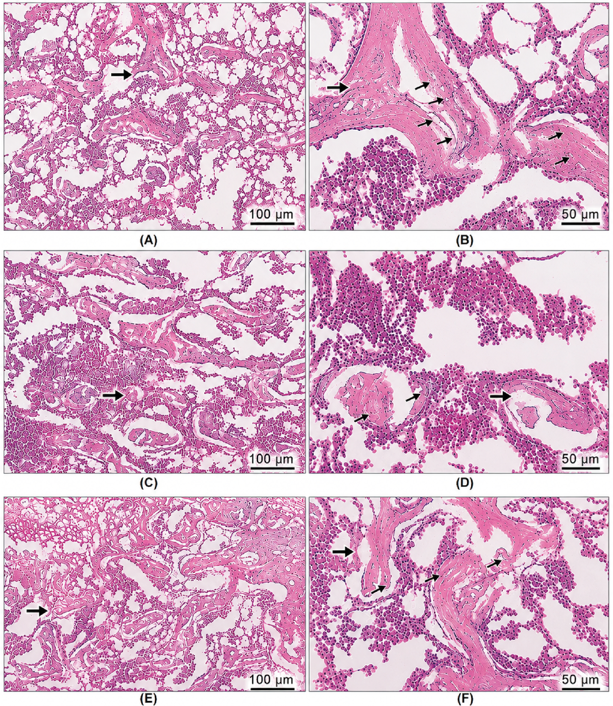
Figure 1. Microscopic pictures of HE stained sections of femur showing marked reduction in trabecular number & thickness with increased distance between trabeculae (thick arrows) (trabecular separation) in OVX group (C, D) when compared to control group (A, B). Numbers of osteocytes (thin arrows) located in lacunae in these trabeculae markedly decreased in OVX group when compared to control normal group. These criteria are reversed in treated groups OVX+Tempol (E, F). Magnifications X100 bar100 and X200 bar50.
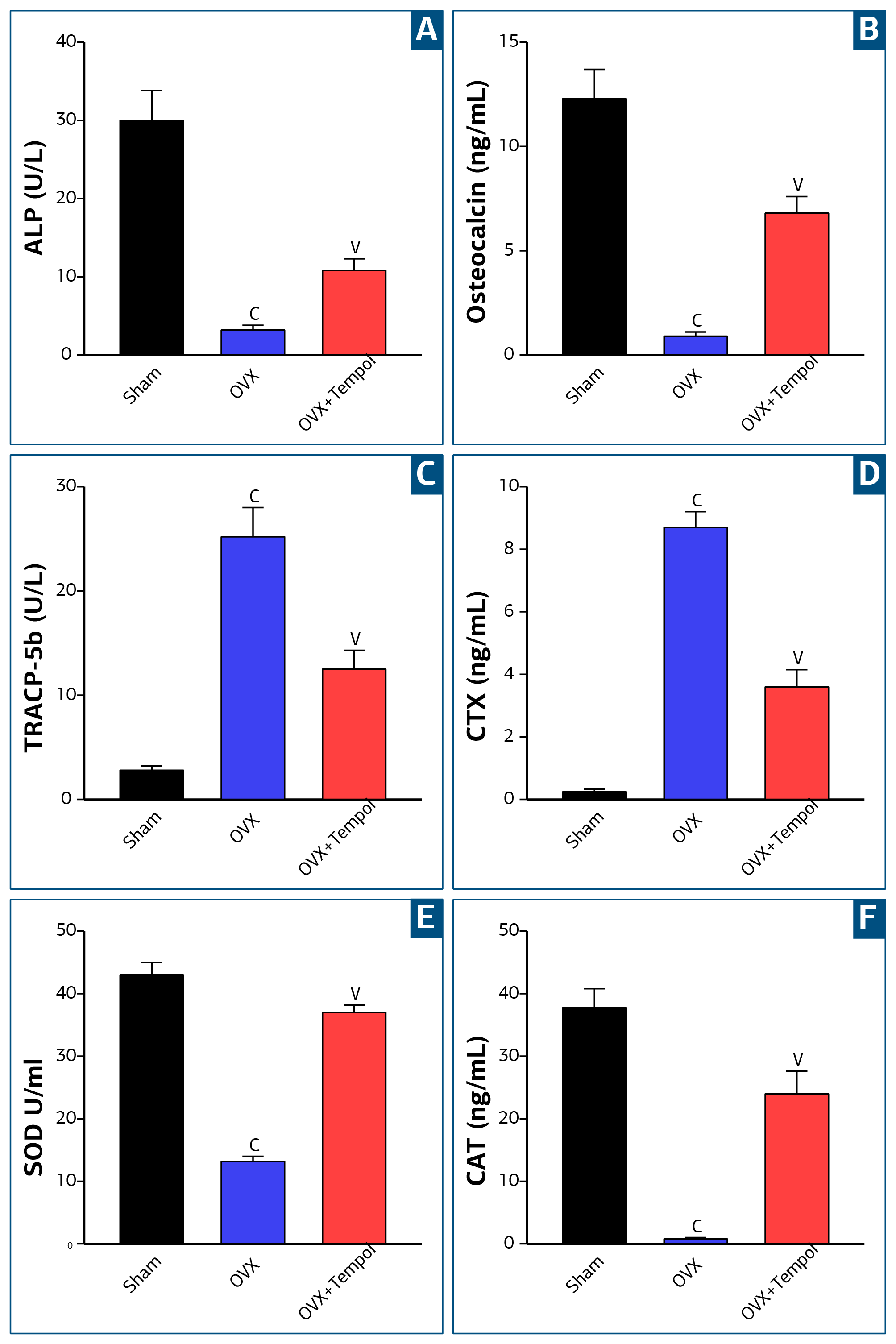
Figure 2. Impact of tempol on osteoporotic markers (A) ALP, (B) osteocalcin (C) TRACP-5b (D) CTX, and oxidative markers (E) SOD, (F) CAT in different female rat groups. cp< 0.05 indicates significance to sham group, vp< 0.05 significant of OVX group.
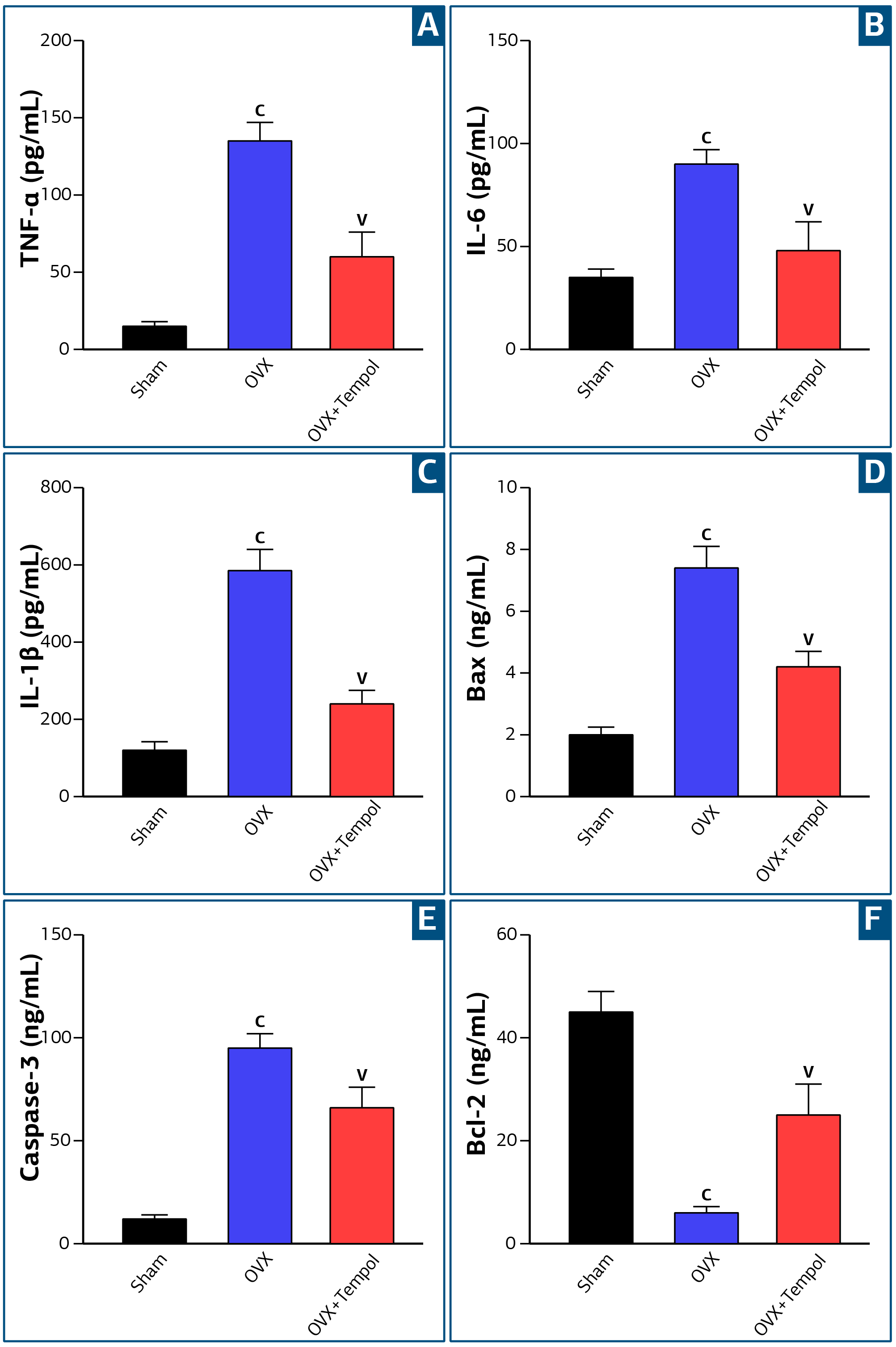
Figure 3. ELISA level of (A) TNF-α, (B) IL-6, (C) IL-1β, (D) Bax, (E) Caspase-3, (F) Bcl-2 in tissue homogenates of all groups. cp< 0.05 vs sham rats, vp< 0.05 vs OVX rats.
Additional Information
Publisher’s Note
Bayrakol MP remains neutral with regard to jurisdictional and institutional claims.
Rights and Permissions

This work is licensed under a Creative Commons Attribution-NonCommercial 4.0 International License (CC BY-NC 4.0). To view a copy of the license, visit https://creativecommons.org/licenses/by-nc/4.0/
About This Article
How to Cite This Article
Alaa Jameel A. Albarakati. Antiosteoporosis effect of Tempol against bone loss in ovariectomized rats, halting oxidative stress, inflammation, and apoptosis. Ann Clin Anal Med. Articles in Press. doi:10.4328/ACAM.22954
Publication History
- Received:
- 20.10.2025
- Accepted:
- 02.12.2025
- Published Online:
- 11.03.2026
- Printed:
- 01.10.2026