
Abstract
AimThe anterolateral thigh (ALT) flap has become a widely used option for soft tissue reconstruction in upper extremity injuries due to its versatility, reliability, and minimal donor site morbidity. This study evaluates the functional and aesthetic outcomes of ALT flap applications in complex hand trauma cases.MethodsA retrospective analysis was conducted on 30 patients who underwent ALT flap reconstruction for upper extremity defects. Patients’ demographics, injury mechanisms, surgical procedures, defect and flap dimensions, postoperative complications, and functional outcomes were assessed. Functional recovery was evaluated using MAYO and Q-DASH scores.ResultsThe mean defect size was 98.67 ± 52.34 cm² (min:20, max:260), and the average flap size was 134.12 ± 71.42 cm² (min:36, max:290). 60% (n = 18) of patients had radial artery anastomosis, while 23.3% (n = 7) had ulnar artery end-to-end anastomosis. No total flap loss was observed, though 46.7% (n = 14) of patients experienced minor complications, such as partial flap necrosis or donor site hematoma. Functional assessment revealed an average MAYO score of 71.82 ± 10.45 and a Q-DASH score of 30.47 ± 7.59, indicating satisfactory functional recovery.ConclusionThe ALT flap provides excellent functional and aesthetic outcomes in upper extremity reconstruction. Compared to other flap options, it offers greater tissue volume, lower donor site morbidity, and enhanced cosmetic outcomes. Despite its technical complexity, it remains a gold standard in complex hand trauma management, with low complication rates and predictable functional recovery.
Keywords
Introduction
Skin flaps are commonly used to cover vital structures such as vessels, nerves, and bones, while fascia flaps are preferred for protecting tendinous tissues. In cases where a flap does not fully close the soft tissue defect or when there is a risk of contamination due to cavity formation in the bone, muscle flaps are employed to eliminate dead space and reduce infection risk. Additionally, in injuries causing vascular defects, the flap pedicle can be utilized for revascularization of the ischemic extremity. Various reconstructive techniques have been described to restore functional structures following extensive upper extremity soft tissue injuries.1-2
The ALT flap presents an excellent reconstructive option for upper extremity injuries, offering advantages such as versatility, low donor site morbidity, and optimal size for large-scale tissue defects. It features a long pedicle with a suitable vessel diameter, allowing for better adaptation to the contours of the hand and upper extremity. Additionally, by incorporating the lateral femoral cutaneous nerve, the flap can be designed as a sensory reconstruction. The procedure can also be performed efficiently under regional block anesthesia, enabling simultaneous work by two surgical teams and reducing operative time. Achieving a flap capable of covering large soft tissue defects is technically challenging. However, as a multi-component flap, the ALT flap effectively meets all the essential requirements for reconstruction, making it a preferred choice in complex upper extremity injuries.3
In this study, we aimed to evaluate the clinical outcomes of ALT flap reconstruction in upper extremity soft tissue defects and discuss its functional and aesthetic results.
Materials and Methods
Study DesignThis study included the patients who had soft tissue defects in addition to upper extremity trauma in AA hospital between the dates of 2022-2024. Patients’ demographic data, surgical procedure, postoperative complications, and functional outcomes of ALT flap surgery of the upper extremity were evaluated and discussed.Surgical TechniqueAll flaps were prepared via the same technique. The operation was started by two surgical teams, the first surgeon prepared the recipient site and the second surgeon prepared the donor site. Recipient site debridement, bone fixation, repair of tendon, muscle, and nerve injuries, and then preparation of recipient vessels were performed. The second surgeon performed the elevation of the flap at the same time. Possible skin perforators were detected by using hand doppler primarily on the line and inferior within a 3 cm diameter circle area from the middle 1/2 point of the line on the line between the rectus femoris and vastus lateralis and it was drawn between the spina iliaca anterior superior and the lateral of the patella of the patient lying in the supine position. The flap size was decided according to the defect area. The drawing was made so that 1/3 of the flap’s length is superior to the drawn line, while 2/3 is inferior, and its width is aligned to the midpoint between the two landmarks. Dissection of the descending branch of the LCFA was completed by considering the flap adaptation in the recipient site and the thickness of the recipient vessels at the planned anastomosis level and the required pedicle length. The artery and 2 veins above the pedicle were separated by at least 3 cm of intrapedic dissection, and the flap was separated by ligating the arteries and veins separately. Then, after the coarse adaptation of the flap and the appropriate position of the pedicle were achieved, 1 artery, and 2 vein anastomoses were performed. If 2 suitable veins were not found in the recipient site or if the pedicle veins were converged to a single vein until they reached the appropriate thickness, a single vein was performed. Except for these cases, 2 vein anastomosis was preferred in every possible case. After anastomoses, hemorrhage control and skin suturing were completed and the operation was completed by applying an appropriate dressing and splint to stabilize the extremity.
Post-operative follow-up
Flap follow-up was performed by closely monitoring variables such as the evaluation of flap skin color, turgor, surface temperature, and presence of bleeding. Patients for whom flaps were applied were followed up frequently in the postoperative period. Early intervention was performed in cases suggestive of venous or arterial insufficiency, vessel thrombosis, and hematoma.Ethical ApprovalThis study was approved by the Clinical Research Ethics Committee of Harran University (Date: 07.02.2022, Decision No:2022/03/15).Statistical AnalysisStatistical analyzes were performed with the Statistics for Social Sciences (SPSS) v18 package program. As descriptive statistics, arithmetic mean ± standard deviation and median (minimum, maximum) were used to summarize numerical data, and numbers and percentages were used to summarize categorical data. The relationship between categorical data was analyzed with the Chi-square (χ2) test. P values below 0.05 were considered statistically significant.Reporting GuidelinesThis study was reported in accordance with the STROBE guideline.
Results
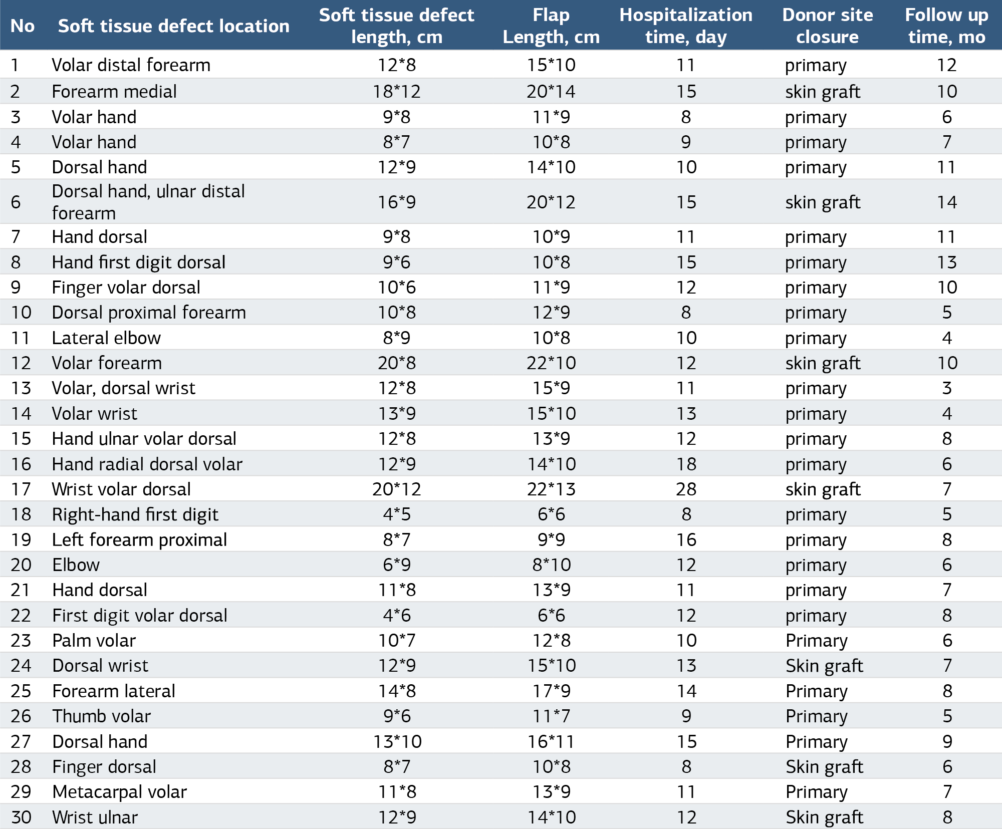
Patients’ demographic data, injury mechanism, and surgical procedure are shown in Table 1. A total of 30 patients were included in the study. 93.3% (n = 28) of the patients were male, and 6.7% (n = 2) were female. The mean age of the patients was 31.2 ± 12.3 years (min:10, max:55). The injury occurred in the right upper extremity in 66.7% of the patients and the left upper extremity in 33.3%. The flap was harvested from the right thigh in 53.3% of the patients and from the left thigh in 46.7%. Gunshot injuries were the most common mechanism, accounting for 36.7% (n = 11) of the cases (Figure 1), followed by occupational injuries in 26.7% (n = 8), traffic accidents in 16.7% (n = 5), electrical burns in 13.3% (n = 4) (Figure 2), and industrial injuries in 6.7% (n = 2). A musculocutaneous flap was applied in 22 patients, while a septocutaneous flap was used in 8 patients (Table 1). Patient’s soft tissue defect location, defect length, flap length, hospitalization time, donor site closure, and follow-up time are shown in Table 2. The mean size of the defect was 98.67 ± 52.34 cm² (min:20, max:260). The smallest defect size was 4×5 cm², and the largest defect size was 20×12 cm². The average size of the flaps applied was 134.12 ± 71.42 cm² (min:36, max:290), with the smallest flap size being 6×6 cm² and the largest being 22×13 cm² (Figure 3). The average time to post-traumatic flap surgery was 9.87 ± 4.21 days (min:1, max:21), and the average operation time was 236.72 ± 33.84 minutes (min:170, max:305). The mean postoperative hospitalization time was 12.73 ± 4.41 days (min:8, max:28), and the mean postoperative follow-up time was 8.12 ± 3.24 months (min:3, max:14). Regarding donor site closure, 80% (n = 24) of the donor sites were sutured primarily, whereas 20% (n = 6) required skin grafting. The mean pedicle size was 10.04 ± 1.85 cm (min:6, max:14) (Table 2). Among the patients, 60% (n = 18) underwent radial artery end-to-end anastomosis, while 23.3% (n = 7) had ulnar artery end-to-end anastomosis. Additionally, 13.3% (n = 4) underwent ulnar artery end-to-end anastomosis with a flow-through flap, and 3.3% (n = 1) had brachial artery end-side suturation.
Regarding venous anastomosis, 36.7% (n = 11) of the patients had one deep and one superficial vein anastomosis, while 26.7% (n = 8) had two superficial vein anastomoses. No total flap loss was observed in any patient postoperatively. In all cases, donor site healing was uneventful in patients who underwent either primary repair or skin grafting. No weakness or movement restriction was noted in the lower extremity from which the flap was harvested.
Complications occurred in 46.7% (n = 14) of the patients during follow-up. Finger necrosis developed in three patients, infection in three patients, finger loss in two patients, hematoma at the donor site in one patient, flexion limitation secondary to flap size in one patient, hematoma under the flap in one patient, partial finger necrosis in one patient, partial flap loss in one patient, and partial flap necrosis in one patient. Finger losses were unrelated to the flap itself; they occurred due to revascularization failure in complex hand injuries.
Among the patients who developed complications, nine (64.3%) underwent debridement, four (28.6%) required finger amputation, and one patient underwent debulking. Additional surgeries were required in four patients during follow-up: tenolysis was performed in three patients, and one patient underwent metacarpal arthrodesis after osteosynthesis. The patient requiring osteosynthesis had an ulna fracture that was initially stabilized with an external fixator. In the first session, debridement and vascular nerve repair were performed, while in the second stage, the ulna fracture was stabilized with a plate, and an anterolateral thigh (ALT) flap was applied.
At the end of postoperative physical therapy, the average MAYO score of the patients was 71.82 ± 10.45 (min:50, max:86), while the average Q-DASH score was 30.47 ± 7.59 (min:18, max:45).
Discussion
The primary goal of limb reconstruction is to restore and maintain limb function. For this purpose, achieving an aesthetic appearance, minimizing donor site morbidity, and ensuring stable soft tissue coverage are essential. Functional reconstruction of large and complex soft tissue defects in the extremities remains a challenging task for hand surgeons.4 Historically, the ALT flap has been used more frequently in head and neck surgery, but it has also become a reliable option for upper extremity reconstructions. The ALT flap serves as an alternative to the groin flap, temporoparietal fascial flap, and lateral arm flap. It provides a larger and more voluminous tissue transfer than these alternatives while causing fewer donor site complications. In large-scale case series, perforators have been reported to supply the flap in each case.5-6-7 However, if a perforator cannot be identified during preoperative planning, it should be considered a contraindication for surgery.
If the flap size exceeds 8 cm, skin grafting may be required for donor site closure. In our study, 24 (80%) donor sites were sutured primarily, while 6 (20%) required skin grafting, which aligns with the literature. Additionally, in agreement with previous studies, we observed that functional limitations at the donor site were minimal, and cosmetic outcomes were more acceptable compared to other flap types.8 Patients with smoking history, diabetes, or peripheral vascular disease are at higher risk for flap failure.9-10 One of our patients was diagnosed with diabetes, and three of our patients were smokers in our study. Contrary to the literature, no complications occurred in the four patients mentioned despite these additional factors.
Although the tissue expansion technique can also be utilized for soft tissue reconstruction, it requires a longer preparation period.11 Moreover, it has been reported that functional recovery following upper extremity tissue expansion reconstruction is inferior to other methods.12 Since our patient cohort primarily consisted of acute injury cases, we opted for ALT flap reconstruction.
The ALT flap can be thinned to a thickness of 3–4 mm, making it cosmetically suitable for dorsal hand reconstruction.13-14-15 In our study, we performed thinning in the ALT flaps prepared for the dorsum of the hand and fingers, achieving improved aesthetic outcomes. Furthermore, for cases involving arterial injury with soft tissue loss, the flow-through flap technique is an option to bridge arterial damage.16-17 Dong et al. reported a 100% flap survival rate and no complications in their series of four patients undergoing flow-through ALT flap reconstruction for severe limb injuries. Similarly, in our study, we observed a 100% flap survival rate with no major complications, aligning with Dong et al.’s findings and supporting the reliability of the flow-through technique in complex upper extremity reconstructions.16 A significant drawback of the ALT flap is the meticulous surgical technique required, particularly for perforator dissection. This procedure is technically demanding, requiring microsurgical expertise. Unexpected pedicle lengths may complicate flap elevation or positioning, which can present intraoperative challenges.18-19
Under optimal conditions, flap failure rates should remain below 5%.5 In our study, no total flap loss was observed, and complications were within acceptable limits, including partial flap necrosis and skin necrosis. Furthermore, no significant restriction in range of motion was detected after ALT flap surgery.20-21 All patients underwent postoperative upper extremity rehabilitation, and we observed that their functional outcomes, as measured by MAYO and Q-DASH scores, were within acceptable ranges.
Limitations
The limited number of patients in our study, short follow-up period, retrospective design of the study, and the nonhomogeneous injury patterns can be considered among the limitations of our study.
Conclusion
In conclusion, our findings support the use of ALT flaps as a reliable option for upper extremity soft tissue reconstruction, particularly in cases with large defects, exposed tendons, and combined vascular injuries requiring revascularization. The versatility of this flap makes it suitable for both functional and aesthetic restoration. However, in patients with smaller defects, cases requiring thin skin coverage, or when donor site morbidity is a concern, alternative flaps such as the radial forearm flap or lateral arm flap should be considered. Future studies with larger cohorts and longer follow-up periods will help further refine patient selection criteria.
Abbreviations
ALT: Anterolateral thigh
LCFA: Lateral circumflex femoral artery
MAYO: Mayo wrist score
Q-DASH: Quick Disabilities of the Arm, Shoulder and Hand
SPSS: Statistical Package for the Social Sciences
References
- Yu JL, Crowe CS, Yesantharao P, Kennedy SA, Keys KA. Soft tissue reconstruction for upper extremity necrotizing soft tissue infections. Ann Plast Surg. 2022;89(6):631-636. doi:10.1097/sap.0000000000003237
- Naalla R, Chauhan S, Dave A, Singhal M. Reconstruction of post-traumatic upper extremity soft tissue defects with pedicled flaps: an algorithmic approach to clinical decision making. Chin J Traumatol. 2018;21(6):338-351. doi:10.1016/j.cjtee.2018.04.005
- Duan A, Xiang Z, Fan Y, Duan W, Wei Q, Duan X. Evaluating the clinical efficacy of the anterolateral thigh flap in lower limb reconstruction surgeries: a systematic review and meta-analysis. Am J Transl Res. 2024;16(7):3326-3337. doi:10.62347/hfve6316
- Ono S, Sebastin SJ, Ohi H, Chung KC. Microsurgical flaps in repair and reconstruction of the hand. Hand Clin. 2017;33(3):425-441. doi:10.1016/j.hcl.2017.04.001
- Xu Q, Li W, Zheng X, Lin L. Clinical outcomes of reconstruction on tissue defects with thin anterolateral thigh flap versus commonly used free soft flaps: a meta-analysis. J Stomatol Oral Maxillofac Surg. 2025;1(1):1-10. doi:10.1016/j.jormas.2025.102225
- Berkane Y, Giorgino R, Ng ZY, Dukan R, Lellouch AG. Alternative flap options for upper extremity reconstruction. Hand Clin. 2024;40(2):291-299. doi:10.1016/j.hcl.2023.08.010
- Coquillard CL, Bai J, Ko JH. Thin and thinned anterolateral thigh flaps for upper extremity reconstruction. Hand Clin. 2024;40(2):199-208. doi:10.1016/j.hcl.2023.12.001
- Kim JH, Yoo H, Eun S. Reconstruction of extensive soft tissue defects of lower extremity with the extended anterolateral thigh flap. Int J Low Extrem Wounds. 2022;21(4):601-608. doi:10.1177/1534734620982238
- Hassan B, Fairchild B, Grant MP, Lamaris GA. The role of the fascia-only anterolateral thigh flap in extremity reconstruction: the fascia-only anterolateral thigh flap. Ann Plast Surg. 2024;92(4):412-417. doi:10.1097/sap.0000000000003878
- Liu F, Yan WQ, Ma Q, Liu YB, Yang ZB. Clinical effect of anterolateral thigh flow-through chimeric perforator free flap transplantation in the treatment of upper limb complex tissue defects with main artery injury. Zhonghua Shao Shang Yu Chuang Mian Xiu Fu Za Zhi. 2024;40(2):172-179. doi:10.3760/cma.j.cn501225-20231103-00176
- Qureshi MR. Study of effectiveness in replacing damaged soft tissue of the hand and forearm by abdominal flap. Res J Med Sci. 2024;18(10):482-486. doi:10.36478/makrjms.2024.10.482.486
- Li X, Fan J, Ma J, Tian J. Reconstruction of circumferential upper extremity defect using a bipedicle expanded flank flap. Ann Plast Surg. 2021;86(3):279-286. doi:10.1097/sap.0000000000002694
- Georgescu AV, Corpodean A, Matei IR. A new understanding of “flow-through” concept in flaps surgery and review of the literature. JOREP. 2023;2(4):1-9. doi:10.1016/j.jorep.2023.100218
- Azoury SC, Kovach SJ, Levin LS. Reconstruction options for lower extremity traumatic wounds. J Am Acad Orthop Surg. 2022;30(16):735-746. doi:10.5435/jaaos-d-21-01081
- Lee ZH, Daar DA, Yu JW, et al. Updates in traumatic lower extremity free flap reconstruction. Plast Reconstr Surg. 2023;152(5):913-918. doi:10.1097/prs.0000000000010404
- Dong K, Zhou Y, Xu YQ, Xu XY, He XQ. Anterolateral thigh flow-through flap: a versatile method for reconstruction of complex extremity defects. Microsurgery. 2023;43(7):685-693. doi:10.1002/micr.31095
- Wang L, Liu H, Ma T, Wu X, Zhang L. Reconstruction of soft tissue defects in the hand with a free anterolateral thigh deep fascia flap. Orthop Surg. 2021;13(3):758-767. doi:10.1111/os.12948
- Zhang N, Liu F, Huang C, et al. Application of flow-through anterolateral thigh perforator flaps in replantation of complex severed limb. Transplant Proc. 2024;56(8):1856-1860. doi:10.1016/j.transproceed.2024.08.013
- Gu Y, Sun Y, Xu K, et al. Chimeric free flaps based on end-to-side anastomosis for complex lower extremity reconstruction. Ann Plast Surg. 2025;94(2):185-191. doi:10.1097/sap.0000000000004180
- Hallock GG. Musculoseptocutaneous perforator of anterolateral thigh flap: a clinical study. Plast Reconstr Surg. 2022;149(1):148-149. doi:10.1097/prs.0000000000008642
- El-Shennawy H, Elzawawy EM, Atta-Allah ES, Omar S. Perforators of the anteromedial thigh skin and possible design of the anteromedial thigh flap: a cadaveric study. Ann Plast Surg. 2022;89(3):312-325. doi:10.1097/sap.0000000000003244
Tables
Table 1. Demographic Characteristics, Injury Mechanism, and Surgical Details of Patients Undergoing ALT Flap Reconstruction
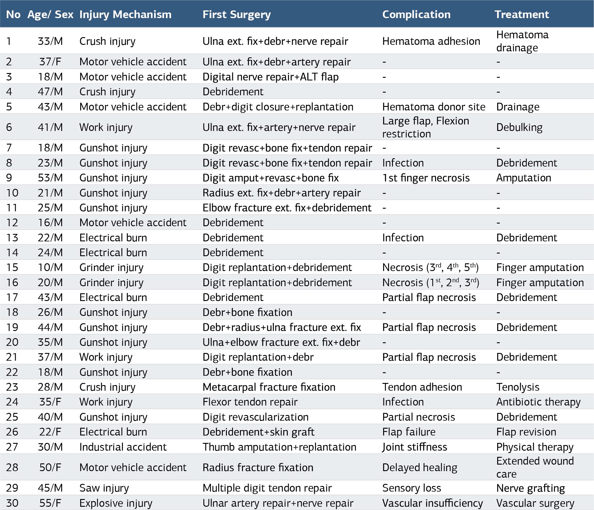
Table 2. Soft Tissue Defect Location, Defect and Flap Dimensions, Hospitalization Duration, Donor Site Closure Method, and Follow-up Time in Patients Undergoing ALT Flap Reconstruction
Figures
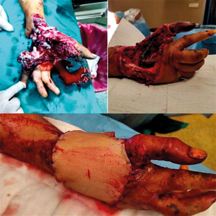
Figure 1. Free ALT flap application to the soft tissue defect in the volar forearm and early postop image. Preoperative image showing a soft tissue defect in the volar forearm following trauma. The intraoperative image demonstrates the preparation and application of the ALT flap for defect coverage. The early postoperative image shows the successful flap adaptation with viable tissue integration
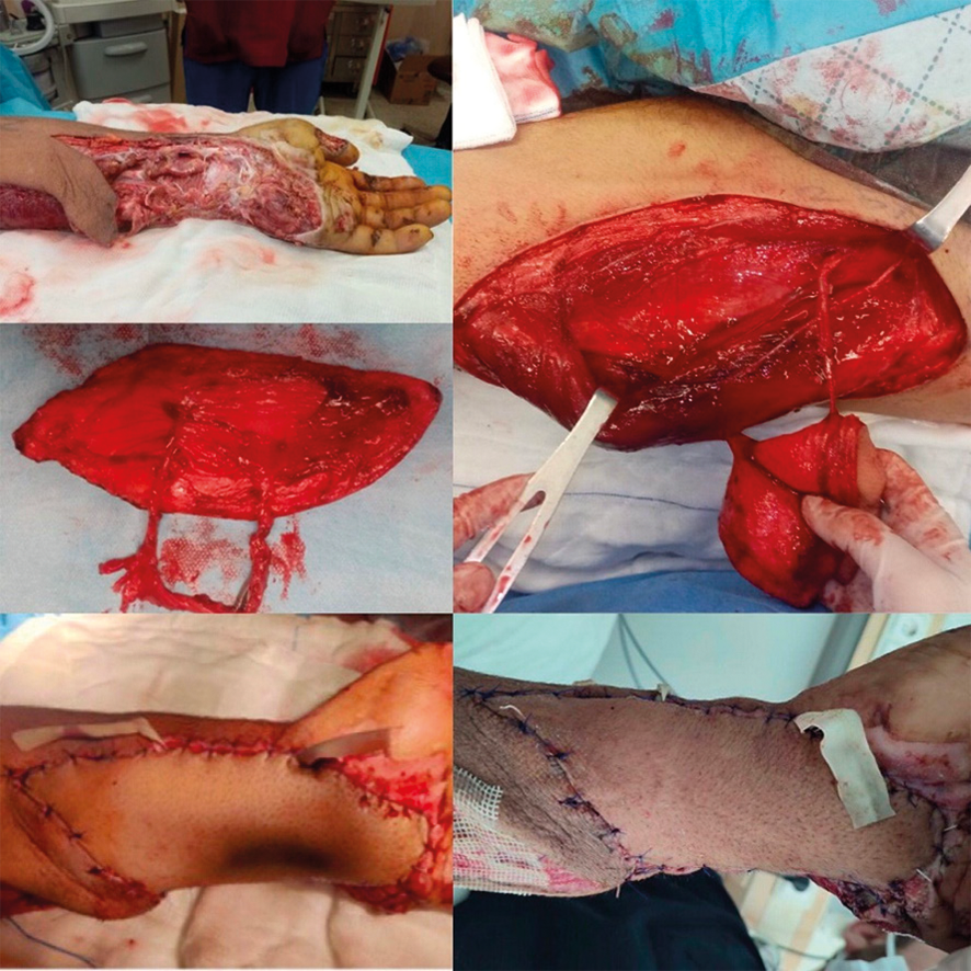
Figure 2. Step-by-step demonstration of ALT flap reconstruction in a patient with a volar forearm tissue defect. (A) Initial debridement of the defect site, (B) Identification and preparation of perforators of the ALT flap, (C) Harvested ALT flap before transfer, (D) Immediate postoperative view of the reconstructed forearm, (E) Long-term postoperative result showing stable coverage and good functional recovery
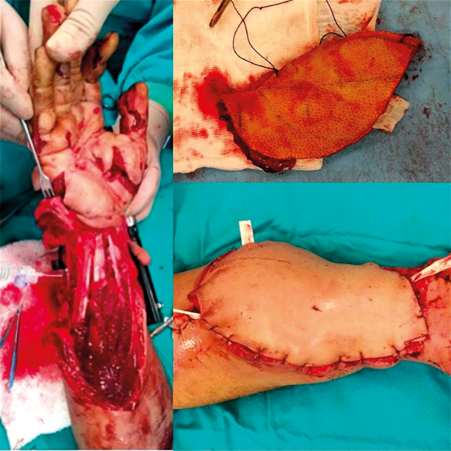
Figure 3. Stepwise representation of ALT flap application for a gunshot-induced soft tissue defect in the dorsal wrist. (A) Initial presentation of the tissue defect, (B) Post-debridement and bone fixation phase, (C) Immediate postoperative appearance following ALT flap reconstruction, (D) Long-term postoperative outcome demonstrating stable coverage and functional preservation
Additional Information
Publisher’s Note
Bayrakol MP remains neutral with regard to jurisdictional and institutional claims.
Rights and Permissions

This work is licensed under a Creative Commons Attribution-NonCommercial 4.0 International License (CC BY-NC 4.0). To view a copy of the license, visit https://creativecommons.org/licenses/by-nc/4.0/
About This Article
How to Cite This Article
Numan Atilgan, Numan Duman, Ozlem Orhan. Optimizing upper extremity reconstruction: outcomes of anterolateral thigh (ALT) flap in complex hand injuries. doi:10.4328/ACAM.22607
Publication History
- Received:
- 16.02.2025
- Accepted:
- 18.03.2025
- Published Online:
- 24.03.2025
- Printed:
- 25.03.2025