
Abstract
Aim Computed tomographic aortography (CTA) is standard for aortic dissection (AD) diagnosis, but not commonly used for acute coronary syndromes. In this study, we aimed to investigate the success of CTA in detecting critical stenosis in coronary arteries in patients with non-ST-elevation myocardial infarction (NSTEMI) diagnosed with suspected AD in the emergency department (ED). Methods This retrospective study was conducted at a tertiary ED from January 2016 to May 2019. Among 6647 acute chest pain patients undergoing CTA for suspected aortic dissection, 50 NSTEMI cases with conventional coronary angiography (CCA) performed were included. Exclusion criteria included age <18, incomplete records, ST elevation on ECG, or confirmed AD on CTA. CTA was performed using a 64-detector CT scanner, and critical stenosis (>50%) in the coronary arteries was evaluated by a radiologist with eight years of cardiothoracic imaging experience. Results In this study, 200 coronary arteries of 50 patients were analyzed. Among the 50 patients included in the study, 29 (58%) were male and 21 (42%) female, with a mean age of 61.4 ± 14.1 years. CTA exhibited a high specificity of 98.71% and sensitivity of 88.89% in detecting critical stenosis within the coronary arteries. A statistically significant positive correlation was observed between the imaging findings of CTA and those of CCA (p<0.001). Conclusion The results indicate that CTA is comparably effective to CCA in identifying critical stenosis within coronary arteries. Moreover, CTA demonstrates potential as an early diagnostic tool for identifying patients with NSTEMI in the ED.Keywords
Introduction
Acute chest pain (ACP) is the most common reason for presentation to the emergency department (ED) after abdominal pain.1,2 Identifying the etiology of ACP is still one of the most important problems for healthcare professionals working in EDs.3-5 Although the causes of ACP are often non-cardiac, its differential diagnosis should include acute coronary syndrome (ACS), pulmonary thromboembolism (PTE), esophageal rupture, pneumothorax, and aortic dissection (AD), which have a mortal course.6,7 Initial evaluation of a patient presenting to the ED with ACP consists of patient history, physical examination, electrocardiography, transthoracic echocardiography, laboratory studies, and imaging modalities. Triple-rule-out coronary CT angiography provides non-invasive visualization of the coronary arteries, pulmonary arteries, and thoracic aorta.8 Although it is efficient in the differential diagnosis of ACP, it is not generally available in most hospitals.
In patients with exertional chest pain radiating to the left arm who also have elevated serum troponin levels, coronary CTA (CCTA) may be requested for the diagnosis of ACS.9-13 However, CCTA is impractical to use in the ED due to disadvantages, including patient compliance, medication to keep the heart rate below 80 beats/minute, and ECG-triggered acquisition.14,15 Computed tomographic aortogram (CTA) is the initial diagnostic imaging modality for AD in patients presenting to the ED with hypertension and chest and back pain. In contrast, it is not in general clinical use for evaluating ACS. Emergent coronary catheterization is essential for high-risk patients with ACS. The laboratory tests and imaging results should be eventuated before conventional coronary angiography (CCA) for patients with non-ST-elevation myocardial infarction (NSTEMI). Further imaging modalities for the diagnosis and risk stratification of ACS, particularly in patients without changes in ECG, might be helpful for physicians in reducing the time to diagnose stenosis.
In this study, we aimed to investigate the success of CTA in detecting critical stenosis in coronary arteries in patients with non-ST-elevation myocardial infarction (NSTEMI) diagnosed with suspected AD in the emergency department (ED).
Materials and Methods
Study Design and ParticipantsThis retrospective study was conducted from January 2016 to May 2019 at a tertiary ED that receives an average of 30,000 patients per month. However, the requirement for informed consent was waived due to the retrospective design of the study.
Patients presenting to the emergency department of our hospital with chest and/or back pain undergo a detailed physical examination, followed by electrocardiography (ECG), cardiac enzyme measurements, D-dimer measurement, chest X-ray (CXR), transthoracic echocardiography (TTE), and CTA when necessary. When STEMI is suspected on ECG, cardiologists are consulted for antithrombotic treatment and coronary angiography. Patients without STEMI are placed on troponin monitoring for NSTEMI. Patients diagnosed with NSTEMI after troponin monitoring undergo coronary angiography and medical treatment within 24 hours. Severe chest and/or back pain, perfusion deficit (unilateral pulse deficit, difference in systolic blood pressure between extremities or neurologic focal signs), enlarged mediastinum on CXR, significant signs on TTE (intimal flap, dilatation of the thoracic aorta, pericardial effusion and aortic valve insufficiency), history of a known AD syndrome (Marfan, vascular Ehlers-Danlos, Loeys-Dietz or Turner) and elevated D-dimer (500 ng/mL), CTA is performed to rule out AD. Patients over 18 years of age who were diagnosed with NSTEMI and underwent coronary angiography were included in the study. Patients younger than 18 years of age who underwent CTA with a prediagnosis of AD, those diagnosed with AD, those with ST elevation on ECG, those who underwent coronary angiography and were diagnosed with a lesion, and those whose records could not be accessed were excluded from the study. Assistive technologies such as artificial intelligence and machine learning are not used in our paper.Data CollectionClinical data were obtained from the hospital’s electronic medical database and patient files. The patients’ age, chronic diseases (diabetes mellitus, hypertension, coronary artery disease, and hypercholesterolemia), vital signs, echocardiography findings (presence or absence of left ventricular wall motion abnormalities), angiography findings, and laboratory results were recorded.Ethical ApprovalThis study was approved by the Ethics Committee of Aksaray University, Faculty of Medicine (Date: 17.10.2023, Decision No: 2023-02/03).Statistical AnalysisAll statistical analyses were performed using the Statistical Package for the Social Sciences version 22 (IBM, Armonk, NY) software. Descriptive statistics were expressed as mean, standard deviation, minimum–maximum, frequency, and percentile values. The results of the CT image analysis of the cases were compared with those of conventional coronary angiography analysis, which is accepted as the gold standard. The sensitivity, specificity, positive predictive, and negative predictive values, and test validity values of all arteries were calculated. Specificity and sensitivity were determined using the Youden index. P values of less than 0.05 were accepted as statistically significant.Reporting GuidelinesThis study was reported in accordance with the STROBE guideline.
Results
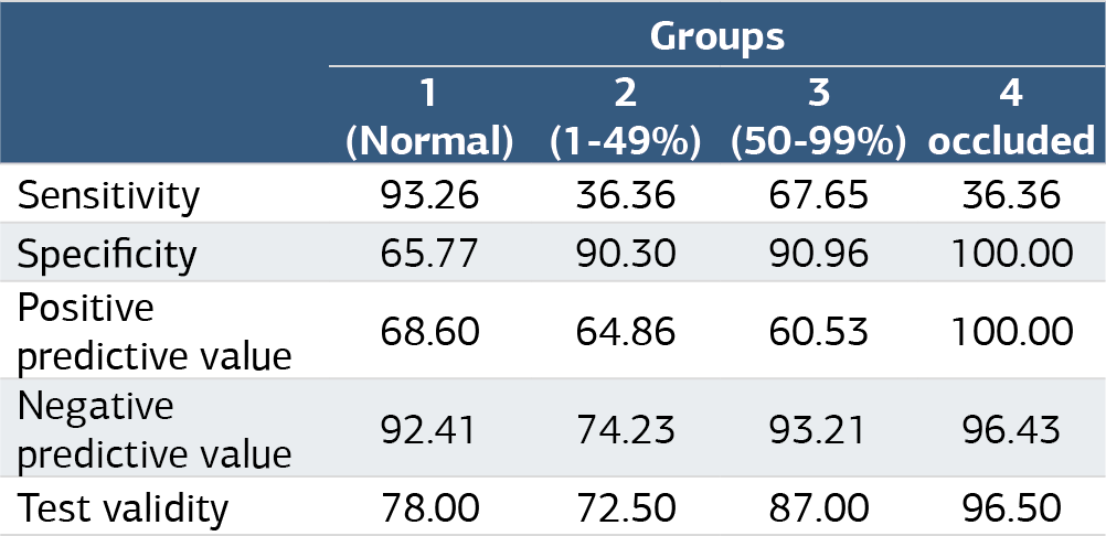
The study included 200 coronary arteries from 50 patients who underwent CTA and CCA at the ED from January 2016 to May 2019. Of the 50 patients included in the study, 29 (58%) were male, and 21 (42%) were female. The mean age of the patients was 61.4 ± 14.1 years. The basic characteristics of the patients are given in Table 1. Before the CTA examination, all patients had a normal ECG. They were diagnosed with non-ST-elevation myocardial infarction (NSTEMI) due to elevated levels of troponin I during their follow-up and underwent CCA within 24 hours. In the coronary artery examination, no critical stenosis was determined in 158 (79%) arteries on the CTA and 155 (77.5%) on the CCA. There was a statistically significant positive correlation between the CTA and CCA findings (p<0.001) (Table 2). The Pearson correlation coefficient was found to be 0.738. It was found that CTA had high specificity (98.71%), sensitivity (88.89%), positive predictive (96.84%), and negative predictive (95.24%) values in the detection of critical stenosis in coronary arteries (Table 3).Computed Tomography (CT) Imaging and AnalysisCTA was performed using a 64-detector row CT scanner (Revolution CT, GE Healthcare, Waukesha, Wisconsin, US). The examination protocol was as follows: 120 kV, 512 x 512 matrix, 64 x 0.6 mm collimation, 0.28 second gantry rotation time, and spiral mode with a pitch of 0.6. Non-ionic contrast medium containing 300 mg of iodine (Omnipaque ®, Nycomed Imaging AS, Oslo, Norway) was administered at a speed of 4–5 mL/s with an automatic injector system. Procedures were successfully completed in all patients without any complications.
All CT images were evaluated by a radiologist (HE) with eight years of experience in cardiothoracic imaging. CT images were evaluated using the Picture Archiving and Communication System (FONET PACS software version 4.1, Ankara, Türkiye). Multiplanar reconstruction images (axial, sagittal, and coronal) were used in all cases. The following four major vascular segments were examined in each patient: the left main coronary artery, the left anterior descending coronary artery, the circumflex coronary artery, and the right coronary artery. A total of 200 vascular segments were examined in 50 patients. The patients were divided into two groups according to whether they had critical stenosis of the coronary arteries (more than 50% stenosis).16 Figure 1, Figure 2 show examples of the CTA and CCA images in patients with critical stenosis, and Figure 3 presents the images of a patient without critical stenosis.
Discussion
Identifying patients with ACP in the ED remains challenging, particularly patients without ECG changes. In order not to overlook a diagnosis of ACS, ED physicians order a 12-lead ECG, laboratory tests (such as troponin I and CKMB), and coronary artery imaging.14,17 It has also been recently suggested that patients with non-specific chest pain in selected centers can be evaluated with a single CTA screening protocol, called “triple rule out” (TRO), to exclude ACS, PTE, and AD.8 However, CCA remains the gold standard for examining coronary arteries. In the current study, we investigated whether CTA taken at the ED was as effective as CCA in detecting stenosis in coronary arteries. To the best of our knowledge, this is the first study to investigate the correlation between CTA and CCA findings for this purpose.
In this study, CTA detected stenosis in 40 of the 45 patients who had critical stenosis in coronary arteries according to CCA, and there was a statistically significant positive correlation between the findings of the two imaging modalities (p<0.001). CTA had a specificity of 98.71% and a sensitivity of 88.89% in detecting critical stenosis in coronary arteries. These findings suggest that CTA is a useful diagnostic imaging modality for detecting critical stenosis in coronary arteries.
In a study conducted with 113 patients with chest pain, Litmanovitch et al. compared TRO-CTA and CCA in the evaluation of coronary arteries and reported that the former detected coronary stenosis with a sensitivity of 93% and a specificity of 88%.18 Similarly, in other studies, Johnson et al. showed that CCTA had a sensitivity of 98% and a specificity of 96% in 109 patients with chest pain, and Stefanini et al. reported 81-99% sensitivity and 64-93% specificity values.19,20 Johnson et al. confirmed stenosis with CCA in 15 of 19 patients who had critical stenosis on CCTA and showed a positive correlation between the two methods.19 In our study, CTA, which we used as a more practical imaging method to exclude AD, was found to have high specificity and sensitivity (98.71% and 88.89%, respectively) in detecting coronary stenosis, consistent with the literature results on TRO-CTA and CCTA. We also found a positive correlation between CTA and CCA in our study and calculated the Pearson correlation coefficient at 0.738. Similar to CCTA, CTA evaluated by an experienced radiologist can provide information about the coronary arteries and help decide whether to perform cardiac follow-up or emergency catheterization in patients with risk factors.
In addition, by reducing the time to diagnose stenosis at the ED, CTA seems to be a more practical method with a lower radiation load but similar safety compared to CCTA and TRO-CTA.
Limitations
This study has several limitations. First, its retrospective and single-center design may limit the generalizability of the findings. The sample size, while sufficient for preliminary analysis, may not capture the full spectrum of NSTEMI presentations or comorbid conditions. Furthermore, potential selection bias cannot be excluded, as patients undergoing both CTA and CCA might represent a more specific clinical subgroup. The diagnostic accuracy and interpretation of CTA could also be influenced by operator expertise and imaging quality, which were not standardized across all cases. Finally, while the results suggest that CTA may facilitate earlier decision-making, the study did not evaluate long-term clinical outcomes or the cost-effectiveness of this approach. Larger, prospective, multicenter studies are needed to confirm these findings and establish clinical guidelines.
Conclusion
Our study revealed that CTA proved to be equally effective to CCA in identifying critical stenosis within the coronary arteries of NSTEMI patients. Evaluating coronary artery stenosis on CTA imaging could also be crucial for patients admitted to the ED with acute chest pain (ACP), despite ECG stability, once AD diagnosis has been ruled out. This approach could potentially prompt immediate coronary catheterization decisions, thereby reducing the time required for NSTEMI diagnosis. Consequently, there could be a reduction in ACS-related morbidity and mortality. To validate our findings, further comprehensive multicenter prospective studies are warranted.
Declarations
Animal and Human Rights Statement
All procedures performed in this study were in accordance with the ethical standards of the institutional and/or national research committee and with the 1964 Helsinki Declaration and its later amendments or comparable ethical standards.
Informed Consent
Written informed consent for CTA imaging was obtained from all patients.
Data Availability
The datasets used and/or analyzed during the current study are not publicly available due to patient privacy reasons but are available from the corresponding author on reasonable request.
Conflict of Interest
The authors declare that there is no conflict of interest.
Funding
None.
Abbreviations
ACP: Acute chest pain
ACS: Acute coronary syndrome
AD: Aortic dissection
CCA: Conventional coronary angiography
CCTA: Coronary computed tomographic angiography
CT: Computed tomography
CTA: Computed tomographic aortography
CXR: Chest X-ray
ECG: Electrocardiography
ED: Emergency department
NSTEMI: Non-ST-elevation myocardial infarction
PTE: Pulmonary thromboembolism
TTE: Transthoracic echocardiography
References
- McCaig LF, Nawar EW. National Hospital Ambulatory Medical Care Survey: 2004 emergency department summary. Adv Data. 2006;(372):1-29.
- Kohn MA, Kwan E, Gupta M, Tabas JA. Prevalence of acute myocardial infarction and other serious diagnoses in patients presenting to an urban emergency department with chest pain. J Emerg Med. 2005;29(4):383-390. doi:10.1016/j.jemermed.2005.04.010
- Gupta M, Tabas JA, Kohn MA. Presenting complaint among patients with myocardial infarction who present to an urban, public hospital emergency department. Ann Emerg Med. 2002;40(2):180-186. doi:10.1067/mem.2002.126396
- Stewart J, Lu J, Goudie A, et al. Applications of machine learning to undifferentiated chest pain in the emergency department: a systematic review. PLoS One. 2021;16(8):e0252612. doi:10.1371/journal.pone.0252612
- Chiang CH, Chiang CH, Lee GH, et al. Safety and efficacy of the European Society of Cardiology 0/1-hour algorithm for diagnosis of myocardial infarction: systematic review and meta-analysis. Heart. 2020;106(13):985-991. doi:10.1136/heartjnl-2019-316343
- Pedersen CK, Stengaard C, Friesgaard K, et al. Chest pain in the ambulance: prevalence, causes, and outcome: a retrospective cohort study. Scand J Trauma Resusc Emerg Med. 2019;27(1):84. doi:10.1186/s13049-019-0659-6
- Litt HI, Gatsonis C, Snyder B, et al. CT angiography for safe discharge of patients with possible acute coronary syndromes. N Engl J Med. 2012;366(15):1393-1403. doi:10.1056/nejmoa1201163
- Wnorowski AM, Halpern EJ. Diagnostic yield of triple-rule-out CT in an emergency setting. AJR Am J Roentgenol. 2016;207(2):295-301. doi:10.2214/ajr.15.15717
- Leber AW, Johnson T, Becker A, et al. Diagnostic accuracy of dual-source multi-slice CT-coronary angiography in patients with an intermediate pretest likelihood for coronary artery disease. Eur Heart J. 2007;28(19):2354-2360. doi:10.1093/eurheartj/ehm294
- Panis V, Donal E. Imaging techniques for cardiac function. Appl Sci. 2021;11(22):10549. doi:10.3390/app112210549
- Budoff MJ, Dowe D, Jollis JG, et al. Diagnostic performance of 64-multidetector row coronary computed tomographic angiography for evaluation of coronary artery stenosis in individuals without known coronary artery disease: results from the prospective multicenter ACCURACY trial. J Am Coll Cardiol. 2008;52(21):1724-1732. doi:10.1016/j.jacc.2008.07.031
- Ngam PI, Ong CC, Chai P, Wong SS, Liang CR, Teo LLS. Computed tomography coronary angiography: past, present, and future. Singapore Med J. 2020;61(3):109-115. doi:10.11622/smedj.2020028
- Athappan G, Habib M, Ponniah T, Jeyaseelan L. Multi-detector computerized tomography angiography for evaluation of acute chest pain: a meta-analysis and systematic review of literature. Int J Cardiol. 2010;141(2):132-140. doi:10.1016/j.ijcard.2008.11.207
- Goldstein JA, Chinnaiyan KM, Abidov A, et al. The CT-STAT trial: coronary computed tomographic angiography for systematic triage of acute chest pain patients to treatment. J Am Coll Cardiol. 2011;58(14):1414-1422. doi:10.1016/j.jacc.2011.03.068
- Hoffmann U, Truong QA, Schoenfeld DA, et al. Coronary CT angiography versus standard evaluation in acute chest pain. N Engl J Med. 2012;367(4):299-308. doi:10.1056/nejmoa1201161
- Hoffmann U, Bamberg F, Chae CU, et al. Coronary computed tomography angiography for early triage of patients with acute chest pain: the ROMICAT trial. J Am Coll Cardiol. 2009;53(18):1642-1650. doi:10.1016/j.jacc.2009.01.052
- Kirk JD, Diercks DB, Turnipseed SD, Amsterdam EA. Evaluation of chest pain suspicious for acute coronary syndrome: use of an accelerated diagnostic protocol in a chest pain evaluation unit. Am J Cardiol. 2000;85(5A):40B-48B. doi:10.1016/s0002-9149(00)00755-4
- Litmanovich D, Zamboni GA, Hauser TH, Lin PJ, Clouse ME, Raptopoulos V. ECG-gated chest CT angiography with 64-MDCT and triphasic IV contrast administration regimen in patients with acute nonspecific chest pain. Eur Radiol. 2008;18(2):308-317.
- Johnson TR, Nikolaou K, Becker A, et al. Dual-source CT for chest pain assessment. Eur Radiol. 2008;18(4):773-780. doi:10.1007/s00330-007-0803-y
- Stefanini GG, Windecker S. Can coronary computed tomography angiography replace invasive angiography? Coronary computed tomography angiography cannot replace invasive angiography. Circulation. 2015;131(4):418-426. doi:10.1161/circulationaha.114.008148
Tables
Table 1. Comparison of age, examination findings, and laboratory parameters according to sex
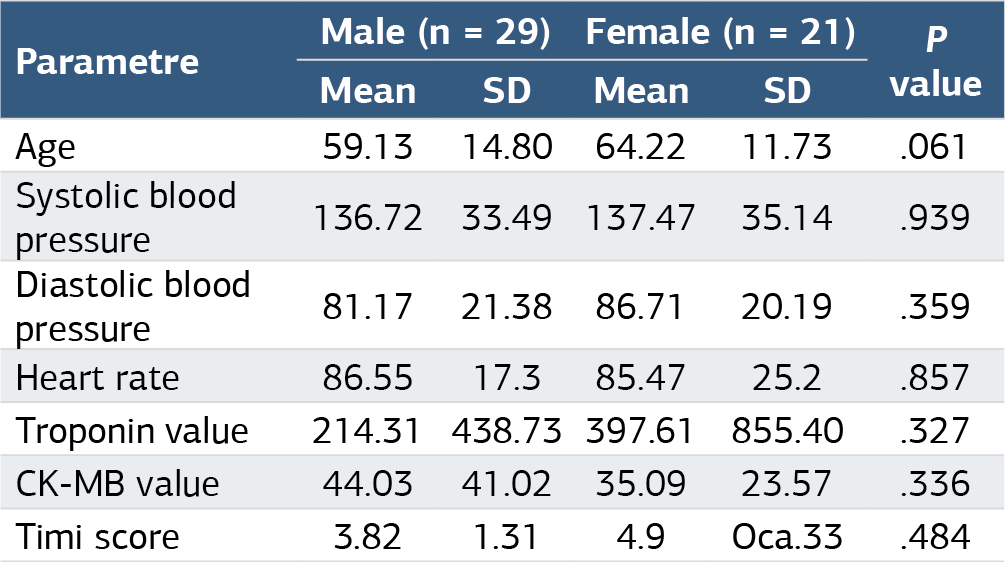
CK-MB: creatine kinase-muscle/brain; Timi: thrombolysis in myocardial infarction; SD: standard deviation
Table 2. Comparison of patient history findings according to sex
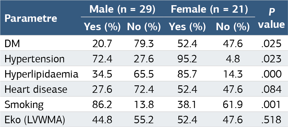
M: Diyabetes mellitus; LVWMA: Left ventricular wall motion abnormality
Table 3. Specificity, sensitivity, positive predictive value, negative predictive value, and test validity values determined according to the stenosis degrees of the patients
Figures
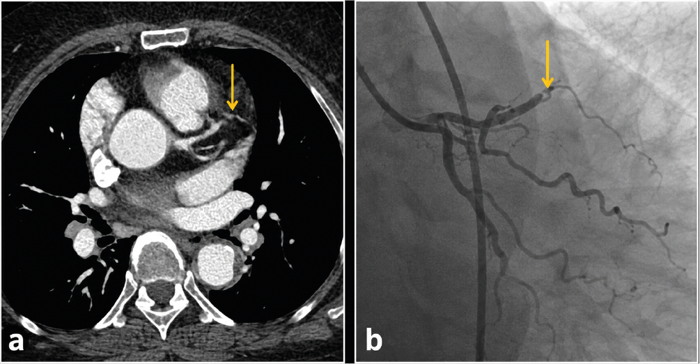
Figure 1. The thoracic computed tomography angiography image shows significant stenosis in the midsection of the left anterior descending (LAD) artery (a). Similarly, conventional coronary angiography image similarly shows 90% stenosis in the middle part of the LAD artery
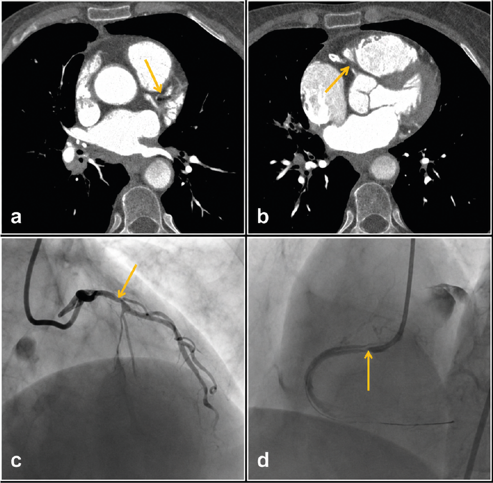
Figure 2. The thoracic computed tomography angiography image shows significant stenosis in the proximal left anterior descending (LAD) artery (a) and total occlusion in the right coronary artery (RCA) (b). Conventional coronary angiography image shows 70% stenosis in the LAD artery (c) and 90% stenosis in the RCA (d)
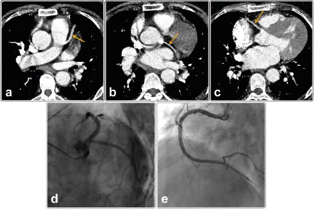
Figure 3. Thoracic computed tomography angiography images show normal findings in the left anterior descending artery (a), circumflex coronary artery (b), and the right coronary artery RCA (c), which are also confirmed to be normal by conventional coronary angiography (d, e)
Additional Information
Publisher’s Note
Bayrakol MP remains neutral with regard to jurisdictional and institutional claims.
Rights and Permissions

This work is licensed under a Creative Commons Attribution-NonCommercial 4.0 International License (CC BY-NC 4.0). To view a copy of the license, visit https://creativecommons.org/licenses/by-nc/4.0/
About This Article
How to Cite This Article
Muhammed Yaşar Sever, Nedim Uzun, Hüseyin Mutlu, Hasan Erdoğan. Detection of critical coronary artery stenosis through computed tomographic aortogram in patients with suspected aortic dissection. Ann Clin Anal Med 2025;16(6):455-459. doi:10.4328/ACAM.22727
Publication History
- Received:
- 28.04.2025
- Accepted:
- 28.05.2025
- Published Online:
- 29.05.2025
- Printed:
- 01.06.2025