
Abstract
AimProstaglandin analogs (PGAs), which are widely used in the treatment of glaucoma, may lead to periorbital changes collectively referred to as prostaglandin-associated periorbitopathy syndrome (PAPS). This study aims to compare the effects of different antiglaucoma eye drops and their durations of use on the periorbital region.MethodsIn this cross-sectional clinical study, patients diagnosed with glaucoma who were using antiglaucoma eye drops were evaluated. The patients were divided into three groups: those using PGAs (Group 1), those using non-PGA medications (Group 2), and those not using any medication (Group 3). Periorbital measurements, including margin reflex distance 1 (MRD1), levator function (LF), inferior scleral show, deepening of the upper eyelid sulcus (DUES), malar edema, and lower eyelid fat herniation, were assessed.ResultsMRD1 was significantly lower in Group 1 than in the other groups (P < .001). LF was lowest in Group 1, higher in Group 2, and highest in Group 3 (P < .001). DUES was most prominent in Group 1, less pronounced in Group 2, and least observed in Group 3 (P < .001). As the duration of medication use increased, a decrease in MRD1 and LF was observed in both Group 1 (P < .001 and P < .001, respectively) and Group 2 (P = .001 and P < .001, respectively). While DUES became more prominent with increased medication duration in Group 1 (P = .007).ConclusionPGAs were associated with a decrease in MRD1 and LF values, leading to ptosis and DUES. Other antiglaucoma eye drops did not cause significant ptosis but were linked to certain periorbital changes.
Keywords
Introduction
Glaucoma is the second leading cause of irreversible blindness worldwide.1 Glaucoma-associated progressive optic neuropathy leads to a reduction in visual acuity and constriction of visual fields.1 The standard treatment for glaucoma is the control of intraocular pressure (IOP) with topical antiglaucoma medications. These topical drugs require multiple dosing and long-term use. However, ocular surface toxicity and tear film abnormalities caused by these medications present a serious issue, affecting the entire ocular surface, including the conjunctiva, cornea, and eyelids.2 Topical prostaglandin analogs (PGAs; also known as prostanoid prostaglandin F receptor agonists) are commonly preferred as first-line medical treatments for glaucoma patients. However, the use of these medications has been reported to lead to progressive periorbital changes and the development of periorbital side effects known as prostaglandin-associated periorbital syndrome (PAPS).3,4
The commonly reported signs and symptoms of PAPS include hyperpigmentation of the periorbital skin, trichomegaly and hypertrichosis of the eyelashes, deepening of the upper eyelid sulcus, flattening of the lower eyelid bags, upper eyelid ptosis, mild enophthalmos, inferior scleral visibility, and involution of dermatochalasis.3 Although the mechanism of PAPS is not completely understood, it is believed that the primary pathophysiology involves orbital fat atrophy, which is thought to occur through the inhibition of adipogenesis via stimulation of Prostaglandin F receptors in adipocytes.5 It has even been found that early-onset periorbopathy can develop within just one month of starting PGA use.6 The most prominent feature of PAPS is the development, deepening of the upper eyelid sulcus (DUES).7-9 While PGAs can cause significant changes in the periorbital tissues, there is limited information regarding such effects with non-PGA glaucoma drops. The periorbital effects of other glaucoma drops are less pronounced and require further investigation.
The aim of our study is to analyze the changes observed in the periorbital region in patients using anti-glaucoma drops, based on the type of medication and duration of use.
Materials and Methods
This study was designed as a single-center, cross-sectional study and was conducted between June 2025 and July 2025 at the Department of Ophthalmology, Dünyagöz Ataşehir Hospital. This study included both eyes of patients diagnosed with glaucoma who had been using at least one anti-glaucomatous eye drop for at least one year. Patients using PGA drops (latanoprost, travoprost, bimatoprost, or tafluprost) either alone or in combination with other drops were classified as Group 1. Patients using non-PGA antiglaucomatous drops (adrenergic agonists, beta-blockers, carbonic anhydrase inhibitors) were classified as Group 2. The control group (Group 3) consisted of glaucoma patients who were not using any eye drops.
The age, gender, treatment duration, and the specific glaucoma medications used by each patient were recorded. As the study was conducted at a tertiary healthcare center and referral hospital, many patients were using multiple anti-glaucoma medications, with the most common being the combination of PGA and other eye drops. Therefore, in the study, the effects of anti-glaucoma drops on the periorbital region were compared between patients using a combination of medications with and without PGA, either as a single or combined treatment. To exclude conditions that could contribute to periorbital findings, a detailed patient history was obtained, and a physical examination was conducted.
Exclusion criteria included individuals with a known allergy to the anti-glaucoma medication used, those who had undergone ocular surgery within the last 6 months before the study, those with a history of any ocular plastic surgery, individuals with a history of eyelid trauma, those with a known history of exophthalmos or enophthalmos prior to anti-glaucoma treatment, patients with orbital and adnexal fractures, contact lens users, those with thyroid orbitopathy, and those with a history of carotid-cavernous fistula or neurological disorders.
The patients enrolled in the study were examined in the Oculoplastic and Orbital Surgery Department. Orbital and adnexal measurements were taken, including the margin-reflex distance 1 (MRD1), levator function (LF), inferior scleral appearance, DUES, malar edema, and lower eyelid fat herniation grades. The lower eyelid position was assessed, and the presence of entropion, ectropion, and trichiasis was recorded. All patients’ photographs were taken with a digital camera (EPL-3 14-42 mm lens; Olympus; China) from a distance of 30 cm, without flash and under ambient light conditions. These photos were analyzed by two oculoplastic specialists (E.S.E., S.H.) to verify the physical examination findings, and when they agreed, the findings were confirmed.
MRD1 measurement was performed to detect blepharoptosis. During the LF measurement, the patient’s eyebrow was stabilized with the researcher’s thumb. The patient was asked to look up and then look down. The eyelid movement range was measured using a ruler. The inferior scleral appearance was assessed by considering the contact between the lower eyelid and the limbus in a smooth contour as 0 mm, while the distance between the lower eyelid and the lower limbus was measured in millimeters. DUES was graded using a modified version of previous methods, based on the relationship of the upper sulcus with the upper orbital rim. A scale from 1 to 3 was used.10 Malar edema was graded according to the classification by Lam and colleagues.11 Lower eyelid fat herniation was classified by Liu and colleagues.12Ethical ApprovalThis study was approved by the Ethics Committee of Bahçeşehir University (Date: 18.06.2025, Decision No: 2025-10/01).Statistical AnalysisStatistical analysis for this study was performed using IBM SPSS version 26.0 software. Descriptive statistics (frequency, percentage, median, interquartile range (IQR), and minimum-maximum values) were calculated for the demographic data and eyelid measurements of the participants, grouped by glaucoma groups and the control group. The Chi-square test was used for comparisons of categorical variables. For intergroup comparisons of all numerical parameters, the Kruskal-Wallis test was applied, as the parameters did not follow a normal distribution. The Mann-Whitney U test was used for post hoc comparisons. The normality of the parameters was assessed using the Shapiro-Wilk test. All statistical analyses were conducted with a 95% confidence interval, and significance was considered at P < .05.
Reporting GuidelinesThis study was reported according to the STROBE guidelines.
Results
A total of 110 eyes using PGA drops for glaucoma (Group 1), 54 eyes using non-PGA anti-glaucoma drops (Group 2), and 192 eyes with no drop usage (Group 3) were included in the study. There was no statistically significant difference between the groups in terms of age (P = .385), gender (P = .682), and visual acuity in LogMAR (P = .657).
There is a statistically significant difference in MRD1 between the groups (P < .001). MRD1 in Group 1 (2.76 ± 0.99) is significantly lower than in Group 2 (3.19 ± 0.77) (P = .008). Furthermore, MRD1 values in Group 1 (2.76 ± 0.99) are significantly lower than those in Group 3 (3.37 ± 0.86) (P < .001) (Table 1).
There is a statistically significant difference in LF between the groups (P < .001). LF is the lowest in Group 1 and the highest in Group 3 (Table 1).
There is a statistically significant difference in inferior scleral appearance between the groups (P < .001). There is a significant difference between Group 1 and Group 3 (P < .001) as well as between Group 2 and Group 3 (P < .001). The inferior scleral appearance in patients using anti-glaucomatous eye drops (Groups 1 and 2) was measured as higher compared to Group 3, which did not use eye drops (Table 1).
There is a statistically significant difference in lower eyelid fat herniation between the groups (P = .006). A significant difference was observed between Group 1 and Group 3 (P = .035) and between Group 2 and Group 3 (P = .003). Lower eyelid fat herniation in patients using anti-glaucomatous eye drops (Groups 1 and 2) was measured as higher compared to Group 3, which did not use eye drops (Table 1).
There is a statistically significant difference in DUES between the groups (P < .001). The highest upper eyelid sulcus deformity values were observed in Group 1, while the lowest values were observed in Group 3. A statistically significant difference was found between all groups (Group 1 vs Group 3, P < .001; Group 2 vs Group 3, P < .001; Group 1 vs Group 2, P < .001) (Table 1).
There is a statistically significant difference in malar edema between the groups (P < .001). A significant difference was found between Group 1 and Group 3 (P < .001) and between Group 2 and Group 3 (P < .001). Malar edema values in patients using antiglaucomatous eye drops (Groups 1 and 2) were higher compared to Group 3, which did not use eye drops (Table 1).
None of the patients showed entropion or trichiasis. However, there was no statistically significant difference between the groups in terms of ectropion (P = .118) (Table 1).
In Group 1, there is a statistically significant difference in MRD1 and LF measurements according to the duration of medication use (P < .001, P < .001, respectively). As the duration of medication use increases, MRD1 and LF values decrease (Table 2).
In Group 1, there is a statistically significant difference in lower eyelid fat herniation and upper eyelid sulcus deformity values according to the duration of medication use (P = .006, P = .007, respectively). Patients who have used PGAs for more than 10 years showed an increase in lower eyelid fat herniation and DUES compared to those who used them for less than 10 years (Table 2).
In Group 1, there is no statistically significant difference in inferior scleral appearance and malar edema values according to the duration of medication use (P = .107, P = .392, respectively) (Table 2).
In Group 1, there is no statistically significant difference in ectropion based on the duration of medication use (P = .201) (Table 2).
In Group 2, there is a statistically significant difference in MRD1 and LF measurements according to the duration of medication use (P = .001, P < .001, respectively). MRD1 and LF measurements are significantly lower in the 6-10 year group compared to the 0-5 year group (P = .001, P < .001, respectively). Similarly, in the 11+ years group, MRD1 and LF values are significantly lower compared to the 0-5 year group (P = .006, P < .001, respectively) (Table 3).
In Group 2, there is a statistically significant difference in the inferior eyelid fat herniation values according to the duration of medication use (P = .008). This difference is due to the higher inferior eyelid fat herniation values in the 6-10 year medication use group compared to the 0-5 year group (P = .003) (Table 3).
In Group 2, there is no statistically significant difference in inferior scleral appearance, DUES, and malar edema values according to the duration of medication use (P = .608, P = .205, P = .686, respectively) (Table 3).
In Group 2, there is no statistically significant difference in ectropion according to the duration of medication use (P = .099) (Table 3).
Discussion
In this study, glaucoma patients using either combined or single PGA, those using other antiglaucomatous drops (excluding PGA), and those not using any eye drops were compared in terms of periorbital changes.
Antiglaucomatous eye drops, especially PGAs, can cause various changes around the eyes. These changes are known as PAPS and are observed as hyperpigmentation, ptosis, deepening of the upper eyelid sulcus, and periorbital fat loss.3,4,7 Periorbital and ocular surface changes have also been observed with other antiglucomatous drops, excluding PGAs.2,13,14 Topical antiglaucoma drops like beta-blockers, apraclonidine, brimonidine, and dorzolamide can cause periorbital dermatitis, allergic edema, and fibrosis, potentially leading to reversible or scarred eyelid ectropion, especially in predisposed individuals.2,13-16 PGAs are characterized by their strong intraocular pressure (IOP) lowering effects, minimal systemic side effects, and a once-daily dosing regimen, which contributes to good patient compliance.7,17 Among these agents are latanoprost (0.005%), travoprost (0.004%), bimatoprost (0.01% and 0.03%), and tafluprost (0.0015%). Various methods have been suggested in the literature for grading PAPS.8,18 In one study, the prevalence of PAPS (defined as patients with deepening of the upper eyelid sulcus + at least three additional clinical signs) was found to be over 40%. It was particularly determined that patients over 60 years old have a threefold increased risk of developing PAPS.19
Factors contributing to the severity of PAPS include the type of PGA and the duration of use. Risk factors for the development of PAPS include age (with a particularly significant relationship in individuals over 60 years old), the technique of drop application, and the duration of use.7,17,19-22
Consistent with the literature, in this study, the MRD1 value was significantly lower in the PGA group compared to the other groups. The use of non-PGA anti-glaucomatous eye drops did not show a significant effect on MRD1 value or ptosis. Similarly, the LF value was found to be lower in patients using antiglaucomatous eye drops, with or without PGA, compared to those not using any drops. No established relationship between LF and anti-glaucomatous drops has been demonstrated in the literature.
In our study, inferior scleral show, lower eyelid fat herniation, and malar edema were more common in patients using antiglaucomatous drops, possibly due to mechanical trauma during drop application—particularly with beta-blockers, dorzolamide, and brinzolamide.13,14 These findings, though not classic components of PAPS, were not observed in non-drop users. Inferior scleral show was not increased in the PGA group, likely due to the lack of distinction between bimatoprost and other PGAs. As expected, upper eyelid sulcus deformity was most prominent in the PGA group and was associated with longer duration of drop use.23
In this study, MRD1 values in the group using PGAs decreased as the duration of use increased. In other words, the longer the duration of drop usage, the greater the development of pitosis. Similarly, LF measurements decreased with increasing drop usage duration. We believe that fibrosis develops in the levator muscle with prolonged use of anti-glaucomatous drops. Supporting this argument, a study demonstrated that acquired blepharoptosis occurs as a combined effect of degenerative changes in the levator palpebrae superioris muscle and its aponeurosis, and a significant relationship was found between the severity of blepharoptosis and the reduction in levator muscle function.24
DUES was found to be significantly more severe in patients using PGA for 11 years or more. Literature reports that upper eyelid sulcus deformity can begin as early as 3-6 months after starting treatment. The reason we observed this finding in the 0-5 year group in our study may be due to the lack of subgrouping by specific PGA medications. As is well known, upper eyelid sulcus deformity has most frequently been reported with bimatoprost.25
Limitations
This study has several limitations. First, the sample size was relatively small, which may have limited the ability to detect certain findings, such as ectropion, or to distinguish between different prostaglandin analog (PGA) derivatives. Second, the lack of subgroup analysis for individual medications may have masked drug-specific effects. Third, due to the cross-sectional design of the study, no causal inferences can be made regarding the relationship between the duration of drop use and periorbital changes. Lastly, in Group 1, some patients were using combination therapies containing prostaglandins along with other agents, which may have introduced confounding effects and limited the ability to attribute the observed changes solely to prostaglandin analogs.
Conclusion
In patients using PGAs, significant reductions in MRD1 and LF values were found, which were associated with ptosis and fibrotic changes in the levator muscle. Upper eyelid sulcus deformity became more pronounced, especially with long-term PGA use, and was observed to be severe in patients using PGAs for more than 11 years.
Declarations
Animal and Human Rights Statement
All procedures performed in this study were in accordance with the ethical standards of the institutional and/or national research committee and with the 1964 Helsinki Declaration and its later amendments or comparable ethical standards.
Informed Consent
Written informed consent was obtained from all participants.
Data Availability
The datasets used and/or analyzed during the current study are not publicly available due to patient privacy reasons but are available from the corresponding author on reasonable request.
Conflict of Interest
The authors declare that there is no conflict of interest.
Funding
None.
Abbreviations
DUES: Deepening of the upper eyelid sulcus
IOP: Intraocular pressure
LF: Levator function
MRD1: Margin reflex distance 1
PAPS: Prostaglandin-associated periorbitopathy syndrome
PGA: Prostaglandin analog
References
- Tham YC, Li X, Wong TY. Global prevalence of glaucoma and projections of glaucoma burden through 2040: a systematic review and meta-analysis. Ophthalmology. 2014;121(11):2081-2090. doi:10.1016/j.ophtha.2014.05.013
- Servat JJ, Bernardino CR. Effects of common topical antiglaucoma medications on the ocular surface, eyelids, and periorbital tissue. Drugs Aging. 2011;28(1):267-282. doi:10.2165/11588830-000000000-00000
- Sakata R, Chang PY, Sung KR, et al. Prostaglandin-associated periorbitopathy syndrome: addressing an unmet clinical need. Semin Ophthalmol. 2022;37(4):447-454. doi:10.1080/08820538.2021.2003824
- Berke SJ. PAP: new concerns for prostaglandin use. Rev Ophthalmol. 2012;19(10):70.
- Taketani Y, Yamagishi R, Fujishiro T. Activation of the prostanoid FP receptor inhibits adipogenesis leading to deepening of the upper eyelid sulcus in prostaglandin-associated periorbitopathy. Invest Ophthalmol Vis Sci. 2014;55(3):1269-1276. doi:10.1167/iovs.13-12589
- Kucukevcilioglu M, Bayer A, Uysal Y. Prostaglandin-associated periorbitopathy in patients using bimatoprost, latanoprost, and travoprost. Clin Exp Ophthalmol. 2014;42(2):126-131. doi:10.1111/ceo.12163
- Sakata R, Shirato S, Miyata K. Incidence of deepening of the upper eyelid sulcus on treatment with a tafluprost ophthalmic solution. Jpn J Ophthalmol. 2014;58(2):212-217. doi:10.1007/s10384-013-0299-8
- Rabinowitz MP, Katz LJ, Moster MR, et al. Unilateral prostaglandin-associated periorbitopathy: a syndrome involving upper eyelid retraction distinguishable from the aging sunken eyelid. Ophthalmic Plast Reconstr Surg. 2015;31(6):373-378. doi:10.1097/iop.0000000000000351
- Sakata R, Shirato S, Miyata K. Recovery from deepening of the upper eyelid sulcus after switching from bimatoprost to latanoprost. Jpn J Ophthalmol. 2013;57(2):179-184. doi:10.1007/s10384-012-0219-3
- Liang L, Sheha H, Fu Y. Ocular surface morbidity in eyes with senile sunken upper eyelids. Ophthalmology. 2011;118(12):2487-2492. doi:10.1016/j.ophtha.2011.05.035
- Lam SM, Glasgold MJ, Glasgold RA. Complementary Fat Grafting. Lippincott Williams & Wilkins; 2006:12-25. doi:10.1007/978-3-540-71097-4_29
- Liu J, Huang C, Song B. A graded approach in East Asian personalized lower blepharoplasty: a retrospective study spanning 12 years. Indian J Ophthalmol. 2022;70(8):3088-3094. doi:10.4103/ijo.ijo_222_22
- Kalavala M, Statham BN. Allergic contact dermatitis from timolol and dorzolamide eye drops. Contact Dermatitis. 2006;54(6):345. doi:10.1111/j.0105-1873.2006.0645b.x
- Armisen M, Vidal C, Quintans R, Castroviejo M. Allergic contact dermatitis from apraclonidine. Contact Dermatitis. 1998;39(3):193. doi:10.1111/j.1600-0536.1998.tb05893.x
- Britt T, Burnstine MA. Iopidine allergy causing lower eyelid ectropion progressing to cicatricial entropion. Br J Ophthalmol. 1999;83(8):992-993. doi:10.1136/bjo.83.8.987f
- Grassberger M, Baumruker T, Enz A, et al. A novel anti-inflammatory drug, SDZ ASM 981, for the treatment of skin diseases: in vitro pharmacology. Br J Dermatol. 1999;141(2):264-273. doi:10.1046/j.1365-2133.1999.02974.x
- Manju M, Pauly M. Prostaglandin-associated periorbitopathy: a prospective study in Indian eyes. Kerala J Ophthalmol. 2020;32(1):36-40. doi:10.4103/kjo.kjo_90_19
- Tanito M, Ishida A, Ichioka S, et al. Proposal of a simple grading system integrating cosmetic and tonometric aspects of prostaglandin-associated periorbitopathy. Medicine (Baltimore). 2021;100(34). doi:10.1097/md.0000000000026874
- Patradul C, Tantisevi V, Manassakorn A. Factors related to prostaglandin-associated periorbitopathy in glaucoma patients. Asia Pac J Ophthalmol (Phila). 2017;6(3):238-242. doi:10.22608/apo.2016108
- Shah M, Lee G, Lefebvre DR, et al. A cross-sectional survey of the association between bilateral topical prostaglandin analogue use and ocular adnexal features. PLoS One. 2013;8(5). doi:10.1371/journal.pone.0061638
- Kim HW, Choi YJ, Lee KW. Periorbital changes associated with prostaglandin analogs in Korean patients. BMC Ophthalmol. 2017;17(1):126. doi:10.1186/s12886-017-0521-4
- Davis SA, Sleath B, Carpenter DM. Drop instillation and glaucoma. Curr Opin Ophthalmol. 2018;29(2):171-177. doi:10.1097/icu.0000000000000451
- Habchane A, Khatem S, Alioua A, et al. Ptosis following the use of dexamethasone-based eye drops. Sch J Med Case Rep. 2024;12(2):215-217. doi:10.36347/sjmcr.2024.v12i02.020
- Pereira LS, Hwang TN, Kersten RC. Levator superioris muscle function in involutional blepharoptosis. Am J Ophthalmol. 2008;145(6):1095-1098. doi:10.1016/j.ajo.2008.02.002
- Li W, Chen X, Chen S. Changes in prostaglandin-associated periorbital syndrome: a self-controlled and prospective study. Cutan Ocul Toxicol. 2025;44(1):35-42. doi:10.1080/15569527.2024.2431570
Tables
Table 1. Group comparisons
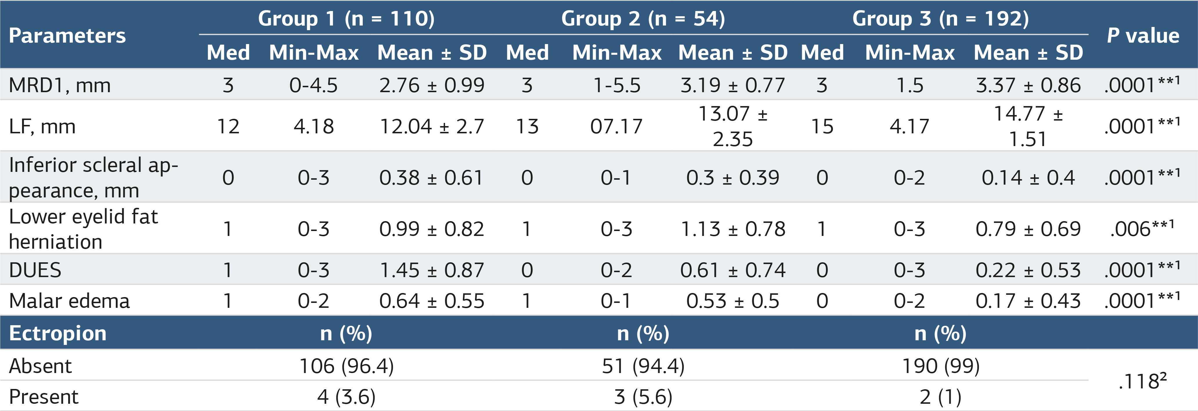
¹: Kruskal-Wallis test; ²: Chi-Square test; **: P < .01, Min-Max: Minimum-Maximum, Mean ± SD: Mean ± Standard Deviation, p: p value, mm: millimeter, MRD1: Marjin-reflex distance 1, LF: Levator Function, DUES: Deepening upper eyelid sulcus.
Table 2. Group 1 – comparison by duration
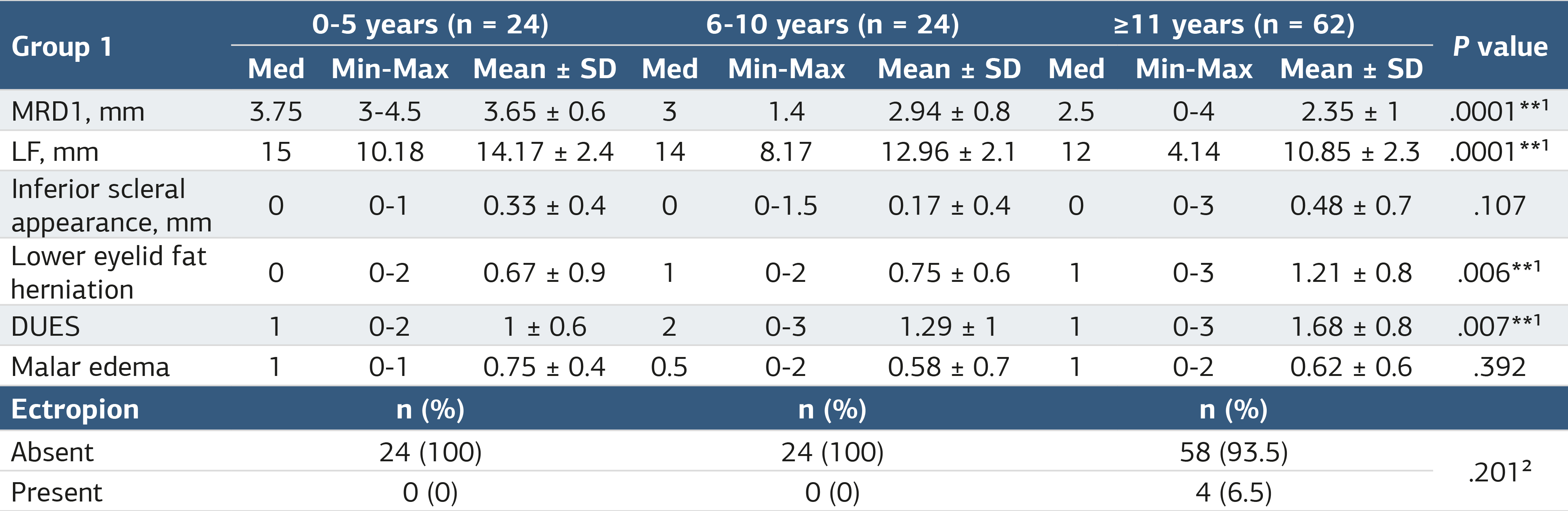
¹: Kruskal-Wallis test; ²: Chi-Square test; **: P < .01, Min-Max: Minimum-Maximum, Mean ± SD: Mean ± Standard Deviation, p: p value, mm: millimeter, MRD1: Marjin-reflex distance 1, LF: Levator Function.
Table 3. Group 2 – comparison by duration
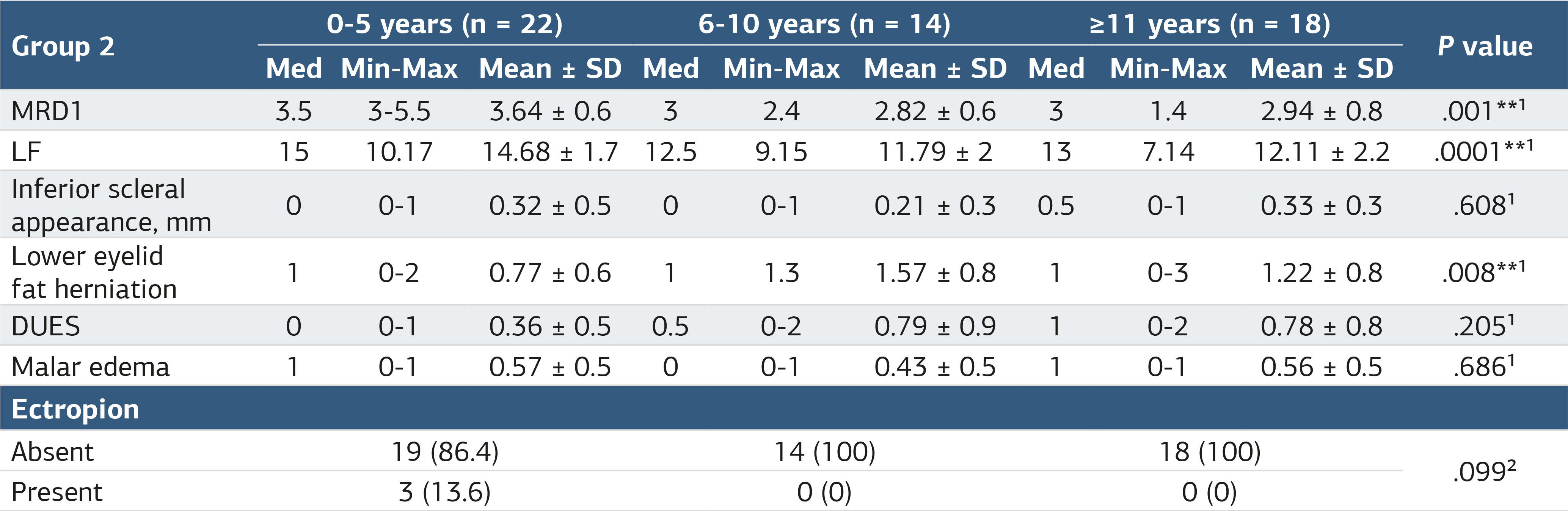
¹: Kruskal-Wallis test; ²: Chi-Square test; *: P < .05, **: P < .01, Min-Max: Minimum-Maximum, Mean ± SD: Mean ± Standard Deviation, p: p value, mm: millimeter, MRD1: Marjin-reflex distance 1, LF: Levator Function, DUES: Deepening upper eyelid sulcus.
Additional Information
Publisher’s Note
Bayrakol MP remains neutral with regard to jurisdictional and institutional claims.
Rights and Permissions

This work is licensed under a Creative Commons Attribution-NonCommercial 4.0 International License (CC BY-NC 4.0). To view a copy of the license, visit https://creativecommons.org/licenses/by-nc/4.0/
About This Article
How to Cite This Article
Emine Savran Elibol, Sezer Hacıağaoğlu. Periorbital effects of antiglaucoma eye drops: a comparative analysis of prostaglandin analogs and other agents. Ann Clin Anal Med 2025;16(9):627-631. doi:10.4328/ACAM.22780
Publication History
- Received:
- 02.07.2025
- Accepted:
- 19.08.2025
- Published Online:
- 30.08.2025
- Printed:
- 01.09.2025