
Abstract
AimPercutaneous catheter drainage has emerged as a less invasive alternative to traditional large-bore chest tubes in the management of pneumothorax. This study aimed to compare the clinical outcomes of 10F percutaneous catheters with 28F chest tubes.MethodsA retrospective analysis was conducted on 78 patients treated for pneumothorax between May 2023 and May 2025. Patients were divided into two groups: those treated with a 10F percutaneous catheter (n = 40) and those treated with a 28F chest tube (n = 38). Demographics, pneumothorax etiology, length of hospital stay, drainage duration, prolonged air leak, and need for video-assisted thoracoscopic surgery (VATS) were evaluated. Statistical analyses included Mann–Whitney U, Fisher’s exact, and Chi-square tests with Monte Carlo simulation where appropriate.ResultsThere were no significant differences between groups in age (P = .1596), sex (P = .1406), pneumothorax side (P = .4383), or COPD prevalence (P = .9382). Etiological distribution showed a statistically significant difference (P = .0051); notably, 10F catheters were not utilized in cases of penetrating trauma. Mean hospital stay and drainage duration were not significantly different between groups (P = .0656 and P = .2709, respectively). The rates of prolonged air leak (P = .3493) and VATS requirement (P = .9495) were also similar.ConclusionThe 10F percutaneous catheter demonstrated comparable efficacy and safety to the conventional 28F chest tube in the management of pneumothorax. Despite being less invasive, it yielded similar clinical outcomes, supporting its use as a safe and effective alternative for treating spontaneous pneumothorax and blunt trauma cases.
Keywords
Introduction
Pneumothorax, defined as the presence of air in the pleural space, can compromise respiratory function and lead to potentially life-threatening consequences if not managed promptly and effectively.1 It may occur spontaneously (primary or secondary), traumatically, or iatrogenically, and its clinical presentation varies from asymptomatic to severe respiratory distress depending on the underlying etiology and patient comorbidities.2,3 The primary goal of treatment is to evacuate intrapleural air, allow lung re-expansion, and prevent recurrence.4
Traditionally, large-bore chest tubes (e.g., 28F–32F (French)) inserted via thoracostomy have been the standard of care for moderate to large pneumothoraces, especially in traumatic or secondary cases.5 However, these tubes are associated with significant patient discomfort, longer hospital stays, and complications such as infection, bleeding, and injury to surrounding structures.6 In recent years, minimally invasive techniques using small-bore catheters, typically inserted percutaneously under imaging guidance, have gained popularity due to their reduced procedural trauma and improved patient comfort.7-9
Several studies have demonstrated that small-bore catheters (≤14F), including pigtail catheters and percutaneous drainage tubes, can achieve similar clinical outcomes to large-bore chest tubes in the treatment of spontaneous or iatrogenic pneumothorax.10-12 In particular, 10F percutaneous catheters have been increasingly adopted as an initial management strategy, especially in cases not related to major trauma.13,14 The American College of Chest Physicians and British Thoracic Society both acknowledge small-bore catheters as appropriate for stable patients with spontaneous pneumothorax, although evidence remains heterogeneous.1,14
Despite growing interest in percutaneous catheters, there remains some reluctance among clinicians to favor them over conventional chest tubes, especially in more complex or traumatic cases.15 Concerns persist regarding prolonged air leaks, higher failure rates, and the potential need for escalation to surgical intervention such as video-assisted thoracoscopic surgery (VATS).16 Furthermore, comparative data on the efficacy and safety of small-bore percutaneous catheters versus standard large-bore chest tubes in mixed patient populations—including trauma cases—remain limited, particularly in real-world clinical settings.
This study aims to address this knowledge gap by retrospectively comparing the clinical outcomes of 10F percutaneous catheters and 28F chest tubes in patients treated for pneumothorax over a two-year period at a tertiary care center. By analyzing parameters such as drainage duration, length of hospital stay, complication rates, and need for surgical intervention, we aim to evaluate whether the less invasive 10F catheter represents a safe and effective alternative to traditional chest tubes across a diverse pneumothorax population.
Materials and Methods
Study Design and SettingThis retrospective comparative study was conducted at a tertiary care university hospital between May 2023 and May 2025. The study protocol was approved by the institutional ethics committee (Date: 02.07.2024, Decision No: 2024-KAEK-24) and conducted in accordance with the Declaration of Helsinki. Informed consent was waived by the ethics committee due to the retrospective nature of the study.Study PopulationPatients aged ≥18 years who were diagnosed with pneumothorax and underwent interventional drainage were included. Both isolated pneumothorax and hemopneumothorax cases were eligible for inclusion. Patients were divided into two groups based on the initial intervention: the 10F percutaneous catheter group and the 28F chest tube group. The choice of drainage method was at the discretion of the attending physician, based on clinical status and radiological findings. Patients with tension pneumothorax, hemopneumothorax, and various etiologies—including spontaneous, traumatic (blunt and penetrating), and iatrogenic pneumothorax—were eligible for inclusion. Clinical decision-making was based on patient stability and radiological findings. Clinically unstable patients were treated with immediate chest tube thoracostomy. In stable patients, 10F percutaneous catheters were preferred when imaging showed >2 cm separation from the lateral chest wall to allow safe percutaneous access. If catheter placement failed or was technically not feasible, conversion to chest tube drainage was performed. In cases with <2 cm separation, chest tube placement was preferred. Additionally, in all cases of penetrating trauma, large-bore chest tubes were used in accordance with standard trauma care protocols. Exclusion CriteriaPatients were excluded if they.1 had incomplete medical records.2 were non-compliant with treatment (e.g., self-discharge or refusal of procedure), or.3 experienced a failed catheter or tube insertion requiring immediate conversion.Operator and StandardizationAll procedures, including both catheter and chest tube placements, as well as any surgical interventions, were performed by a single experienced thoracic surgeon, ensuring consistency in technique and clinical decision-making throughout the study period.Diagnostic and Procedural ApproachAll diagnoses were confirmed via thoracic computed tomography (CT). In patients considered for catheter placement, a wireless Clarius ultrasound device (Clarius Mobile Health Corp., Vancouver, Canada) was used to confirm the presence and extent of pneumothorax and to assess thoracic anatomy in order to determine suitability for catheter thoracostomy (Figure 1). In the 10F catheter group, procedures were performed under real-time ultrasonography (USG) guidance at the 4th or 5th intercostal space along the mid-axillary line, within the “safe triangle”. Chest tubes (28F) were placed via standard blunt dissection using the same anatomical landmarks. All procedures were carried out under sterile conditions. A post-procedural chest radiograph was obtained to confirm the correct positioning of the catheter and to assess lung re-expansion (Figure 2).Surgical InterventionPatients who did not respond to drainage or developed complications were evaluated for video-assisted thoracoscopic surgery (VATS). Surgical indications were based on standard clinical criteria including persistent air leak, incomplete lung expansion, or recurrence, at the discretion of the surgeon.Data CollectionDemographic and clinical variables recorded included age, sex, comorbidities (especially chronic obstructive pulmonary disease (COPD)), etiology (spontaneous, traumatic, iatrogenic), side of pneumothorax, length of hospital stay, drainage duration, presence of prolonged air leak (defined as air leak >7 days), and need for VATS. Data were collected from electronic medical records by two independent researchers and verified for consistency.Sample Size CalculationUsing G*Power (version 3.1; Heinrich Heine University, Düsseldorf, Germany), a sample size of 72 patients (36 per group) was calculated to detect a moderate effect size (d=0.6) in hospital stay with α=0.05 and power=0.80. The final study population consisted of 78 patients (40 in the 10F group and 38 in the 28F group), satisfying the power requirement.Ethical ApprovalThis study was approved by the Ethics Committee of Kastamonu University (Date: 02.07.2024, Decision No: 2024-KAEK-24).Statistical AnalysisStatistical analyses were performed using IBM SPSS Statistics for Windows, version 26.0 (IBM Corp., Armonk, NY, USA). The Shapiro-Wilk test was used to assess normality of continuous variables. Non-normally distributed continuous data were compared using the Mann-Whitney U test. Categorical variables were analyzed using the chi-square test or Fisher’s exact test, with Monte Carlo simulation applied where appropriate. A two-tailed P value <0.05 was considered statistically significant. Reporting GuidelinesThis study was reported according to the STROBE guidelines.
Results
A total of 91 patients were assessed for eligibility during the study period. Thirteen patients were excluded for the following reasons: five patients died due to non-pneumothorax-related causes before treatment could be completed, three were referred to external centers during follow-up, two patients were initially managed with percutaneous catheter insertion but were switched to chest tube drainage due to massive air leak and failure of lung re-expansion, two had incomplete medical records, and one patient was non-compliant and intentionally removed the catheter. In two patients, although a percutaneous catheter was initially planned, the procedure could not be successfully completed due to technical failure in advancing the catheter. As a result, these patients were managed with standard chest tube thoracostomy and included in the 28F group for outcome analysis. Ultimately, 78 patients were included in the final analysis: 40 in the 10F catheter group and 38 in the 28F chest tube group (Figure 3).
There were no statistically significant differences between the two groups in terms of mean age (10F: 43.9 ± 22.7 years vs. 28F: 36.0 ± 17.4 years; P = .1596) or sex distribution (male: 75.0% in 10F vs. 89.5% in 28F; P = .1406). The side of pneumothorax (right-sided: 60.0% in 10F vs. 68.4% in 28F) did not significantly differ between groups (P = .4383), nor did the presence of COPD (10.0% vs. 10.5%; P = .9382) (Table 1). The etiology of pneumothorax showed a significant difference between the two groups (P = .0051). The 10F group included more patients with primary spontaneous pneumothorax (PSP) (65.0% vs. 47.4%) and blunt trauma (22.5% vs. 15.8%), while penetrating trauma was observed exclusively in the 28F group (0% vs. 21.1%) (Table 1).
The mean hospital stay was 7.4 ± 6.7 days in the 10F group and 4.6 ± 2.3 days in the 28F group (P = .0656). The mean drainage duration was 5.8 ± 4.4 days in the 10F group and 4.4 ± 2.1 days in the 28F group (P = .2709). Although both durations were longer in the 10F group, these differences were not statistically significant (Table 2).
In terms of outcomes, prolonged air leak (defined as >7 days) was observed in 8 patients (20.0%) in the 10F group and 4 patients (10.5%) in the 28F group (P = .3493). The number of patients requiring video-assisted thoracoscopic surgery (VATS) was equal in both groups (n = 3, P = .9495) (Table 2). No additional complications were observed in the 10F percutaneous catheter group other than prolonged air leak. However, in two patients, the procedure was unsuccessful due to technical difficulties in advancing the catheter, and these patients were subsequently treated with standard chest tube thoracostomy. Additionally, one patient with tension pneumothorax was managed successfully with a 10F percutaneous catheter without the need for further intervention. Overall, the two groups exhibited comparable clinical outcomes across all measured parameters. The use of a 10F percutaneous catheter appears to be a safe and effective alternative to large-bore chest tubes, particularly in spontaneous pneumothorax and blunt traumatic cases.
Discussion
This study compared the clinical outcomes of 10F percutaneous catheters and 28F chest tubes in the treatment of pneumothorax. The findings demonstrated that, despite being less invasive, the 10F catheter achieved clinical outcomes comparable to those of the traditional large-bore chest tube, particularly in cases of spontaneous pneumothorax and blunt trauma. These results are consistent with previous studies suggesting that small-bore drainage systems can be a safe and effective alternative in selected patient populations.17,18 The use of small-bore catheters has gained traction due to advantages such as reduced pain, shorter procedure times, and fewer complications related to insertion trauma.19,20 In our study, although hospital stay and drainage duration were slightly longer in the 10F group, the differences were not statistically significant. This aligns with findings from previous trials that showed no major difference in clinical outcomes between small- and large-bore drainage devices in terms of air leak resolution and hospital course.
Etiologically, our study observed a significant difference between groups, primarily because penetrating trauma was exclusively managed with large-bore chest tubes. This reflects standard clinical practice, as penetrating injuries carry a higher risk of massive air leak or hemothorax, which may not be adequately managed with smaller devices.21 However, in cases of primary spontaneous pneumothorax (PSP) and blunt trauma, the 10F catheter performed safely and effectively, consistent with reports recommending its use in stable, nontraumatic or closed trauma scenarios.22 The rate of prolonged air leak was similar in both groups. Although small-bore catheters are sometimes associated with higher recurrence or leak persistence, our findings suggest no clinically meaningful disadvantage in this regard, especially when patient selection is appropriate. The equal need for VATS in both groups further supports the non-inferiority of percutaneous catheter use in uncomplicated cases.
Another strength of our study is that all interventions were performed by a single thoracic surgeon, ensuring procedural consistency. Additionally, thoracic CT and ultrasound guidance were used systematically for diagnosis and catheter placement, enhancing safety and standardization. Our results support growing evidence that challenges the dogma favoring large-bore chest tubes in all pneumothorax cases. Although randomized controlled trials are limited, numerous retrospective and prospective cohort studies report comparable efficacy between small- and large-bore devices when used under appropriate clinical indications.23
In our study, no additional complications were observed in patients treated with the percutaneous catheter apart from prolonged air leak. However, in two cases, the procedure was unsuccessful due to technical difficulties in advancing the catheter, necessitating conversion to standard chest tube drainage. This highlights that percutaneous techniques may not always be feasible, emphasizing the importance of anatomical suitability and operator experience in patient selection. The slightly longer hospital stay and drainage duration observed in the 10F group may be attributed to two main factors. First, because air leaks could not be visually monitored through the 10F catheter, a more conservative discharge approach was adopted in some cases. Second, due to the percutaneous nature of catheter insertion, we preferred this method in patients with more than 2 cm of lung separation from the lateral chest wall to ensure safe placement. Consequently, patients with relatively larger pneumothoraces were more frequently included in the 10F group. These factors may have contributed to the observed differences, although they did not reach statistical significance, as confirmed by the study’s power analysis.
In our setting, the cost of a 28F chest tube with a closed underwater drainage system is approximately 2 USD, compared to 9 USD for a 10F pleural catheter set. While the catheter is more expensive upfront, it may offer advantages in patient comfort and ease of use. However, the inability to visually monitor air leaks led to a more cautious discharge approach in some cases, potentially increasing hospital stay and related costs. These factors highlight the importance of future studies evaluating the overall cost-effectiveness and patient-centered outcomes of small-bore catheter use.
Finally, a number of recent analyses report outcomes similar to ours. For example, Chang et al.13 observed comparable rates of lung re-expansion, complications, and hospital stay between small-bore pigtail catheters and large-bore chest tubes in various pneumothorax settings. In addition, Lyons et al.21 found in a multicenter cohort of traumatic pneumothorax cases that 8–10F catheters and 24–28F tubes demonstrated similar efficacy and conversion rates. These reports suggest that, under appropriate patient selection, small-bore catheters may offer outcomes broadly equivalent to those of traditional chest tubes and support their consideration as a less invasive alternative in suitable patients.
Limitations
This study has limitations. First, its retrospective nature may introduce selection bias. Second, the sample size, although adequately powered, was modest. Third, long-term recurrence was not evaluated. Future prospective, multicenter studies with larger populations and long-term follow-up are needed to confirm these findings and better delineate the optimal indications for percutaneous catheter use in pneumothorax.
Conclusion
In conclusion, the 10F percutaneous catheter represents a safe and effective alternative to the conventional 28F chest tube in the treatment of pneumothorax, particularly in patients with spontaneous pneumothorax and blunt trauma. Its less invasive approach, combined with comparable clinical outcomes, supports its wider use in carefully selected cases.
Declarations
Animal and Human Rights Statement
All procedures performed in this study were in accordance with the ethical standards of the institutional and/or national research committee and with the 1964 Helsinki Declaration and its later amendments or comparable ethical standards.
Informed Consent
Informed consent was waived by the ethics committee due to the retrospective design of the study.
Data Availability
The datasets used and/or analyzed during the current study are not publicly available due to patient privacy reasons but are available from the corresponding author on reasonable request.
Conflict of Interest
The authors declare that there is no conflict of interest.
Funding
None.
Abbreviations
COPD: Chronic obstructive pulmonary disease
PSP: Primary spontaneous pneumothorax
SSP: Secondary spontaneous pneumothorax
VATS: Video-assisted thoracoscopic surgery
References
- DeMaio A, Semaan R. Management of pneumothorax. In: Feller-Kopman D, Maldonado F, eds. Pleural Disease. Elsevier; 2021:729-738. doi:10.1016/j.ccm.2021.08.008
- Baumann MH, Strange C, Heffner JE, et al. Management of spontaneous pneumothorax: an American College of Chest Physicians Delphi consensus statement. Chest. 2001;119(2):590-602. doi:10.1378/chest.119.2.590
- MacDuff A, Arnold A, Harvey J; BTS Pleural Disease Guideline Group. Management of spontaneous pneumothorax: British Thoracic Society Pleural Disease Guideline 2010. Thorax. 2010;65(suppl 2). doi:10.1136/thx.2010.136986
- Tülüce K, Türüt H. Management of primary spontaneous pneumothorax: our single-center, 5-year experience. Turk Gogus Kalp Damar Cerrahisi Derg. 2022;30(1):75-82. doi:10.5606/tgkdc.dergisi.2022.21242
- Etoch SW, Bar-Natan MF, Miller FB. Tube thoracostomy: factors related to complications. Arch Surg. 1995;130(5):521-525. doi:10.1001/archsurg.1995.01430050071012
- Viftrup A, Laussten S. Pain during and after insertion of chest drains. Nord Sygeplejeforsk. 2020;10(2):127-138. doi:10.18261/issn.1892-2686-2020-02-05
- Horsley A, Jones L, White J, et al. Efficacy and complications of small-bore, wire-guided chest drains. Chest. 2006;130(6):1857-1863. doi:10.1378/chest.130.6.1857
- Wilson PM, Rymeski B, Xu X. An evidence-based review of primary spontaneous pneumothorax in the adolescent population. J Am Coll Emerg Physicians Open. 2021;2(3). doi:10.1002/emp2.12449
- Rasihashemi SZ, Ramouz A, Amini H. Comparison of the therapeutic effects of a pigtail catheter and chest tube in the treatment of spontaneous pneumothorax: a randomized clinical trial study. Turk Thorac J. 2021;22(6):459-465. doi:10.5152/turkthoracj.2021.20281
- Contou D, Razazi K, Katsahian S, et al. Small-bore catheter versus chest tube drainage for pneumothorax. Am J Emerg Med. 2012;30(8):1407-1413. doi:10.1016/j.ajem.2011.10.014
- Korczyński P, Górska K, Nasiłowski J. Comparison of small-bore catheter aspiration and chest tube drainage in the management of spontaneous pneumothorax. Adv Exp Med Biol. 2015;866:15-23. doi:10.1007/5584_2015_146
- Lin YC, Tu CY, Liang SJ, et al. Pigtail catheter for the management of pneumothorax in mechanically ventilated patients. Am J Emerg Med. 2010;28(4):466-471. doi:10.1016/j.ajem.2009.01.033
- Chang SH, Kang YN, Chiu HY. A systematic review and meta-analysis comparing pigtail catheter and chest tube as the initial treatment for pneumothorax. Chest. 2018;153(5):1201-1212. doi:10.1016/j.chest.2018.01.048
- Roberts ME, Rahman NM, Maskell NA, et al. British Thoracic Society guideline for pleural disease. Thorax. 2023;78(11):1143-1156. doi:10.1136/thorax-2023-220304
- Kwiatt M, Tarbox A, Seamon MJ, et al. Thoracostomy tubes: a comprehensive review of complications and related topics. Int J Crit Illn Inj Sci. 2014;4(2):143-155.
- Dugan KC, Laxmanan B, Murgu S. Management of persistent air leaks. Chest. 2017;152(2):417-423. doi:10.1016/j.chest.2017.02.020
- Mehra S, Heraganahally S, Sajkov D. The effectiveness of small-bore intercostal catheters versus large-bore chest tubes in the management of pleural disease: a systematic review. Lung India. 2020;37(3):198-203. doi:10.4103/lungindia.lungindia_229_19
- Ramírez-Giraldo C, Rey-Chaves CE, Rodriguez Lima DR. Management of pneumothorax with 8.3-French pigtail catheter: description of the ultrasound-guided technique and case series. Ultrasound J. 2023;15(1):1. doi:10.1186/s13089-022-00303-4
- Anderson D, Chen SA, Godoy LA. Comprehensive review of chest tube management: a review. JAMA Surg. 2022;157(3):269-274. doi:10.1001/jamasurg.2021.7050
- Boccatonda A, Tallarico V, Venerato S. Ultrasound-guided small-bore chest drain placement: a retrospective analysis of feasibility, safety, and clinical implications in internal medicine ward. J Ultrasound. 2025;28(2):389-396. doi:10.1007/s40477-025-01000-3
- Lyons NB, Abdelhamid MO, Collie BL, et al. Small versus large-bore thoracostomy for traumatic hemothorax: a systematic review and meta-analysis. J Trauma Acute Care Surg. 2024;97(4):631-638. doi:10.1097/ta.0000000000004412
- Le KDR, Wang AJ, Sadik K. Pigtail catheter compared with formal intercostal catheter for the management of isolated traumatic pneumothorax: a systematic review and meta-analysis. Complications. 2024;1(3):68-78. doi:10.3390/complications1030011
- Orlando A, Cordero J, Carrick MM, et al. Comparing complications of small-bore chest tubes to large-bore chest tubes in the setting of delayed hemothorax: a retrospective multicenter cohort study. Scand J Trauma Resusc Emerg Med. 2020;28(1):56. doi:10.1186/s13049-020-00754-5
Tables
Table 1. Baseline demographic and clinical characteristics of patients treated with 10F percutaneous catheter and 28F chest tube
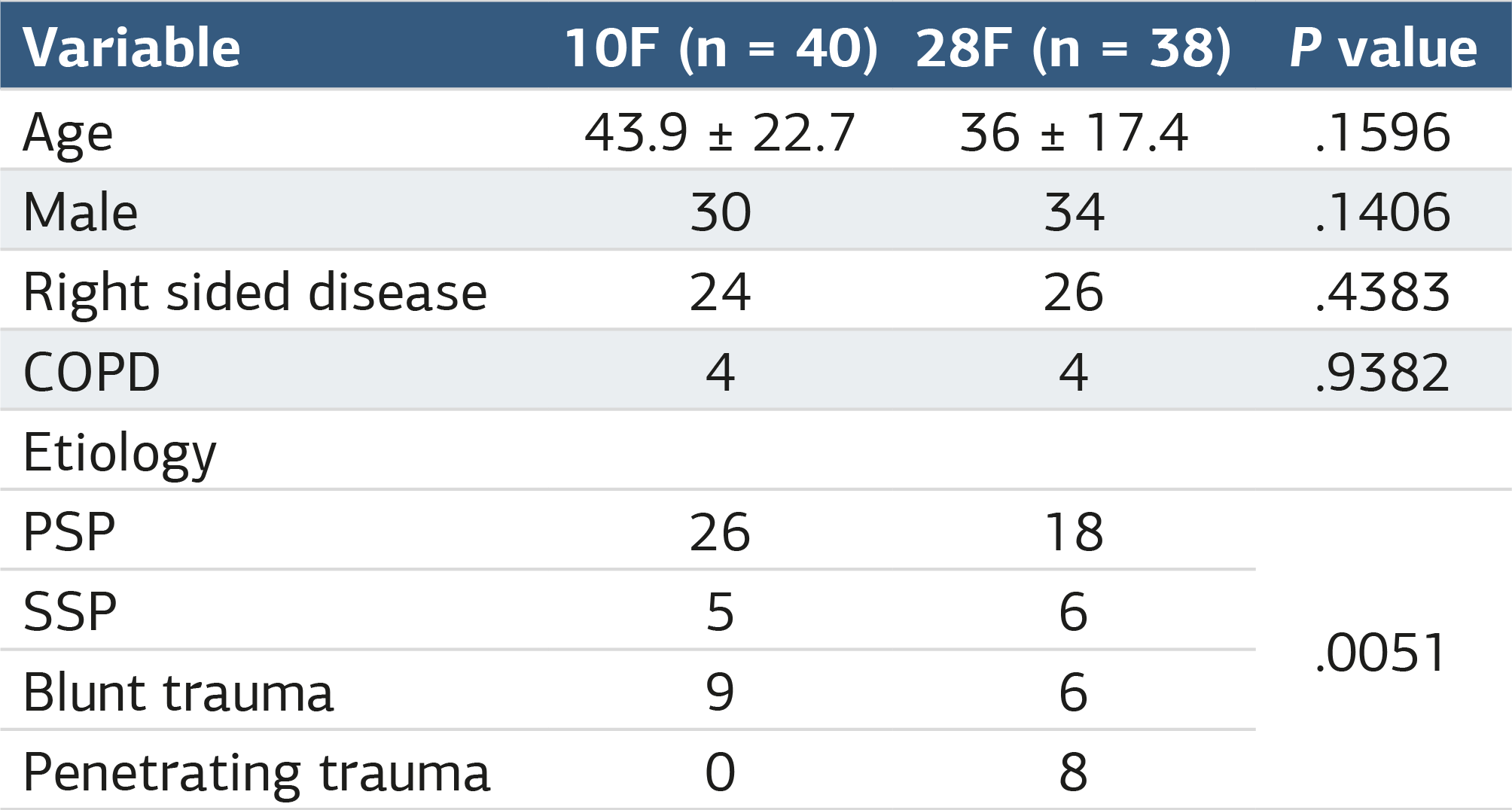
COPD, chronic obstructive pulmonary disease; PSP, primary spontaneous pneumothorax; SSP, secondary spontaneous pneumothorax.
Table 2. Comparison of clinical outcomes between 10F catheter and 28F chest tube groups
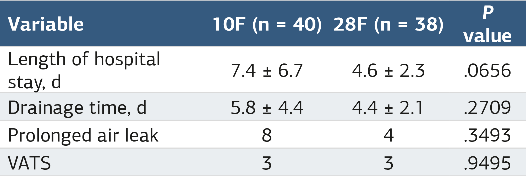
VATS, video assisted thoracic surgery.
Figures
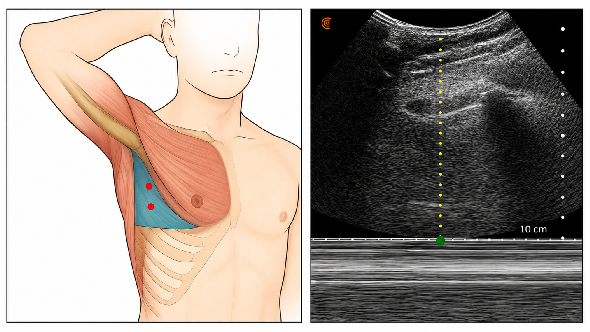
Figure 1. Anatomical landmark and thoracic ultrasound imaging demonstrating the ‘barcode sign’ indicative of pneumothorax with corresponding appropriate intervention site
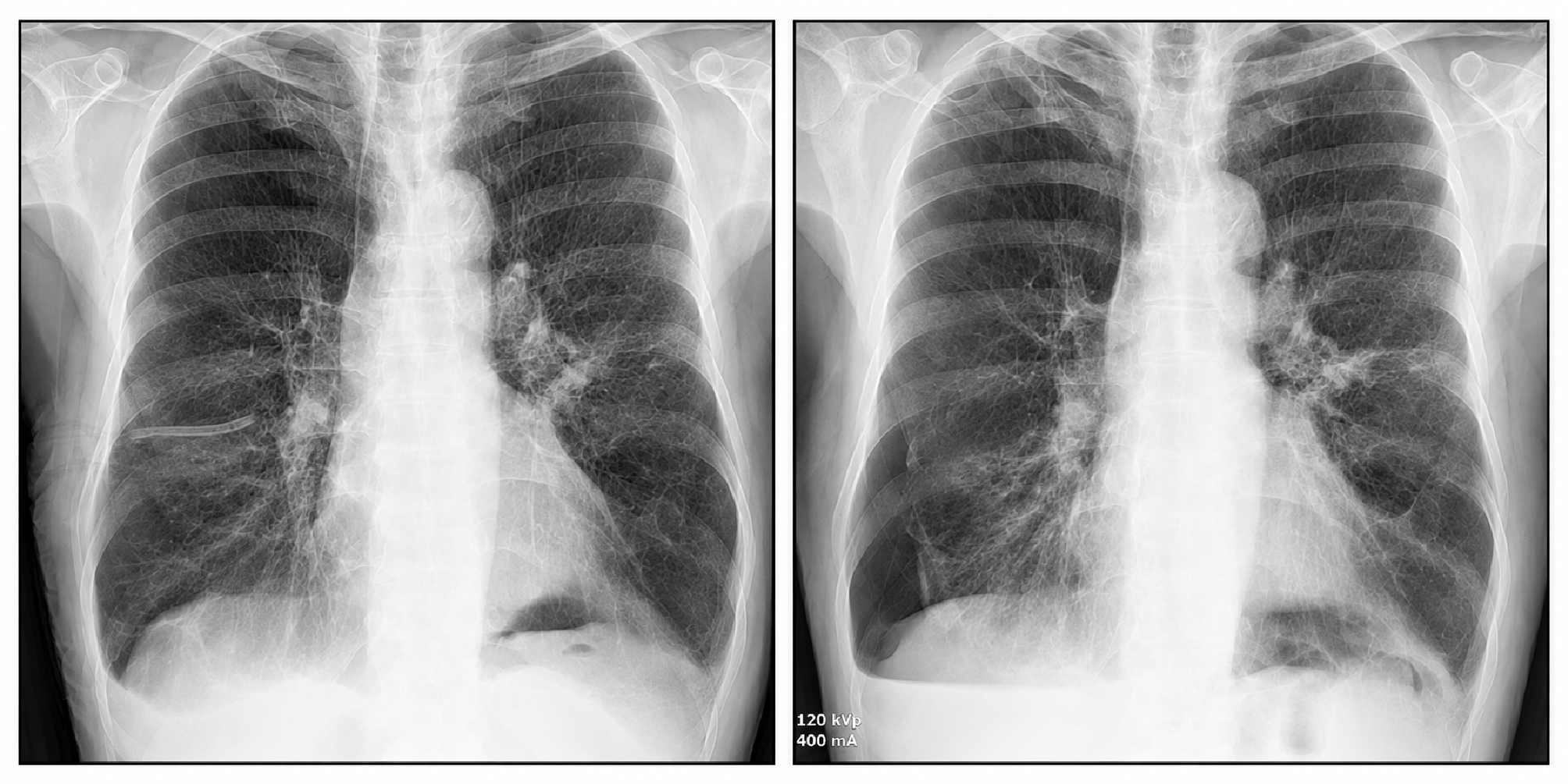
Figure 2. Right-sided pneumothorax: pre- and post-catheter insertion chest radiographs
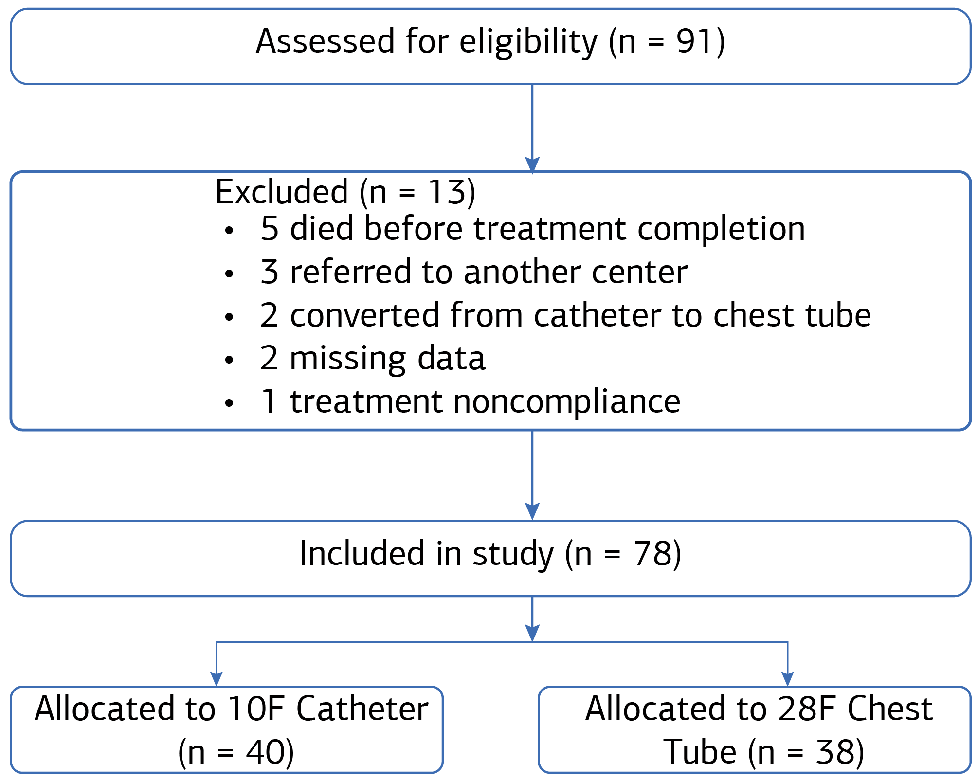
Figure 3. Consort diagram
Additional Information
Publisher’s Note
Bayrakol MP remains neutral with regard to jurisdictional and institutional claims.
Rights and Permissions

This work is licensed under a Creative Commons Attribution-NonCommercial 4.0 International License (CC BY-NC 4.0). To view a copy of the license, visit https://creativecommons.org/licenses/by-nc/4.0/
About This Article
How to Cite This Article
İsmail Dal. Efficacy and safety of 10F percutaneous catheter versus 28F chest tube in pneumothorax: a retrospective comparative study. Ann Clin Anal Med 2025;16(9):637-640. doi:10.4328/ACAM.22800
Publication History
- Received:
- 03.07.2025
- Accepted:
- 04.08.2025
- Published Online:
- 12.08.2025
- Printed:
- 01.09.2025