
Abstract
IntroductionThis work reports the case of a large cyst of the extra hilar common bile duct with a paraduodenal location in a female child, and takes stock of this malformative pathology of the bile ducts which can compromise the patient’s functional hepato-bilio-pancreatic and vital prognosis mainly due to multi organ failure and/or serious septic conditions. Case PresentationThe symptoms are generally progressive with the possibility of inflammatory, cirrhotic and neoplastic complications. Surgery is the treatment of choice. ConclusionThis case study sheds light on this malformative pathology in order to become familiar with its clinical and radiological picture as well as its post-therapeutic prognosis.
Keywords
Introduction
Described for the first time in 1723 by VATER, cystic bile duct disease is part of congenital dilatations of the extrahepatic bile ducts.1 This is a rare, predominantly female condition, which exists before birth since more and more cases are currently diagnosed antenatally during periodic medical visits for pregnancy monitoring. The dilatation is saccular or fusiform in character with or without blockage of biliary drainage. A choledochal cyst is generally suspected in cases of extra hepatic cholestasis in children with triad: mass, pain, and jaundice. Pancreatic and cholangitis are frequently associated with risk of biliary cirrhosis and long-term malignant degeneration, hence the interest in reviewing this entity through this case study in order to acquire the elements necessary to investigate this pathology and prevent its complications. Bile duct cysts (BDC) is a rare congenital disorder, with an estimated prevalence ranging from 1 in 13,500 to 1 in 200,000 live births in Western countries, while the disease is more frequent in Asia.2,3 A marked female predominance has been widely recognized (female to male ratio 3:1).4
Case Presentation
This is a 6-year-old girl, with no particular pathological history, admitted for symptoms that had been developing for five days previously, consisting of moderately painful abdominal bloating with pallor. The clinical examination reveals jaundice with abdominal arching next to the right hypochondrium, the flank and the iliac fossa on the same side with sensitivity to palpation, all evolving in a febrile context measured at 38.2°.
The biological assessment reveals cholestasis without hepatic cytolysis, with hyperleukocytosis on the blood count predominantly neutrophils, and normal levels of lipase and amylase.
Ultrasound reveals a cystic formation projecting opposite the arch described above clinically, occupying the area of the hepatic hilum and extending downward to the right iliac fossa, with a thin wall and finely echogenic content, having intimate relationships with the liver and digestive system. The cholangio MRI complement reveals a cystic formation at the expense of the common bile duct with paraduodenal location (extra and intramural portions) compressing the normal lumen of the common bile duct which appears flattened with minimal dilation of the proximal intrahepatic bile ducts and infiltration of fat local peritoneal effusion without significant intraperitoneal effusion. A reflex paralytic ileus is also evidenced by an attraction of the digestive structures towards the right iliac fossa with the appearance of stasis of the intestinal contents. The pancreas and Wirsung are without any abnormality detectable by magnetic resonance. (Figure 1 , Figure 2 and Figure 3).
A laparotomy is performed on the little girl with the establishment of a bilio-digestive diversion by raising the jejunal loop, associated with drainage of the cystic formation (difficulty of resection given the intimate adhesion to the digestive tract and the risk of duodenal wound) and washing of the peritoneal cavity with the release of agglutinated loops.
The operative consequences are simple, marked by regression of the abdominal arch with normal recoloration of the skin and mucosa, resumption of transit and regression of the clinical-biological infectious state.
An MRI check is carried out 6 months and one year after the operation showing a functional bilio-digestive diversion with no sign of hepatic overload and persistence of residual dilatation of the proximal intrahepatic bile ducts. The biology was free of abnormalities during the two checks described above.Ethical ApprovalNot applicable.Statistical AnalysisNot applicable.
This case report was prepared in accordance with the CARE guidelines.
Discussion
The common bile duct cyst is the most common malformative cystic anomaly of the bile ducts in children. It is an anomaly detectable after the appearance of suggestive hepatobiliary symptoms or even during pregnancy in the antenatal stage.
The pathophysiology is essentially based on the malformative component of the duodenal intramural junction of the bile and pancreatic ducts, responsible for the erosion of the mucous layer by reflux of bile and pancreatic juice. The flow therefore expands the weakened wall in the zone of mucosal failure and thus forms a pseudo-reservoir whose contents promote compression, concretion and lithiasis pathology as well as superinfection. The evolution revealing the malformation is generally in the inflammatory direction causing pancreatitis or even cholangitis. However, early biliary cirrhosis can set in if the picture is blurred and the risk of malignant transformation is described in the long-term pictures.
A significant association between BDC is unanimously reported with a clear increasing age-related incidence. Cholangiocarcinoma (CC) is the most frequent histological type encountered. The incidence of synchronous CC associated with BDC is estimated to be 2.5–30%.4 and was 6% in the largest reported Western experience.5
The diagnosis is based on mapping the biliopancreatic ducts using cholangio-MRI, capable of providing a complete morphological assessment of the bile ducts, an analysis of their contents and an evaluation of the nearby local intraperitoneal state. The role of cholangio-MRI is also essential in monitoring patients in the medium and long term to investigate a possible anomaly at the early stage.
The treatment is based on the complete excision of all extrahepatic biliary structures, with the reestablishment of bilio-digestive continuity being ensured by a jejunal anastomosis, either in a Y shape or on the duodenum.
The added value of our case study lies in the dual-component management of the problem. The patient benefited from the implementation of a bilio-digestive diversion, which halted the bile replacement. On the other hand, the drainage of the bulky cystic bile duct dilatation was performed to eliminate the compression exerted by the stagnant bile contents.
Limitations
The main limitation of this case is that the experience is based on a single patient. In addition, the follow-up period was limited to one year, which does not allow evaluation of possible long-term complications.
Conclusion
Through this case study, it turns out that it is important to screen for malformations of the bile ducts in the antenatal period or before any sign of hepato-bilio-pancreatic call during the first weeks of life, in order to diagnose early cystic choledochal malformations instead of seeing them late at the complication stage.
Declarations
Animal and Human Rights Statement
All procedures performed in this study were in accordance with the ethical standards of the institutional and/or national research committee and with the 1964 Helsinki Declaration and its later amendments or comparable ethical standards.
Informed Consent
Written informed consent was obtained from the patient’s parent or legal guardian for publication of this case report.
Data Availability
The datasets used and/or analyzed during the current study are not publicly available due to patient privacy reasons but are available from the corresponding author on reasonable request.
Conflict of Interest
The authors declare that there is no conflict of interest.
Funding
None.
Abbreviations
BDC: Bile duct cyst
CC: Cholangiocarcinoma
MRI: Magnetic resonance imaging
References
- Vater A. Dissertation in Auguralis Medica. Poes Diss Qua Scirrhis Viscerum Dissert CS Ezlerus. University Library; 1723;70:19.
- Wiseman K, Buczkowski AK, Chung SW, Francoeur J, Schaeffer D, Scudamore CH. Epidemiology, presentation, diagnosis, and outcomes of choledochal cysts in adults in an urban environment. Am J Surg. 2005;189(5):527-531.
- Fishman D, Isenberg DA. Splenic involvement in rheumatic diseases. Semin Arthritis Rheum. 1997;27(3):141-155. doi:10.1016/s0049-0172(97)80013-3
- Söreide K, Körner H, Havnen J, Söreide JA. Bile duct cysts in adults. Br J Surg. 2004;91(12):1538-1548. doi:10.1002/bjs.4815
- Edil BH, Cameron JL, Reddy S, et al. Choledochal cyst disease in children and adults: a 30-year single-institution experience. J Am Coll Surg. 2008;206(5):1000-1008. doi:10.1016/j.jamcollsurg.2007.12.045
Figures
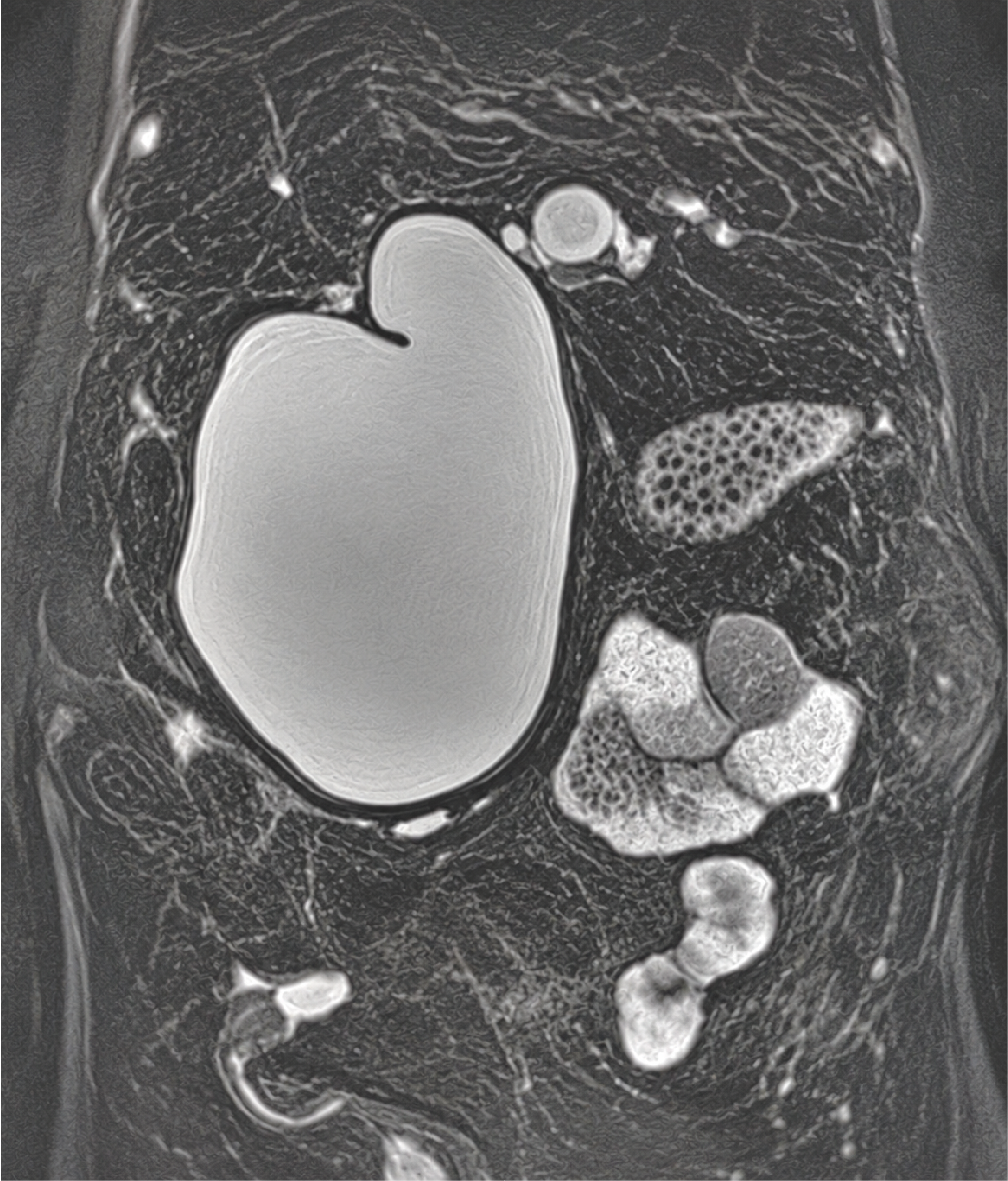
Figure 1. Coronal Section of the 3D BILI Sequence on MRI
Showing a large paraduodenal choledochal biliary cyst.
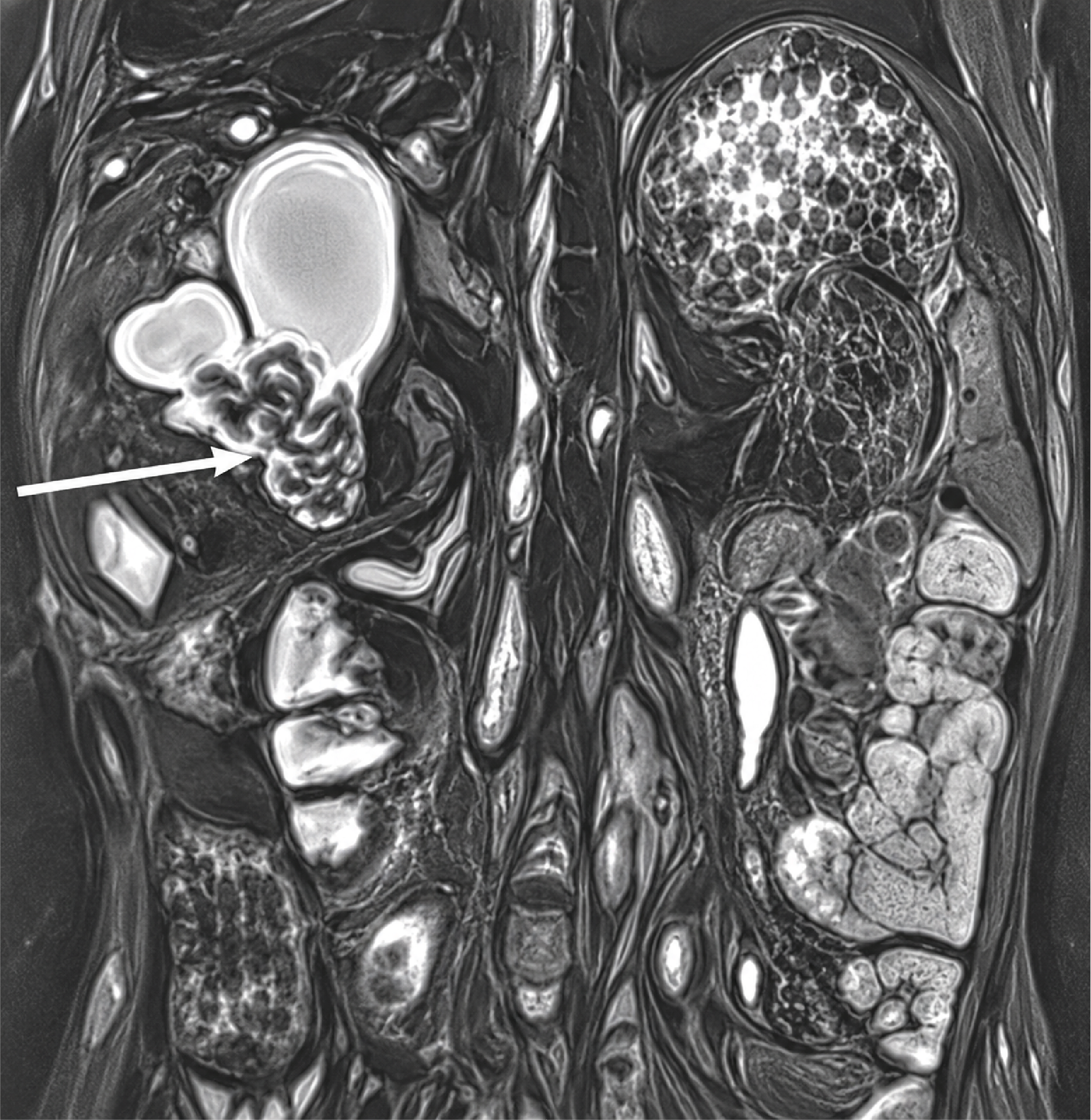
Figure 2. Coronal Section of the 3D BILI Sequence on MRI
Showing a functional bilio-digestive diversion with regression of the volume of the large biliary cyst after drainage.
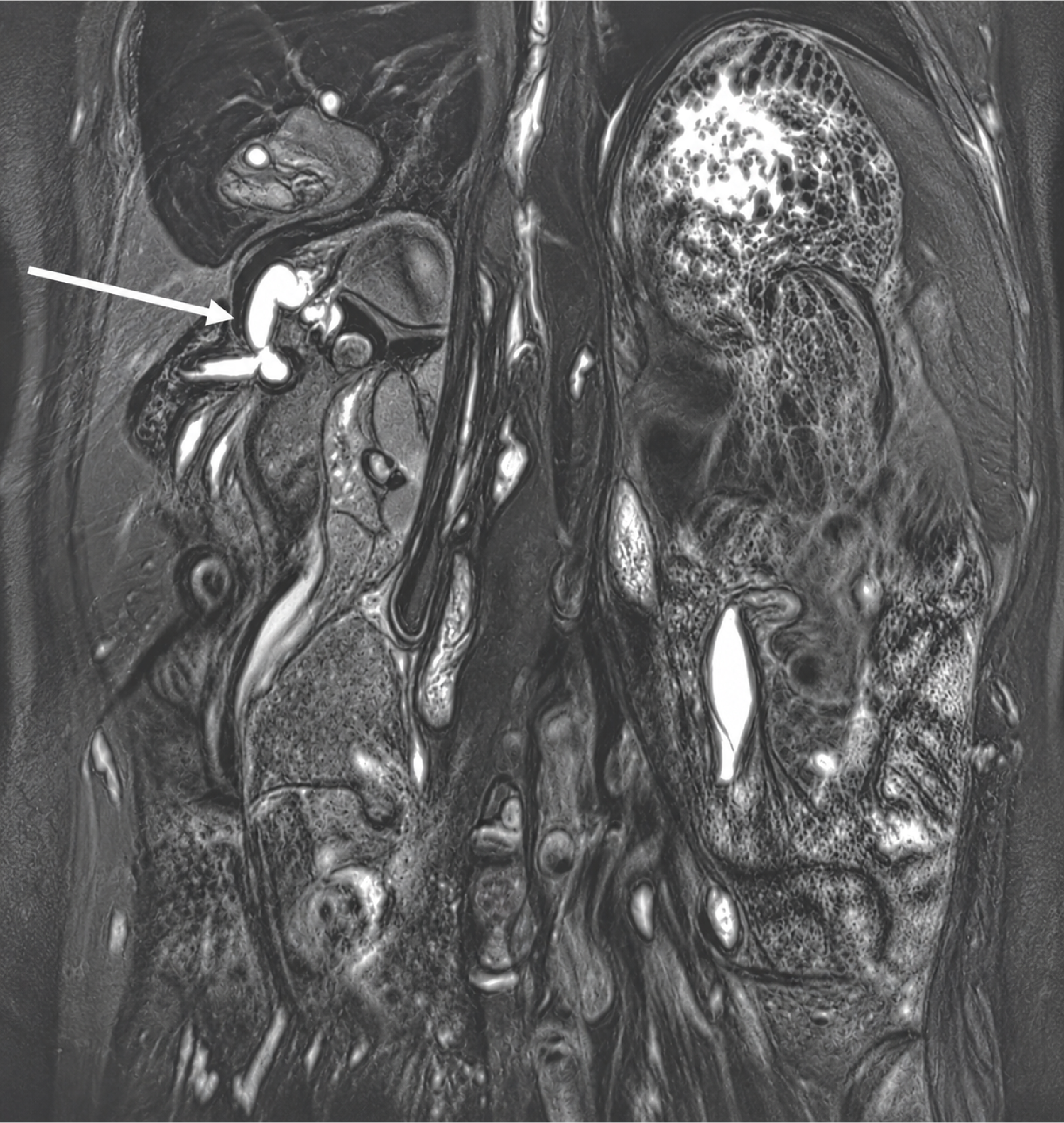
Figure 3. Coronal Section of the 3D BILI Sequence on MRI
Showing the persistence of a discreet ectasia of the proximal intrahepatic bile ducts.
Additional Information
Publisher’s Note
Bayrakol MP remains neutral with regard to jurisdictional and institutional claims.
Rights and Permissions

This work is licensed under a Creative Commons Attribution-NonCommercial 4.0 International License (CC BY-NC 4.0). To view a copy of the license, visit https://creativecommons.org/licenses/by-nc/4.0/
About This Article
How to Cite This Article
Soufiane Bigi, Mohamed Amine Baba, Ahmed Kharbach, Soukaina Wakrim. Biliary cyst of the paraduodenal bile duct: report of a case: A Case Report. Ann Clin Anal Med 2024;15(Suppl 2):79-81. doi:10.4328/ACAM.22171
Publication History
- Received:
- 08.03.2024
- Accepted:
- 13.05.2024
- Published Online:
- 11.09.2024
- Printed:
- 20.10.2024