
Abstract
AimIn this study, we aimed to analyze the expression of the Phosphatase and Tensin Homologous gene (PTEN) by interphase-fluorescence in situ-hybridization (iFISH), and localization and immunoreactivity of PTEN by immunocytochemistry method in bronchoalveolar lavage (BAL) fluid of patients with non-small cell lung cancer (NSCLC).MethodsBAL samples collected from 30 patients diagnosed with NSCLC and from 10 control subjects without any malignancy were included. iFISH was performed with a 10q23.3 PTEN DNA dual color FISH probe, analyzing multiple cells. Immunocytochemistry was based on the Streptavidin-Biotin-Peroxidase method and PTEN levels were semi-quantitatively scored.ResultsThe percentage of cells with 2G/2R was 86.8% which was significantly lower than that of the control group (P < .001). Loss of the PTEN gene was 6.43% among all patients. The percentage of cells with monoallelic deletion (P = .0246) and atypical signal (P = .001) and the percentage of monosomic cells (P = .001) were significantly higher in patients compared to controls. PTEN immunopositivity was significantly increased in patients compared to controls (P < .001). ConclusionIncreased abnormal signals and mutational cells in BAL samples of NSCLC patients suggest that the investigation of the PTEN gene in BAL by using iFISH may guide clinicians in differential diagnosis and therapeutic management of NSCLC. Being faster, easier and cost-effective in routine testing, the immunocytochemistry method can also be combined as a diagnostic tool with molecular methods.
Keywords
Introduction
Lung cancer, the most common cancer in the world, accounts for approximately a third of cancer-related deaths.1 Mostly derived from the primary bronchus tissue, the tumor is located centrally in the lung. Lung cancers are divided into two main groups according to the appearance of the cells observed under a microscope: small cell lung cancer (SCLC) and non-small-cell lung cancer (NSCLC). The NSCLC group is responsible for 80–85% of all lung cancer cases and is divided into three major subtypes: Squamous cell carcinoma (SCC), adenocarcinoma and large cell lung carcinoma (LCLC).2
The main molecular alterations observed in lung cancer are mutational activation of oncogenes, inactivation of tumor suppressor genes, alterations manifested in genes involved in cell cycle regulation and DNA repair.3,4 Loss, insertion or mutation of different gene loci responsible for the cell proliferation in cancer cells leads to initiation and progression of the tumor.
One of the genetic alterations in cancers is associated with Phosphatase and Tensin Homologous gene (PTEN) that is the best-known tumor suppressor gene, which has a key role of a complex intra-cellular phosphoinositide signaling network.5 PTEN is a critical regulator gene located in the q23.2 region of the long arm of the 10th chromosome and is involved in the regulation of multiple types of cancer.6 This gene is associated with familial cancer syndromes, as one of the alleles is inherently inactive, and the increased risk of cancer can be inherited from generation to generation in the form of an autosomal dominant.7
PTEN is involved in several processes, including aging, angiogenesis, apoptosis, cell cycle progression, cell contraction, and response to DNA damage.8 Loss of function of PTEN as a tumor suppressor protein is one of the most common events observed in a lot of types of cancer.9 Li et al. investigated the PTEN expression and its prognostic effects in primary NSCLC patients and reported that increased expression in NSCLC patients was associated with a good prognosis. They also found that a high level of the PTEN expression was correlated with a 43% reduction in mortality risk among all NSCLC patients.10
In our study, we aimed to analyze PTEN gene expression using the interphase Fluorescent in situ Hybridization (iFISH) method and localization and immunoreactivity levels of PTEN by immunocytochemistry (ICC) method in bronchial lavage (BAL) fluid of NSCLC patients.
Materials and Methods
Patient SelectionAmong the patients admitted to the Department of Chest Diseases in Istanbul University Cerrahpasa Medical Faculty due to suspicion for lung diseases, a total of 30 cases diagnosed with NSCLC were selected in a blind manner independent of age, gender and tumor stage, and these cases were designated as the patient group. The inclusion criteria were adequate samples with a diagnosis of NSCLC. The control group consisted of 10 individuals who had no viral disease, no histopathologically confirmed malignancy in the lung, no smoking, but who underwent bronchoscopy for any other indication. Bronchial lavage fluids were collected from the individuals selected for the patient and control groups, after signing the ‘Informed Consent Form’ for the study.
The demographics including age and gender were recorded. Cancer staging in patients was performed by a specialist pulmonologist, by using computerized tomography (CT) and positron emission tomography (PET) scans of the lungs, and according to the new 8th TNM staging system for lung cancer.11Sample CollectionThe BAL samples selected for the study were secretions aspirated through a bronchoscopic tube after injecting 20 ml of cold and sterile saline to the suspicious region for a lesion during a bronchoscopy. The first two tubes were used for iFISH analysis, and the third tube was used for the ICC method.IFISH Method10q23.3 PTEN DNA dual color FISH probe (MyProbes Powered by Cytocell, UK), which was specially designed and checked for quality was applied to the cells obtained from the BAL samples, according to the manufacturer’s instructions. The smears were analyzed under a fluorescence microscope with 100x objective, using Texas red, FITC (green) and DAPI filters, and visualized with the “Isis FISH Imaging System” program.Immunocytochemistry (ICC)ICC staining based on the Streptavidin-Biotin-Peroxidase method was applied by using Histostain-Plus Bulk Kit (Mouse and Rabbit Specific HRP Zymed Lab. Ins. San Francisco CA, USA) containing AEC (ABC) Detection IHC Kit (ab93705). The specificity of the immunostaining was confirmed by preparing a negative control slide in which all steps of ICC were applied except for the primary antibody, which was replaced by PBS. Positive immunostaining of PTEN in cells selected from ten random fields on slides was semi-quantitatively determined under a light microscope (LEICA DM500) at x40 magnification (-; no staining, +; poor staining, ++; moderate staining, +++; severe staining). According to this system, the scores were determined as (-): 0, (+): 1, (++): 2 and (+++): 3.Ethical ApprovalEthical approval for this study was obtained from the Ethics Committee of Istanbul University-Cerrahpasa (Date: 19.08.2016, Decision No. 66018902-050.99-306809).Statistical AnalysisFor all statistical analysis, GraphPad InStat Software (version 3.06) was used. The Kolmogorov-Smirnov test was used to test the normality of variables. Paired T-test was used to compare the two groups. One-way ANOVA test was used for multiple group comparisons. As a multiple comparison test for intragroup and intergroup, the Tukey-Kramer Multiple Comparisons test was applied. The significance level was chosen as P < .05.
Reporting GuidelinesThis study was reported in accordance with the STROBE guideline.
Results
PatientsThe patient group had 21 cases diagnosed with SCC (70%), 6 cases with adenocarcinoma (20%) and 3 cases with unclassified subtypes (10%) of NSCLC. 90% of patients were males (n = 27) with a mean age of 65.7 years for males and 54.6 years for females. All cases included in the control group (n = 10) were males, and the mean age was 60.6 years (Table 1).
According to the 8th staging guideline for NSCLC, 23.3% of the cases in the patient group were diagnosed at Stage IV (n = 7), 56.7% at Stage III (n = 17), 3.3% at Stage II of NSCLC (n = 1), however 16.7% (n = 5) could not be staged due to inadequate amount of sample or low differentiation of the tumor.iFISH FindingsThe chromosome insertions or losses were detected independently for each case. The microscopical images of the iFISH analysis were given in Figure 1. These images showed a cell containing a 3G3R signal in a case with adenocarcinoma (Figure 1A), an atypical cell containing a 6G3R signal in a case with adenocarcinoma (Figure 1B), a number of cells containing an atypical 1G2R signals in the case with squamous cell lung cancer (Figure 1C & 1D), a number of cells with monoallelic 2G1R, monosomy (1G1R) and normal (2G2R) signals in a case with squamous cell lung cancer. In the control group, a number of cells containing 2G2R signals and one cell with monoallelic deletion (2G1R) were observed.
Table 2 presents the comparison of the percentage of signals and cells in the samples of NSCLC and control cases. The median percentage of cells with normal signal in the control group was 93.7%, with monoallelic deletions 1.67%, the percentage of monosomic cells was 1.9% and with an atypical signal 2.68%, but no biallelic deletion was detected in the control group (Table 2). The median percentage of cells containing normal signal (2G/2R) in the patient group was 86.8%, while the median percentage of loss of PTEN gene was 6.43% in total, the median of monoallelic deleted cells was 2.67% and monosomic cells 3.76%. The percentage of total atypical signals was 5.67% in the patient group. In addition, we found that the percentage of cells containing normal signals in the patient group decreased by 11.2% compared to the control group (Table 2). The number of cells with atypical signals increased among patients at higher stages of lung cancer.
In the patient group, the median percentage of cells with biallelic deletion, monoallelic deletion, and percentage of monosomic cells and cells with atypical signals were significantly lower than the number of cells with normal signal (P < .001). This has also been observed in subtypes of NSCLC. In the control group, the median percentage of cells with monoallelic deletion, monosomic cells and atypical signals was significantly lower than the number of cells with normal signals (P < .001). The median percentage of normal cells in the patient group was significantly lower than that of the control group (P < .001). The number of cells with monoallelic deletion (P = .0246), monosomic cells (P = .001) and cells with atypical signal (P = .001) was significantly higher in the patient group compared to those of the control group (Table 2).Immunocytochemical FindingsThe micrographs of ICC staining for PTEN immunoreactivity observed in the BAL samples obtained from the patients are given in Figure 2. Epithelial cells in the control group had apparent nuclei with occasionally PTEN (+) heterochromatin regions and cytoplasm. PTEN (+) hyperplastic cells were observed around the epithelial cells (Figure 2A). The epithelial cells from the patients with SCC had PTEN (+) nucleus and dense cytoplasm or PTEN (+) epidermoidal-derived tumor cell accompanied with leukocytes. In the vicinity, PTEN (+) round macrophages with a granular cytoplasm were observed in adenocarcinoma cases (Figure 2B). Active PTEN (+) macrophages with very clear cell boundaries or damaged and aged PTEN (+) macrophages filled with granules or vacuolized juvenile macrophage cell were remarkable in the samples of patients with SCC.
The statistical comparison of the immunostainings for PTEN in the control and patient groups is presented in Table 3. PTEN immunopositivity was significantly increased in the patient group compared to the control group (P < .001). The immunopositivity was between moderate to severe in the patient group, while it was between poor to moderate in the control group. According to the subtypes of NSCLC diagnosed in the patient group (SCC, adenocarcinoma, and unclassified subtype), there was a significant increase in PTEN immunopositivity in samples of patients with SCC and unclassified subtypes compared to that of the control group (P < .001) (Table 3).
Discussion
Studies of the PTEN tumor suppressor gene have accelerated in the last decade, and it has been reported to have an important role in the progression of many cancers.12,13 The present findings of iFISH and ICC analysis of PTEN expression in BAL samples from the patients with NSCLC showed that the signal characteristics of cells can alter among cancer patients. To the best of our knowledge, our study is the first report showing a significantly higher percentage of cells with biallelic deletion, atypical signal, monoallelic deletion and a higher percentage of monosomic cells in the BAL samples of NSCLC patients than those of the control group. In contrast, the immunoreactivities of PTEN were significantly higher in samples of NSCLC patients than in the control group, and each patient had different intensities of cytoplasmic and nuclear PTEN expression.
Loss of PTEN expression was significantly more common in SCC cells, but PTEN expression was shown to have a significant effect on disease-free survival rate only in adenocarcinomas.14 In the present study, the percentage of normal signals for PTEN expression in the samples of patient with NSCLC was significantly lower than in control cases. This decrease was observed in both subtypes of SCC and adenocarcinoma and even in unclassified NSCLC. More to the point, the percentages of cells with atypical signals, monoallelic deletions and of monosomic cells for PTEN expression were significantly higher than in the control group. These increases were observed in both subtypes of SCC and adenocarcinoma and even in unclassified NSCLC.
To study copy number variations of PTEN expression in NSCLC, previous studies have used FISH method in biopsies of resected or directly harvested cells from lung tissues of patients or on cell lines commercially purchased.15 Nowadays, short-read and long-read DNA sequencing technologies are integrated into molecular techniques to find out the alterations in the PTEN gene in NSCLC.16 We found that the total number of cells obtained from all samples decreased significantly compared to the control group, but no statistical significance was detected. We also demonstrated that the rate of monoallelic deletion, monosomic cell and atypical signaling decreased significantly in the control group compared to the NSCLC group.
As a prognostic biomarker, loss of PTEN has been reported to be useful for identifying low-risk cancer patients who are prone to progression of the disease and therefore require treatment. Xiao et al. demonstrated that decreased expression of PTEN correlated with poor overall survival in NSCLC patients and was indicative of a poor prognosis for disease-free survival and progression-free survival in patients with NSCLC.15 In the present study, we observed the percentage of loss of PTEN gene as 6.74% in all patients with advanced stage of NSCLC, namely Stage-III and IV patients.
In the current study, we observed a significant increase in PTEN expressions immunocytochemically in the bronchial lavage fluids of NSCLC patients.
Limitations
As a limitation of present study, we did not follow-up patients and could not correlate the iFISH and ICC findings with the prognosis of the disease. Unfortunately, it is still unclear how nuclear and cytoplasmic PTEN expression correlates with disease prognosis exactly and how it affects patient survival. Although the expression of nuclear PTEN represents a new observation with interesting results of our study, it is undoubted that more precise and comprehensive studies including a large number of cases are needed to confirm these results.
Conclusion
Increased abnormal signals and mutational cells in BAL samples of NSCLC patients suggest that the investigation of the PTEN gene in BAL by using iFISH may guide clinicians in differential diagnosis and therapeutic management of NSCLC. Being faster, easier and cost-effective in routine testing, the immunocytochemistry method can also be combined as a diagnostic tool with molecular methods. In light of our findings of ICC showing the altered immunoreactivity of PTEN in cells collected from BAL samples, it is suggested that ICC staining method can be used as a diagnostic tool in terms of being faster, easier and more economical in routine testing. Acknowledging the molecular value of the iFISH technique, which provides more sensitive results than ICC, the investigation of cells that transferred from the lung tissue to BAL by using molecular cytogenetic methods will guide the clinicians in the management of the treatment of NSCLC. Indeed, more comprehensive and detailed studies planned with more parameters will allow a better understanding of the differentiation of lung cells in the tumorigenesis process at the molecular level focusing on whether PTEN expression could be a biomarker in terms of predicting the clinical stage and prognosis of NSCLC patients.
Declarations
Animal and Human Rights Statement
All procedures performed in this study were in accordance with the ethical standards of the institutional and/or national research committee and with the 1964 Helsinki declaration and its later amendments or comparable ethical standards.
Informed Consent
Written informed consent was obtained from all participants before enrollment in the study.
Data Availability
The datasets used and/or analyzed during the current study are not publicly available due to patient privacy reasons but are available from the corresponding author on reasonable request.
Conflict of Interest
None of the authors received any type of financial support that could be considered potential conflict of interest regarding the manuscript or its submission.
Funding
This study was supported by Istanbul University Scientific Research Projects Unit. Project No: 23004.
Acknowledgements
The authors are grateful to Dr. Şükriye Yılmaz and Dr. Ayşe Cirakoglu for their excellent FISH cell counting. The author would like to thank Dr. Elif Yaprak for the English language review and contribution to the experimental Immunocytochemistry design.
Abbreviations
AC: Adenocarcinoma
AEC: 3-amino-9-ethylcarbazole
BAL: Bronchoalveolar lavage
CT: Computed tomography
DAPI: 4′,6-diamidino-2-phenylindole
DNA: Deoxyribonucleic acid
FITC: Fluorescein isothiocyanate
HRP: Horseradish peroxidase
ICC: Immunocytochemistry
LCLC: Large cell lung carcinoma
NSCLC: Non-small cell lung cancer
PBS: Phosphate-buffered saline
PET: Positron emission tomography
PTEN: Phosphatase and tensin homolog
SCC: Squamous cell carcinoma
SCLC: Small cell lung cancer
SD: Standard deviation
TNM: Tumor, node, metastasis
References
- Wong MCS, Lao XQ, Ho KF, Goggins WB, Tse SLA. Incidence and mortality of lung cancer: global trends and association with socioeconomic status. Sci Rep. 2017;7(1):1-9.
- Zappa C, Mousa SA. Non-small cell lung cancer: current treatment and future advances. Transl Lung Cancer Res. 2016;5(3):288-300. doi:10.21037/tlcr.2016.06.07
- Schramm M, Wrobel C, Born I, et al. Equivocal cytology in lung cancer diagnosis: improvement of diagnostic accuracy using adjuvant multicolor FISH, DNA-image cytometry, and quantitative promoter hypermethylation analysis. Cancer Cytopathol. 2011;119(3):177-192. doi:10.1002/cncy.20142
- Wadowska K, Bil-Lula I, Trembecki L, Sliwinska-Mosson M. Genetic markers in lung cancer diagnosis: a review. Int J Mol Sci. 2020;21(13):1-22. doi:10.3390/ijms21134569
- Chow JT, Salmena L. Recent advances in PTEN signalling axes in cancer. Fac Rev. 2020;9:1-22.
- Bazzichetto C, Conciatori F, Pallocca M, et al. PTEN as a prognostic/predictive biomarker in cancer: an unfulfilled promise? Cancers (Basel). 2019;11(4):1-14. doi:10.3390/cancers11040435
- Yamamoto H, Yatabe Y, Toyooka S. Inherited lung cancer syndromes targeting never smokers. Transl Lung Cancer Res. 2018;7(4):498-504. doi:10.21037/tlcr.2018.06.01
- Abdulkareem IH, Blair M. Phosphatase and tensin homologue deleted on chromosome 10. Niger Med J. 2013;54(2):79-86. doi:10.4103/0300-1652.110033
- Jiang N, Dai Q, Su X, Fu J, Feng X, Peng J. Role of PI3K/AKT pathway in cancer: the framework of malignant behavior. Mol Biol Rep. 2020;47(6):4587-4629. doi:10.1007/s11033-020-05435-1
- Li X, Yang Y, Zhang H, et al. High levels of phosphatase and tensin homolog expression predict favorable prognosis in patients with non-small cell lung cancer. Cell Biochem Biophys. 2015;73(3):631-637. doi:10.1007/s12013-015-0671-z
- Choi HS, Jeong BK, Jeong H, et al. Application of the new 8th TNM staging system for non-small cell lung cancer treated with curative concurrent chemoradiotherapy. Radiat Oncol. 2017;12(1):1-8.
- Lotan TL, Gurel B, Sutcliffe S, et al. PTEN protein loss by immunostaining: analytic validation and prognostic indicator for a high-risk surgical cohort of prostate cancer patients. Clin Cancer Res. 2011;17(20):6563-6573. doi:10.1158/1078-0432.ccr-11-1244
- Rolfo C, Mack P, Scagliotti GV, et al. Liquid biopsy for advanced NSCLC: a consensus statement from the International Association for the Study of Lung Cancer. J Thorac Oncol. 2021;16(10):1647-1662. doi:10.1016/j.jtho.2021.06.017
- Yanagawa N, Leduc C, Kohler D, et al. Loss of phosphatase and tensin homolog protein expression is an independent poor prognostic marker in lung adenocarcinoma. J Thorac Oncol. 2012;7(10):1513-1521. doi:10.1097/jto.0b013e3182641d4f
- Xiao J, Hu CP, He BX, et al. PTEN expression is a prognostic marker for patients with non-small cell lung cancer: a systematic review and meta-analysis of the literature. Oncotarget. 2016;7(36):832-840. doi:10.18632/oncotarget.11068
- Sakamoto Y, Xu L, Seki M, et al. Long-read sequencing for non-small-cell lung cancer genomes. Genome Res. 2020;30(9):1243-1257. doi:10.1101/gr.261941.120
Tables

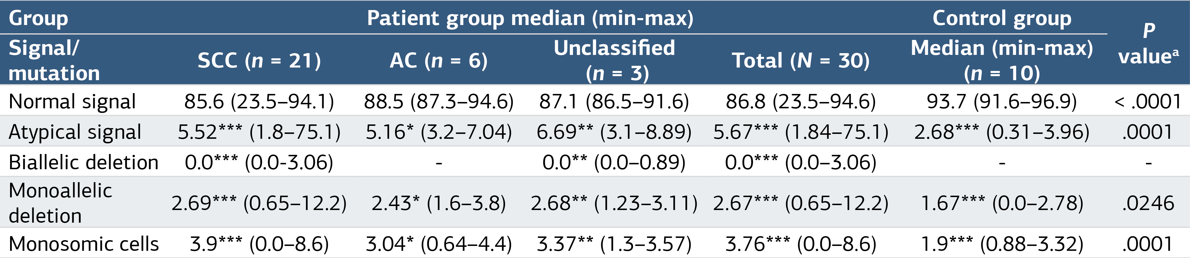
SCC, Squamous cell carcinoma; NSCLC, non-small cell lung cancer; AC, Adenocarcinoma; All values are given in Median [Range]. *P < .05, *P < .01, ***P < .001 vs corresponding normal signal. a Data of all patient groups were compared with the data of the control group
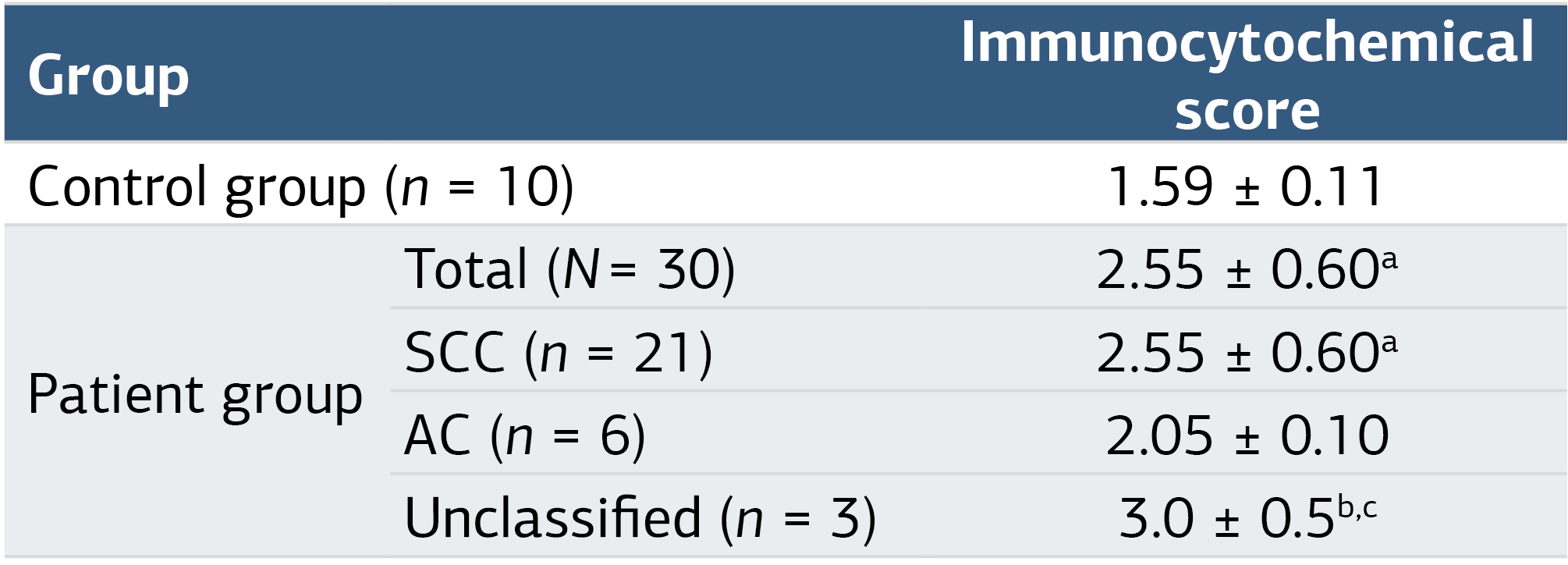
SCC, Squamous cell carcinoma; NSCLC, non-small cell lung cancer; AC, Adenocarcinoma aP < .01, bP < .001 vs the control group; cP < .05 vs adenocarcinoma
Figures
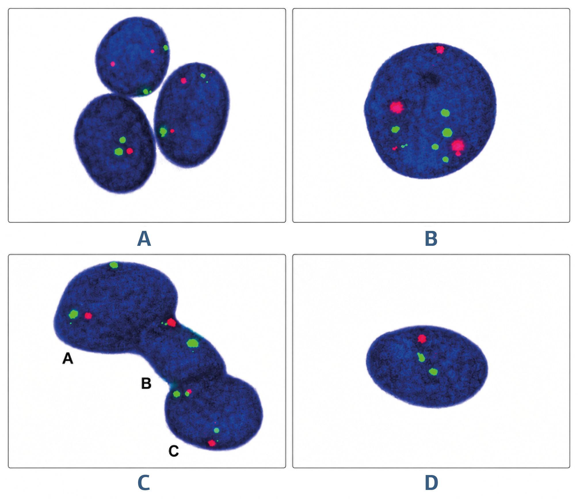
Figure 1. iFISH images of the samples of bronchoalveolar lavage fluids collected from the patient group diagnosed with non-small cell lung cancer (A-D). A) One cell containing a 3G3R signal from a case with adenocarcinoma. B) One atypical cell containing a signal (6G3R) from a case with adenocarcinoma. C) One cell containing an atypical signal (1G2R) from a case with squamous cell lung cancer. D) A case with squamous cell lung cancer showing most cells with atypical signals (1G2R). Magnification: x100.
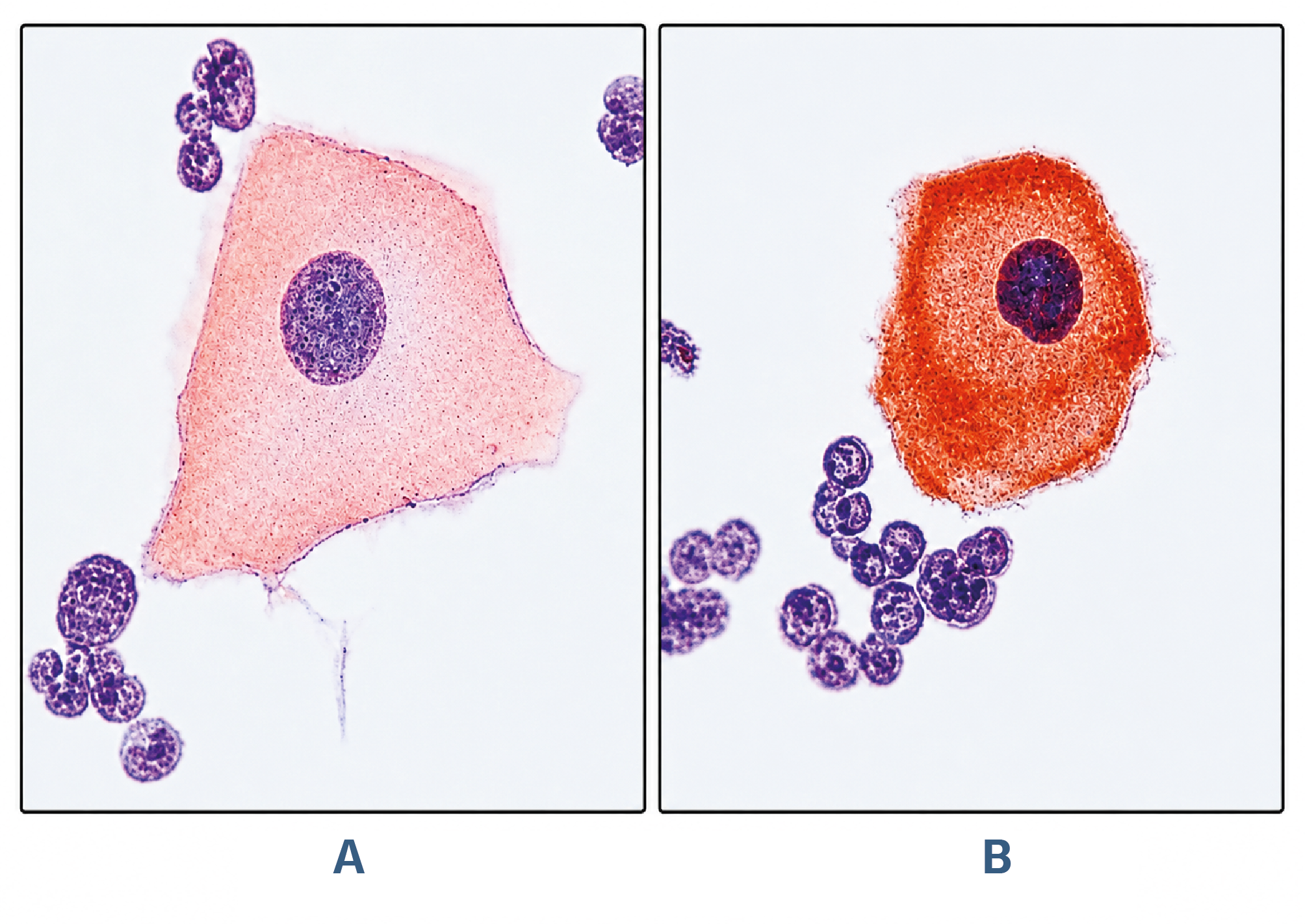
Figure 2. Micrographs of immunostaining for PTEN in the samples of bronchoalveolar lavage fluids collected from the control group (A) and the patient group diagnosed with nonsmall cell lung cancer (B). A) An epithelial cell from the control group, having an apparent nucleus and cytoplasm. B) PTEN (+) staining is observed in occasional parts of the heterochromatin regions in the nucleus (→). Counterstaining: Hematoxylin (x100).
Additional Information
Publisher’s Note
Bayrakol MP remains neutral with regard to jurisdictional and institutional claims.
Rights and Permissions

This work is licensed under a Creative Commons Attribution-NonCommercial 4.0 International License (CC BY-NC 4.0). To view a copy of the license, visit https://creativecommons.org/licenses/by-nc/4.0/
About This Article
How to Cite This Article
Hilal Sahin, Dilhan Kuru, Nail Yilmaz, Ersan Atahan, Ayhan Deviren, Matem Tuncdemir. Alterations in expression of phosphatase and tensin homologous (PTEN) gene analyzed in the bronchial lavage samples of non-small cell lung cancer patients. Ann Clin Anal Med 2024;15(1):11-15. doi:10.4328/ACAM.21695
Publication History
- Received:
- 17.03.2023
- Accepted:
- 05.06.2023
- Published Online:
- 20.11.2023
- Printed:
- 01.01.2024