
Abstract
AimThe aim of this study was to retrospectively investigate the perioperative features and clinical outcomes in cesarean section (C/S) patients who underwent endotracheal intubation using direct laryngoscopy and Glidescope® videolaryngoscopy (GSVL).MethodsThis retrospective study was performed using data gathered from the medical files of 179 C/S patients who underwent C/S under general anesthesia. After the induction of anesthesia with intravenous injection of propofol 2 mg/kg, and vecuronium 0.1 mg/kg, orotracheal intubation was performed using either direct laryngoscopy or GSVL. The patients underwent C/S after endotracheal intubation using either direct laryngoscopy or GSVL. Group I (n = 47) was intubated via direct laryngoscopy, while Glidescope® was used in Group II (n = 132).ResultsBaseline descriptives, craniofacial morphological measurements, duration of intubation and number of attempts for intubation, Cormack Lehane and Mallampati scores, as well as hemodynamic and respiratory parameters including blood pressure, heart rate, oxygen, and carbon dioxide levels were compared between two groups. The interincisal mouth opening (P = .003) and CO₂ levels (P = .023) were increased in Group II. In Group I, the number of patients with protruding front teeth was higher than that in Group II (P = .043).ConclusionOur results demonstrated that the GSVL could be a safe, effective, and practical device for endotracheal intubation in patients scheduled for C/S. Our data imply that GSVL can be incorporated into routine clinical practice in obstetric anesthetic practice for C/S since it allows improved visualization of the larynx in pregnants without bringing any significant burden.
Keywords
Introduction
Challenges encountered in the management of the airway and its associated risks of aspiration, maternal, and fetal hypoxia are the significant causes of morbidity and mortality associated with general anesthesia.1 Pregnant women are known to have a failure rate for endotracheal intubation that is 5 to 7 times higher than that of non-pregnant women.2
When a pregnancy is planned for a cesarean section (C/S), access to the airway can be challenging, especially when the typical anatomy is distorted and prevents the upper airway from being properly directly visualized with a laryngogram. In order to improve intubation, fiberoptic and videoscopic techniques can make it easier to image the laryngeal inlet. For patients who need tracheal intubation, videolaryngoscopy has the potential to boost the success rate of the procedure. The first videolaryngoscope to be sold commercially was the Glidescope®. The camera on the hyperangulated blade is linked to a video screen, which enhances larynx visibility. The utility of Glidescope® videolaryngoscopy (GSVL) must be considered in case of failure of direct laryngoscopy and blind intubation.3
When compared to direct laryngoscopy, tracheal intubation performed by doctors with little intubation experience (10 intubations per year) using the Glidescope® videolaryngoscope has a greater first-pass success rate and takes less time to complete.3 Video technology has recently been introduced to facilitate the management of the upper airway since it provides a better vision for identification of the upper airway. Videoscopic intubation is contraindicated in high-grade upper airway obstructions and excessive secretions, which hinder the visualization of the upper airway. Therefore, anesthesiologists must be aware of the advantages and restrictions of GSVL for orotracheal intubation in pregnants scheduled for C/S.3,4
The purpose of the present study was to compare the clinical features and perioperative findings in C/S patients who underwent orotracheal intubation utilizing either direct laryngoscopy and GSVL in our academic center.
Materials and Methods
This retrospective study was performed by the Anesthesiology & Reanimation and Obstetrics & Gynecology Departments of our tertiary care center after the approval of the local institutional review board (10.03.2021/86). Data were extracted from medical files of 179 patients who underwent C/S under general anesthesia following orotracheal intubation with either GSVL (Group I, n=47) or direct laryngoscopy (Group II, n=132). Information regarding the age, height, weight, body mass index (BMI), pre-existenting or pregnancy-related diseases, American Society of Anesthesiologists (ASA) physical status, Mallampati, and Cormack Lehane laryngoscopic scores, number of attempts for intubation, duration of intubation and complications encountered during surgery were gathered from the hospital database. Intubation procedures were performed by two senior anesthesiologists with ≥ 5 years of anesthetic experience.
Exclusion criteria were known airway pathology, cervical spine injury or other contraindication to neck extension, small mouth opening, ASA score of III or higher, ischemic heart disease, cerebrovascular disease, respiratory disease, and body mass index (BMI) > 35 kg/m2. Patients planned for C/S with ASA scores of I or II were considered eligible for this study. According to routine practice, all patients were pre-oxygenated for 5 minutes before the induction of anesthesia. Anesthesia was induced with fentanyl 2 µg/kg, vecuronium 0.1 mg/kg, and propofol 2 mg/kg injected through intravenous route.5
The vast majority of patients (161, 89.9%) had a fasting period of 4 to 6 hours prior to C/S. The remaining 18 patients (10.1%) were anesthetized and operated without waiting for the fasting period due to the need for emergent C/S. Emergency C/S was performed due to fetal indications such as non-reassuring fetal status, fetal bradycardia, and prolapse of the umbilical cord or fetal limbs or maternal causes like placenta previa, ablatio placenta, eclampsia, previous uterine surgery, trauma, and uterine rupture. Failed tracheal intubation is described as unsuccessful attempts for placement of a tracheal tube using either direct laryngoscopy or alternative intubating equipment, the need to proceed with surgery with a non-elective unsecured airway or the need to abort intubation or surgery and awaken the patient before the surgery.6,7
The Glidescope® GSVL was prepared according to the manufacturer’s instructions. The surface of the GSVL blade was adequately lubricated with a silicone-based lubricant. We have used the Glidescope® (Saturn Biomedical, Burnaby, BC, Canada), which has been commercially available after 2002. It is used both in adult and pediatric orotracheal intubation practice. It is comprised of a MacIntosh type laryngoscope blade and a curved laryngoscope blade with a fixed micro-video camera. The video image is transmitted to a high-quality LCD monitor with a cable. Currently, GSVL is often used in intensive care units and emergency departments where unique airway challenges may often occur. It can be rapidly performed and it is resistant to fogging and does not necessitate force for lifting. The Glidescope® allows a visualized control of insertion and advancement of an endotracheal tube. It facilitates the insertion of nasogastric tubes and transesophageal echocardiographic probes. The technique for GSVL is similar to direct laryngoscopy, it can be readily performed by the experienced anesthesiologist and it is excellent for teaching purposes. The disadvantages of Glidescope® involve high cost compared to direct laryngoscopic devices, the need for training, the difficulty to overcome barriers for visualization of the laryngeal opening and need for adequate mouth opening.3 After successful intubation, the teeth, and oral cavity were examined for any signs of trauma. In case any bloodstains were detected on GSVL blade, direct laryngoscopy was performed to determine its source. GSVL intubation was omitted if patients were in Cormack Lehane grades III and IV and spO₂ decreased to < 95% due to prolonged intubation procedure. After recognition of the failure of intubation procedure with GSVL, 100% oxygen was supplied using a facemask.
All patients were followed up for 24 hours postoperatively. During follow-up, the mouth, pharynx, and larynx were examined using indirect laryngoscopy and a tongue blade. Complications were recorded. Baseline descriptives (age, body-mass index, history of smoking and previous surgery), craniofacial morphologic features (tooth morphology, inter incisor distance, thyromental distance, mandibulohyoid distance, neck range of motion), American Society of Anesthesiologists (ASA) scores as well as Mallampati and Cormack Lehane classes, postoperative dysphagia and sore throat, blood pressure, pulse rate, peripheral oxygen (O₂) saturation and carbon dioxide (CO₂) levels were recorded and compared between two groups.
Anthropometric criteria used to predict difficult airway involved thyromental distance, mandibulohyoid distance, and interincisor distances. Thyromental distance was described as the distance from the mentum to the thyroid notch, while mandibulohyoid distance was measured from the chin to hyoid. The interincisor distance was defined as the distance between the upper and lower incisors.8Ethical ApprovalThis study was approved by the local institutional review board (Date: 10.03.2021; Decision No: 86).Statistical AnalysisOur data were analyzed using the Statistical Package for Social sciences program version 21.0 (SPSS Inc., Chicago, IL, USA). Descriptive data have been expressed as mean, standard deviation, median, minimum and maximum for qualitative variables, while categorical variables have been given as numbers and percentages. Normality was assessed using the Kolmogorov-Smirnov test. The significance of the difference between the 2 groups was tested with either the T-test or the Mann-Whitney U test. The Chi-square test was employed to analyze the relationship between categorical variables. Analysis of variance (ANOVA) was performed to compare repeated measurements between groups and various time intervals. A P value <0.05 was considered statistically significant.Reporting GuidelinesThe study was reported in accordance with the STROBE guidelines.
Results
Our series consisted of 179 women (average age: 31.54±5.67 years). The body mass index was 32.04 ± 5.06. ASA status was I in 122 (68.2%) patients, while 57 patients (31.8%) had ASA status II. Comorbidities (hypertension, diabetes mellitus, etc.) were evident in 37 (20.7%) patients. C/S was primary in 54 (30.2%) cases, while 125 patients had repeat C/S (69.8%). An overview of baseline descriptive data and clinical features is presented in Table 1. Patients were allocated into 2 groups according to the method used for orotracheal intubation. Group I (n = 47) involved patients intubated using direct laryngoscopy, while Group II (n = 132) included patients who underwent GSVL for intubation.
In 4 patients who were planned for GSVL, 6 attempts for intubation failed. In one of these patients, tonsil laceration was detected. In one patient, the initial attempt for intubation with direct laryngoscopy resulted in failure, and intubation was successfully performed using GSVL.
All patients who underwent emergent C/S were successfully extubated after surgery, and no patients exhibited signs of aspiration of gastric contents. The establishment of a surgical airway was not needed and no maternal mortality was detected due to general anesthesia for C/S in this series.
Table 2 displays a comparative presentation of baseline descriptives and clinical features in Groups I and II. Mouth opening was found to be significantly higher in Group II (P = .003). Overbite deformity was more common in Group I (P = .043). The other variables under investigation did not exhibit any remarkable differences between patients intubated via direct laryngoscopy and GSVL. Hemodynamic and respiratory parameters including blood pressure, pulse rate, peripheral oxygen saturation, and carbon dioxide levels before and after intubation are demonstrated in Table 3. Blood pressure and pulse rate values before and after intubation did not exhibit any noteworthy differences between the 2 groups. Peripheral oxygen saturation after intubation was found to be increased compared to that before intubation for the whole study population (n = 179). Carbon dioxide levels in Group II were significantly higher than that in Group I (P = .023).
Discussion
The treatment of anesthesia for various types of surgery has changed as a result of recent advancements in general anesthesia. The prioritization of avoiding general anesthesia in C/S led to insufficient efforts being made to optimize and improve emerging procedures in this subgroup.9 Inability to breathe or oxygenate after general anesthesia was produced and problems with the airways associated with intubation failure are the leading causes of anesthesia-related mortality in pregnant women. Due to the anatomical and physiological changes that take place during pregnancy, pregnant women are more prone to desaturation, difficult mask breathing, and failed intubation. Currently, videolaryngoscope is the preferred method in the event of a failed intubation. Videolaryngoscopes increase the success rate of unexpectedly difficult intubation in patients who have failed traditional direct laryngoscopy by giving appropriate glottis and vocal cord visualization.1
In the operating theatre, the incidence of difficult tracheal intubations ranges from 1.2% to 3.8% in routine clinical practice, and increases to 5.3% in emergency situations. Up to 600 people are thought to have died from tracheal intubation-related problems. Particularly in patients with anatomical traits that can make tracheal intubation challenging, direct laryngoscopy does not always permit optimal viewing of the glottis.5,10 The Glidescope can be used for routine intubations and difficult or failed intubations both in children and adults. It can be suitable for the high anteriorly positioned larynx where direct laryngoscopy has failed and in conditions where cervical immobilization is necessary or mouth opening is limited or in case of airway trauma with blood or secretions in the airway.5
The anesthesiologists’ ability to manage the airway in pregnant patients planned for C/S is hampered by conditions such as shorter apnea times without desaturation, a higher risk of aspiration, tissue edema, mucosal hyperemia, and large breasts. Additionally, in a peripartum environment, the effects of intubation failure are more severe.11,12
Complications of videoscopic intubation involve palatal perforation and hemorrhage. GSVL must be remembered as an option in cases of failure of ventilation and oxygenation. Mouth opening is an important parameter to be remembered during the selection of the method for intubation. Orotracheal intubation using a GlideScope® offers advantages of an easy and simple operation, and satisfactory laryngeal view in patients with a difficult laryngoscopy.13 Trauma-related to intubation using the GlideScope is rare.13 Our results support that GSVL is a favorable and safe way to establish an airway in selected C/S patients.
We observed that mouth opening was significantly increased in patients who underwent intubation via GSVL. This difference may be attributed to the fact that GSVL cannot be performed in patients with a mouth opening < 18 mm. Similarly, overbite deformity may be a factor that restricts mouth opening, and its incidence may be in patients who underwent intubation with direct laryngoscopy. An increase in carbon dioxide levels in patients intubated with GSVL may be linked with the delay due to the failed initial attempt via direct laryngoscopy.
Hoshijima et al. reported that the hemodynamic response following tracheal intubation was not diminished by the GlideScope in comparison to the Macintosh laryngoscope.14 Our results yielded that there was no difference between the two groups concerning neither the blood pressure nor pulse rates. This may be due to effective suppression of the pressure response to orotracheal intubation by general anesthesia. Practice and familiarity with the equipment are critical points for the successful use of GSVL in case of failure of direct laryngoscopic intubation. The key to successful use is proper training and anticipation of situations when their use may be necessary for C/S patients.
Limitations
Our limitations include retrospective design, information confined to the experience of a single center, and possible variations in the grading of Mallampati and Cormack Lehane scores, and ASA status. Since our institution is tertiary care with teaching facilities, an anesthesia resident may initially try the first attempt intubation and senior residents or attending anesthesiologists may take charge of the intubation procedure. The personal preferences for GSVL as the initial method of intubation may have led to an underestimation of the incidence of intubation failures.
Conclusion
Increased awareness of possible risks, anticipation with challenging situations, familiarity with the new devices and improvement of skills with effective training programs are key points to optimize outcomes with general anesthesia and intubation procedures performed in C/S patients. The results of the present study yielded that orotracheal intubation via GSVL provides a safe, secure and practical method in C/S patients which may have additional risks and difficulties for the management of the upper airway.
Declarations
Animal and Human Rights Statement
All procedures performed in this study were in accordance with the ethical standards of the institutional and/or national research committee and with the 1964 Helsinki Declaration and its later amendments or comparable ethical standards.
Informed Consent
Written informed consent was waived due to the retrospective study design.
Data Availability
The datasets used and/or analyzed during the current study are not publicly available due to patient privacy reasons but are available from the corresponding author on reasonable request.
Conflict of Interest
The authors declare that there is no conflict of interest.
Funding
None.
Abbreviations
ASA: American society of anesthesiologists
BMI: Body mass index
C/S: Cesarean section
GSVL: Glidescope® videolaryngoscopy
SPSS: Statistical package for the social sciences
References
- Howle R, Onwochei D, Harrison SL, Desai N. Comparison of videolaryngoscopy and direct laryngoscopy for tracheal intubation in obstetrics: a mixed-methods systematic review and meta-analysis. Can J Anaesth. 2021;68(4):546-565. doi:10.1007/s12630-020-01908-w
- Lim MJ, Tan HS, Tan CW, et al. The effects of labor on airway outcomes with Supreme laryngeal mask in women undergoing cesarean delivery under general anesthesia: a cohort study. BMC Anesthesiol. 2020;20(1):213. doi:10.1186/s12871-020-01132-5
- Van Schuppen H, Wojciechowicz K, Hollmann MW, Preckel B. Tracheal intubation during advanced life support using direct laryngoscopy versus GlideScope videolaryngoscopy by clinicians with limited intubation experience: a systematic review and meta-analysis. J Clin Med. 2022;11(21):6291. doi:10.3390/jcm11216291
- Meshram TM, Ramachandran R, Trikha A, Rewari V. Haemodynamic responses following orotracheal intubation in patients with hypertension: Macintosh direct laryngoscope versus GlideScope videolaryngoscope. Indian J Anaesth. 2021;65(4):321-327. doi:10.4103/ija.ija_417_20
- Pascarella G, Caruso S, Antinolfi V, Costa F, Sarubbi D, Agrò FE. The VL3 videolaryngoscope for tracheal intubation in adults: a prospective pilot study. Saudi J Anaesth. 2020;14(3):318-322. doi:10.4103/sja.sja_145_20
- Govender I, Nzaumvila DK, Maphasha OM. Failed tracheal intubation in primary health care. S Afr Fam Pract (2004). 2022;64(1). doi:10.4102/safp.v64i1.5532
- Joffe AM, Aziz MF, Posner KL, et al. Management of difficult tracheal intubation: a closed claims analysis. Anesthesiology. 2019;131(4):818-829. doi:10.1097/aln.0000000000002815
- Rao KVN, Dhatchinamoorthi D, Nandhakumar A, Selvarajan N, Akula HR, Thiruvenkatarajan V. Validity of thyromental height test as a predictor of difficult laryngoscopy: a prospective evaluation comparing modified Mallampati score, interincisor gap, thyromental distance, neck circumference, and neck extension. Indian J Anaesth. 2018;62(8):603-608. doi:10.4103/ija.ija_162_18
- Choi SU. General anesthesia for cesarean section: are we doing it well? Anesth Pain Med (Seoul). 2022;17(3):256-261. doi:10.17085/apm.22196
- Tsan SEH, Lim SM, Abidin MFZ, Ganesh S, Wang CY. Comparison of Macintosh laryngoscopy in bed-up-head-elevated position with GlideScope laryngoscopy: a randomized controlled noninferiority trial. Anesth Analg. 2020;131(1):210-219. doi:10.1213/ane.0000000000004349
- Louis A, Tiwary MK, Sharma P, Nair AS. Comparison of postoperative pulmonary outcomes in patients undergoing cesarean section under general and spinal anesthesia: a single-center audit. Anesth Essays Res. 2021;15(4):439-442. doi:10.4103/aer.aer_6_22
- Dongare PA, Nataraj MS. Anaesthetic management of obstetric emergencies. Indian J Anaesth. 2018;62(9):704-709. doi:10.4103/ija.ija_590_18
- Turkstra TP, Turkstra DC, Pavlosky AW, Jones PM. Simultaneous en bloc endotracheal tube insertion with GlideScope Titanium video laryngoscope use: a randomized controlled trial. Can J Anaesth. 2020;67(11):1515-1523. doi:10.1007/s12630-020-01778-2
- Hoshijima H, Maruyama K, Mihara T, Boku AS, Shiga T, Nagasaka H. Use of the GlideScope does not lower the hemodynamic response to tracheal intubation more than the Macintosh laryngoscope: a systematic review and meta-analysis. Medicine (Baltimore). 2020;99(48). doi:10.1097/md.0000000000023345
Tables
Table 1. Baseline Descriptives Data
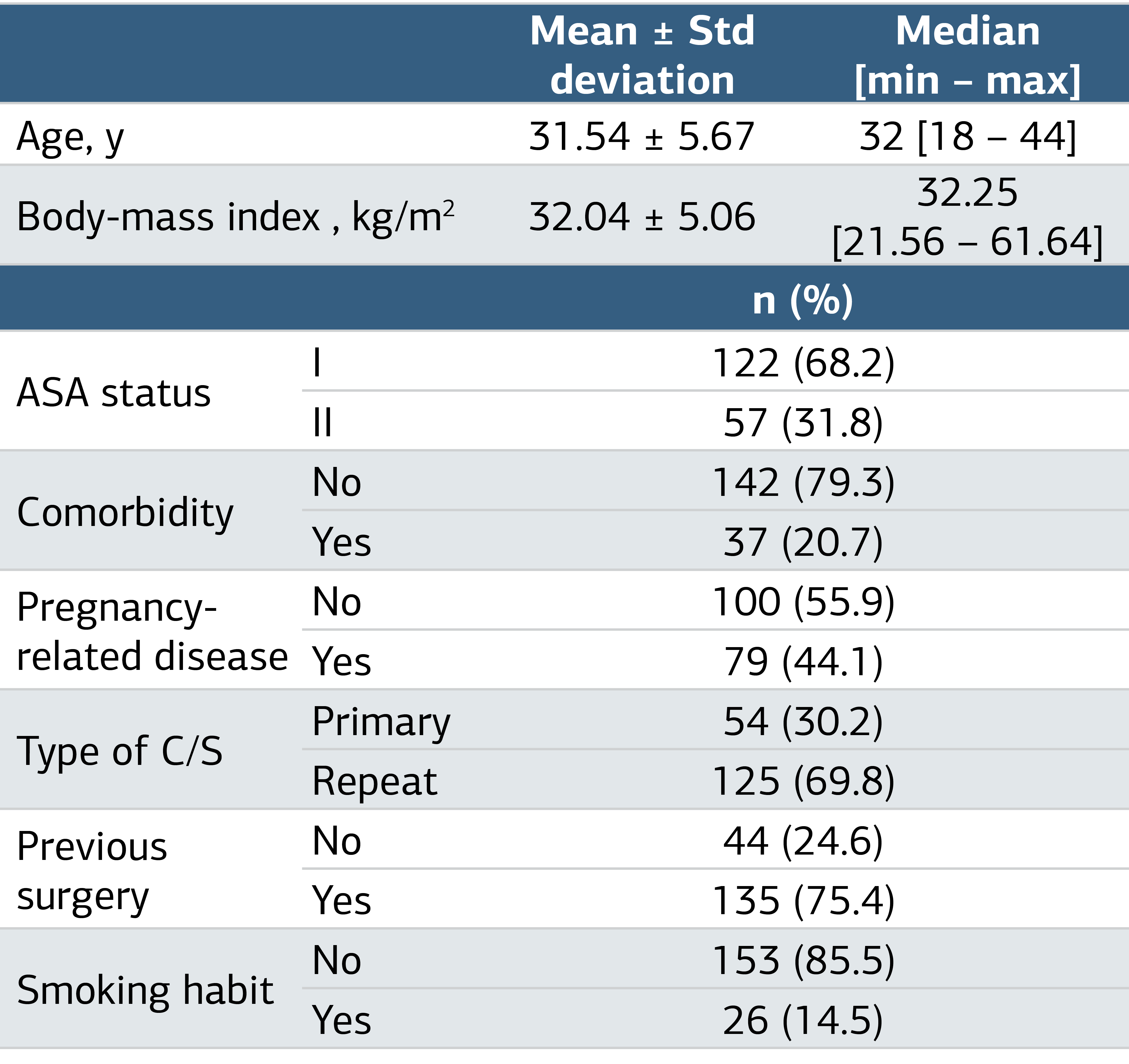
C/S: cesarean section; ASA: American Society of Anesthesiologists
Table 2. Comparison of Descriptive and Clinical Features Between 2 Groups
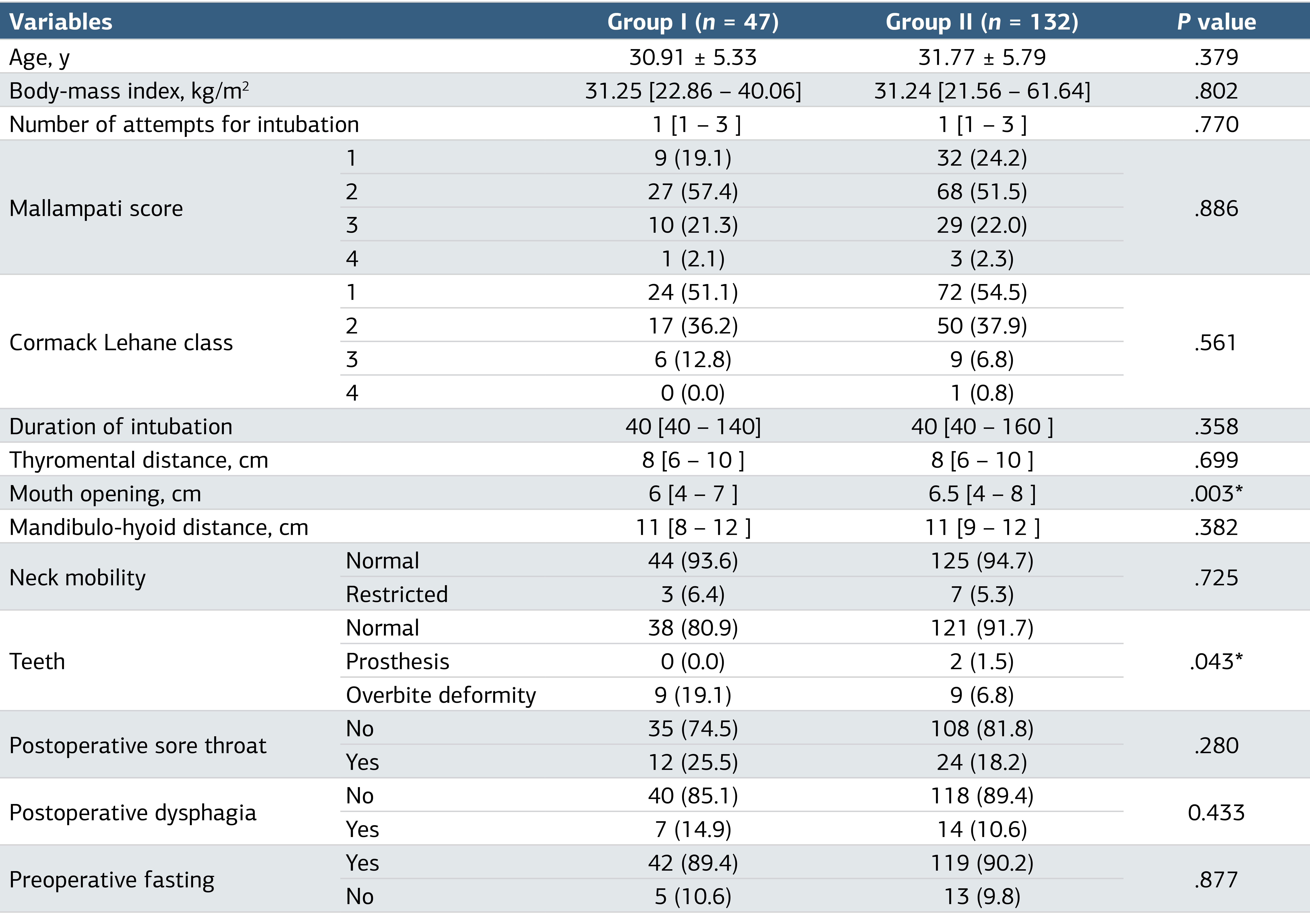
(Hint: *: statistically significant)
Table 3. Comparison of Hemodynamic and Respiratory Measurements with Respect to Groups and Time Interval
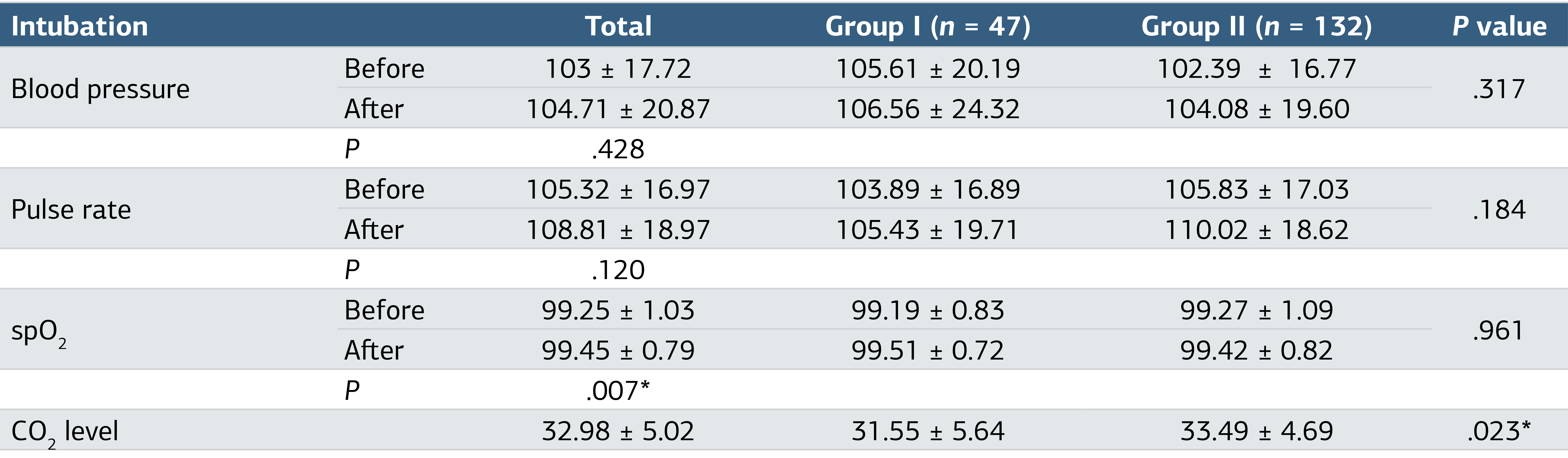
spO₂ : peripheral oxygen saturation; CO₂: carbon dioxide; *: statistically significant)
Additional Information
Publisher’s Note
Bayrakol MP remains neutral with regard to jurisdictional and institutional claims.
Rights and Permissions

This work is licensed under a Creative Commons Attribution-NonCommercial 4.0 International License (CC BY-NC 4.0). To view a copy of the license, visit https://creativecommons.org/licenses/by-nc/4.0/
About This Article
How to Cite This Article
Nevin Aydın, Osman Esen. Comparison of videolaryngoscopy (Glidescope®) and direct laryngoscopy for tracheal intubation for cesarean section. Ann Clin Anal Med 2024;15(2):71-75. doi:10.4328/ACAM.21310
Publication History
- Received:
- 12.07.2022
- Accepted:
- 07.02.2023
- Published Online:
- 07.12.2023
- Printed:
- 01.02.2024