
Abstract
AimIn this study, we used silver nanoparticles (AgNPs/Sw) synthesized from Salvia willeana to investigate the plant’s antioxidant properties and protective activities against aging, Alzheimer, obesity, and diabetes.MethodsPost-synthesis characterisations of AgNPs/Sw were confirmed using UV, FTIR, XRD and SEM methods. Firstly, aqueous, methanol and chloroform extracts of S. willeana were prepared, and the methanol extract was found to have the highest phenolic content. Therefore, methanol extract was used in all analyses of our study. We analysed the lipid peroxidation inhibitory activity, 1,1-Diphenyl-2-picrylhydrazyl (DPPH) free radical scavenging capacity, and iron reducing antioxidant power (FRAP) of AgNPs/Sw to determine their antioxidant properties. Acetylcholinesterase (AChE), butyrylcholinesterase (BChE), and amyloid-beta (Aβ) aggregation inhibition assays were performed to evaluate the anti-Alzheimer effects of AgNPs/Sw. Additionally, α-amylase, pancreatic lipase, and α-glucosidase inhibition activity tests were conducted to assess the protective effects of AgNPs/Sw against obesity and diabetes.ResultsSilver nanoparticles synthesised using S. willeana extract enhance the antioxidant, anti-aging, anti-Alzheimer, anti-obesity, and antidiabetic activities of the plant extract.ConclusionAgNPs/Sw can be applied for therapeutic purposes in medicine.
Keywords
Introduction
Salvia willeana grows endemically only in Cyprus and has been used for years as a functional tea that provides health benefits to prevent disease by strengthening human immunity. It is also used for abdominal pain, colds, and nausea. Compounds such as caffeic acid oligomers, phenolic, flavonoid and fatty acid derivatives have been isolated from Salvia willeana. Salvia species have been characterised as having antioxidant, antibacterial, anti-inflammatory, antidiabetic, anti-aging, and tumour suppressor properties due to their isolated phenolic and flavonoid chemical compositions.1-3 The phytochemical content of Salvia willeana extract has not been extensively analysed.
Recent scientific studies about nanoscience can make important contributions to medicine, including the diagnosis, treatment, and prevention of diseases, as well as drug development. Nanoparticles have been found to be effective as antimicrobial, antioxidant, antifungal, anticancer, and antidiabetic agents in the treatment of diseases. Green synthesis is preferred over chemical reactions for obtaining nanoparticles due to its cost-effectiveness, environmental friendliness, and ease of preparation. Silver nanoparticles are commonly used in health studies due to their unique optical, electrical, and thermal properties, as well as their antioxidant, antimicrobial, antifungal, and high catalytic activities.4
Natural herbal products are commonly used to treat diseases, including Alzheimer’s disease, obesity and diabetes. One treatment strategy for these diseases is to inhibit the enzymes, and Salvia species are a promising research topic due to their potential inhibitory effects on enzymes. Thus, our study aims to examine the in vitro antioxidant activity of Salvia willeana extract and silver nanoparticles and their protective effects against aging, Alzheimer’s disease, obesity, and diabetes.
Materials and Methods
All chemicals and silver-nitrate (AgNO3 ) used in the study were obtained from Merck (Germany). S. willeana was collected in May 2023 from the Troodos Mountain in Nicosia. Some of them were kept at the University Pharmaceutical Research Institute. They were dried and grounded with an electric grinder.Preparation Of S. Willeana ExtractFirstly, aqueous, methanol and chloroform SwE were prepared.5 For aqueous extraction, 50 g powdered S. willeana was added to a balloon flask and diluted with 250 mL distilled water. The mixture was boiled (15 min), cooled (20°C), and filtered through Whatman No. 1 filter paper. It was stored in the refrigerator (+4°C).
For methanol extraction, 50 g S. willeana powder was extracted with 95% methanol and filtered 5-7 times. It was then filtered through a Whatman No. 4 filter paper and concentrated using a rotary evaporator (IKA RV 10, Germany) at 50°C. It was dried and stored in a refrigerator (+4°C).
For chloroform extraction, 50 g S. willeana powder was placed in a balloon flask, and 300 mL chloroform was added. It was boiled (4 h) and filtered using Whatman No. 40 filter. It was concentrated in a rotary evaporator at 50°C, washed with warm hexane, yielding 4.55%.Phytochemical Analysis Of SwEThe phytochemical analysis of SwE, they were resuspended in sterile phosphate-buffered saline (pH 7.2) to 200 mg/mL final concentration.
The total phenolic content of SwE was determined using the Folin-Ciocalteu method.5 A blue solution was obtained by adding 50 μL each SwE to 5 mL Folin-Ciocalteu reagent and 5 mL Na2CO3. Then it was incubated (20°C/1 h), and the absorbance was measured at 760 nm using UV spectrophotometer (Shimadzu UV 1800 UV, Japan). Gallic acid was used as reference material, and the result was expressed as mg-gallic acid/g-SwE.
The total flavonoid content of the SwE extracts was determined using the aluminium-chloride method.5,6 Briefly, 60 μL SwE were mixed with 4 mL distilled water and 20 μL aluminium-chloride reagent. The final volume was adjusted to 5 mL with pure methanol. It was incubated (20°C/30 min), and the absorbance was measured at 430 nm, spectrophotometrically. Quercetin was used as a reference chemical, and the result was expressed as mg-quercetin/g-SwE.
The alkaloid content of SwE was analysed using the bromocresol-green (BCG) method and produced yellow-coloured solution.5,6 200 μL SwE was diluted with 2 N HCl to 2000 μL final volume. It was washed triplicate with 10 mL chloroform in separating funnel, and then neutralised with 0.1 N NaOH. After 10 min, 5 mL BCG solution and 5 mL phosphate-buffer were added. After shaking thoroughly, we made 10 mL chloroform dilutions of SwE. We measured its absorbance at 470 nm, spectrophotometrically. The results were calculated used the formula [alkaloids=(Abs-0.048)/0.021], expressed as mg-SwE/mL.
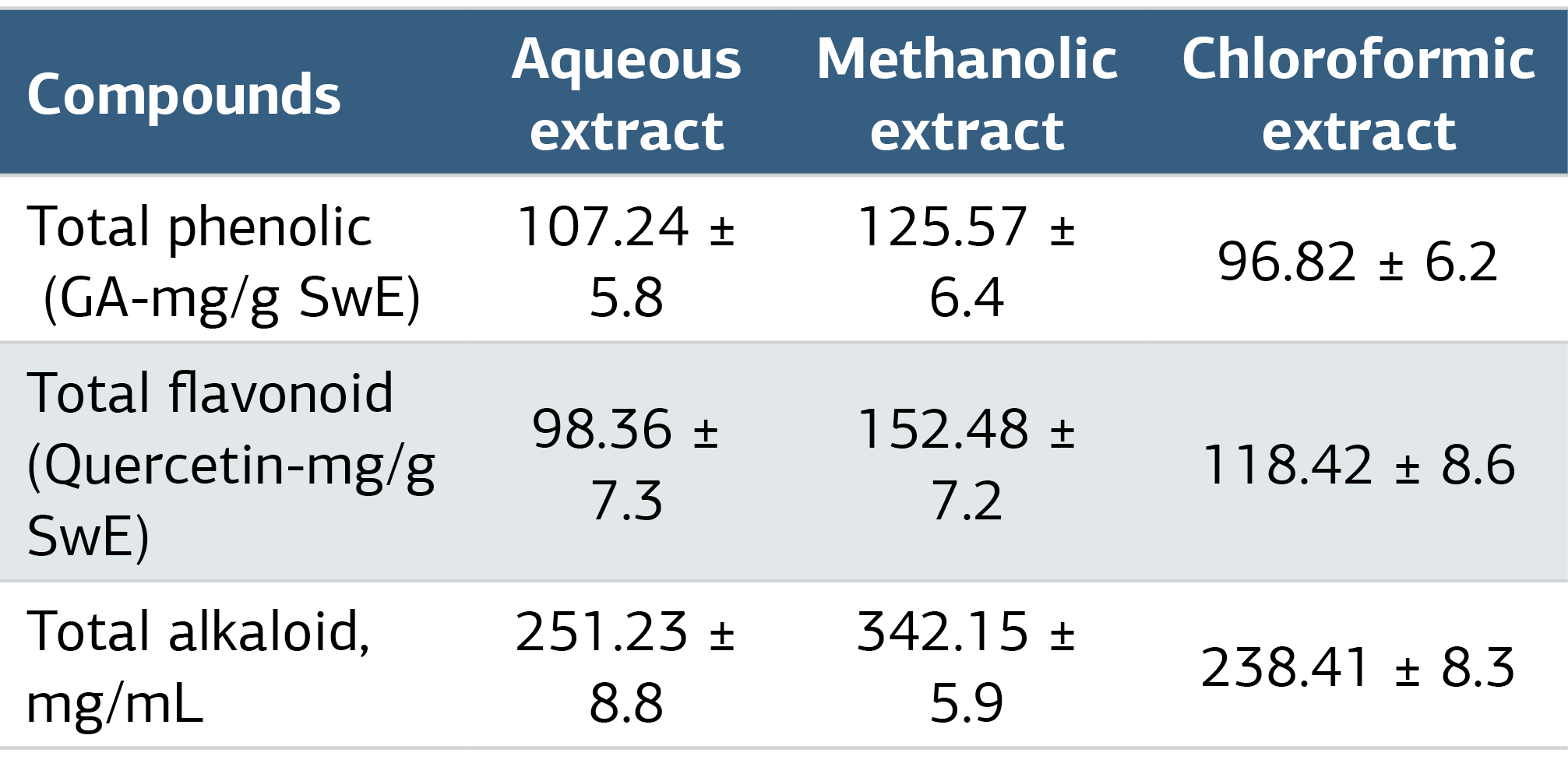
In our study, as well as in another study.5 the highest phenolic content was found in the methanol extract (Table 1). Therefore, we used the methanol extract for all analyses.Synthesis of AgNPsCommercial AgNO3 was purchased to synthesise AgNPs. In a 1000 mL beaker, 500 mL AgNO3 and 50 mL SwE were added and stirred using magnetic stirrer (40-60°C/30 min). It was treated with ultrasound (3 h). As a result of the synthesis, the colour was changed from colourless to brown. It was then centrifuged with distilled water 10000×g/5 min, filtered, and dried in an oven (50°C/48 h). Absorbance was measured at 350-700 nm spectrophotometrically.7Evaluation of Antioxidant ActivityWe analysed the lipid peroxidation inhibition, DPPH, and FRAP capacity of AgNPs/Sw.5,6 The lipid peroxidation inhibition as MDA was determined by the thiobarbituric acid (TBA) test. Concentration series of AgNPs/Sw (50-100-150-250-500 mg/L) were prepared with 97% ethanol. A mixture 200 μL each of AgNPs/Sw, FeCl3, EDTA, H2O2 and ascorbic acid was added to a test tube and vortexed. It was then incubated (37°C/1.5 h), 1.2 mL TBA (28%) was added, and centrifuged (3000×g/15 min). The pellet was mixed with 1.2 mL TBA and boiled (100°C/10 min). After cooling on ice, the supernatant was discarded, and the absorbance was measured at 532 nm, spectrophotometrically. The results were expressed as pmol/mg-AgNPs/Sw.
To analyse the DPPH, first concentration series of AgNPs/Sw described above was prepared in 3.9 ml ethanol with DPPH. It was then vortexed (1 min), and incubated (20°C/1 h). The absorbance was measured at 517 nm, spectrophotometrically. Vitamin C was used as a reference sample. Each measurement was repeated three times. Equation 1 (Eq-1) was used to calculate the %inhibition DPPH. %inhibition=[(Acontrol-Asample)/Acontrol]×100.
The FRAP assay was performed preparing concentration series of AgNPs/Sw described above. 3.9 ml FRAP reagent (acetate buffer (pH 3.6)/tripyryltriazine in HCl/FeCl3) and 0.1 mL AgNPs/Sw were added in a test tube. After incubating (20°C/30 min), we measured the absorbance at 593 nm, spectrophotometrically. Trolox (TEAC) was used as a reference chemical, and FRAP activity was expressed as mg-TEAC/g-AgNPs/Sw.Evaluation of Anti-Aging ActivityThe anti-aging effects of AgNPs/Sw were determined by assaying their anti-collagenase/elastase/hyaluronidase/tyrosinase activities.
In anti-collagenase assay.8 0.5 mg azo dye-impregnated collagen, 400 μL Tris-HCl (0.1 M/pH 7), and 50 μL AgNPs/Sw were vortexed in a tube. Then, 50 µl collagenase (200 U/mL) was added, incubated (43°C/1 h), and centrifuged (3000×g/10 min). Its absorbance was measured at 550 nm, spectrophotometrically. Epicatechin gallate was used as a reference chemical.
To determine anti-elastase levels.8 in a test tube, 20 μL AgNPs/Sw was mixed with 80 μL Tris-HCl (0.1 M/pH 8) and 40 μL elastase. It was incubated (37°C/10 min. The reaction was initiated by adding 60 µL N-succinyl-(Ala)-3-nitroanilide. The absorbance was measured at 410 nm, spectrophotometrically. Oleanolic acid was used as reference chemical.
In anti-hyaluronidase assay.8 20 μL AgNP/Sw and 20 μL hyaluronidase (8 mg/mL) were put in a test tube and then incubated (37°C/20 min). Next, 40 μL calciumchloride was added and incubated again (37°C/20 min). Then, 100 μL hyaluronic acid was added and incubated (37°C/40 min). 40 μL potassium tetraborate tetrahydrate and 4 μL NaOH were added to the solution. It was heated in a water bath (100°C/3 min). Finally, 1200 μL dimethylaminobenzaldehyde was added, cooled (20°C), and incubated (37°C/20 min). The absorbance was measured at 585 nm, spectrophotometrically. Indomethacin was used as reference chemical.
To determine anti-tyrosinase.8 20 μL AgNP/Sw, 220 μL sodiumphosphate buffer (0.1 M/pH 6.8), 20 μL tyrosinase (1500 U/mL), and 40 μL L-tyrosine were added to the wells of a microtiter plate, and incubated (37°C/12 min). The reaction was stopped by placing the plate on ice. The absorbance was measured at 490 nm with microplate reader (Epoch2 Microplate Spectrophotometer, BioTech, USA). Alpha-kojic was used as reference compound.Evaluation of Anti-Alzheimer ActivityThe anti-Alzheimer effect of AgNPs/Sw was determined using acetylcholinesterase (AChE)/butyrylcholinesterase (BChE)/amyloid-beta (Aβ) aggregation inhibition assays.
In the determination of AChE/BChE inhibitions.9 hydrolysis of acetylthiocholine iodide (ATCh1) by AChE/BChE yields a 5-thio-2-nitrobenzoate anion as yellow complexes with 5,5’-dithiobis-2-nitrobenzoic acid (DTNB). 20 μL AgNPs/Sw, 300 μL sodiumphosphate buffer (100 mM/pH 8.0), 20 μL DTNB and 40 μL AChE (0.22 U/mL)/BChE (0.1 U/mL) were added to a test tube. The mixture was vortexed and incubated (37°C/10 min), and then 50 μL ATChl was added to initiate the reaction. After incubation (37 °C/15 min) the absorbance was recorded at 412 nm, spectrophotometrically. Galantamine hydrobromide was used as reference chemical.
For the Aβ-aggregation assay, thioflavin T (ThT) fluorescence test was performed.10 Briefly, 40 μL AgNPs/Sw, 500 μL Aβ42 and 500 μL sodiumphosphate buffer (50 mM/pH 7.4) were added to a test tube, and incubated (37°C/48 h). 10 μL ThT-containing glycine/NaOH buffer (50 mM/pH 9.2) was added. Fluorescence intensities were measured at excitation-446 nm/emission-490 nm with a microplate fluorometer (Twinkle LB970, Germany). Curcumin was used as reference chemical. Fluorescence intensities were recorded and the percentage inhibition of Aβ-aggregation was calculated: %inhibition=(1-IFi/IFc)×100, where IFi and IFc were the fluorescence intensities at absorbances in the presence and absence of AgNPs/Sw, respectively.Evaluation of Anti-Obesity Activityα-Amylase/pancreatic lipase inhibition assays were used to determine anti-obesity effects of AgNPs/Sw.
In α-amylase inhibition assay using 3,5-dinitrosalicylic acid (DNSA) method.9 DNSA reagent was prepared by mixing 12 g sodiumpotassiumtartratetetrahydrate in 8.0 mL NaOH and 20 mL DNSA. In a test tube, 500 μL AgNPs/Sw and 500 μL α-amylase (0.5 mg/ml) in sodiumphosphate buffer (0.02 M/pH 6.9) were mixed, and incubated (30°C/10 min). Then, 400 μL starch (1%) was added, and incubated again. The reaction was initiated adding 400 μL DNSA reagent in water-bath (85-90°C/10 min). It was cooled (20°C), and diluted in 10 mL distilled water. The absorbance was measured at 540 nm, spectrophotometrically. Acarbose was used as reference chemical and the results were calculated using Eq-1.
In pancreatic-lipase inhibition assay using p-nitrophenyl butyrate (PNPB) method.11 PNPB solution was prepared mixing 8.4 μL PNPB stock solution and 10 mL acetonitrile final volume. 250 μL AgNPs/Sw, 100 μL pancreatic-lipase (0.1 mg/mL) and 50 μL PNPB, and incubated (37°C/30 min). The hydrolysis amount of p-nitrophenol from PNPB by lipase enzyme was measured at 405 nm, spectrophotometrically. Orlistat was used as standard chemical, and the results were calculated using Eq-1.Evaluation of Antidiabetic ActivityTo determine the anti-diabetic effects of AgNP/Sw, α-amylase and α-glucosidase were determined (α-amylase enzyme assay was performed in the anti-obesity tests, above).
In α-glucosidase assay.9,12 using maltose as substrate. 80 μL AgNP/Sw, 2 mL maltose (2%) as subtrate and 2 mL α-glucosidase (1 U/mL) in phosphate buffer (10 mM/pH 8.0) were placed in a test tube, and incubated (37°C/5 min). The reaction was initiated by adding 100 µL methanol. The mixture was then placed in a water bath (2 min) and cooled (20°C). Absorbance values were measured at 695 nm, spectrophotometrically. α-Glucosidase inhibition was calculated using Eq-1.Ethical ApprovalEthical approval was not required because no human participants or animals were used in this study.Statistical AnalysisSPSS (Statistical Package for Social Sciences) 25.0 was used, and data were analysed and compared by one-way analysis of variance (ANOVA) followed by post-hoc Tukey test. P < .05 was considered statistically significant. Data are expressed as mean±SD.
Reporting GuidelinesNot applicable.
Results
The assays to determine the total phenolic/flavonoid and/or alkaloid content in SwE were performed and the results are presented in Table 1. In our study, UV, FTIR, XRD and SEM techniques were used to control the green synthesis and catalytic activity of AgNPs/Sw. UV spectrophotometer was used to check the synthesis of AgNPs/Sw in at 300-700 nm, nm, and a sharp peak at 455 nm was observed (Figure 1A). FTIR spectroscopy was used to identify the secondary metabolites formed by the reaction of SwE with Ag ions. FTIR analysis revealed significant absorption peaks for AgNPs/Sw (Figure 1B).
The crystal structure and nanostructure of AgNPs/Sw were confirmed by XRD analysis. Confirmation of the crystal structure of 5 peaks for AgNPs/Sw presented in Figure 2A. They were possible due to the presence of Bragg reflection planes (111), (200), (220), (222) and (311). The morphology of the green synthesised AgNPs/Sw was confirmed by SEM. As shown in Figure 2B, they had a polymorphic appearance as granular clusters. SEM analysis for AgNPs/Sw revealed that their size was around 40 nm.
To determine their antioxidant properties of AgNPs/Sw, the in vitro effects of AgNPs/Sw on the inhibition of lipid peroxidation (MDA), DPPH, and FRAP capacity were analysed (Table 2). The anti-aging effects of AgNPs/Sw on collagenase/elastase/hyaluronidase/tyrosinase enzymes and the anti-Alzheimer/anti-obesity/antidiabetic inhibitory activities of AgNPs/Sw were shown in Table 3.
Discussion
Herbal medicines have become an integral part of complementary health care in the prevention and treatment of disease. Green synthesis of metal nanoparticles, a method of producing nanoparticles from medicinal plants, is a promising strategy in the medical world. In our research, silver nanoparticles were green synthesised using S. willeana in a highly efficient, cost-effective and simple process.
Phenolic compounds, which occur as secondary metabolites in plants, have been reported with their antioxidant, anti-aging, anti-Alzheimer’s, anti-obesity and anti-diabetic properties, help to reduce the development of many chronic diseases and cancer.3,4 Ercetin et al.13 investigated the cholinesterase inhibition of S. willeana, and reported high flavonoid/phenolic contents in the plant leaves (547.55±3.12 and 300.24±1.23 mg/g, respectively). We and El Hajaji et al.5 found that methanolic SwE had the highest phenolic content. Therefore, the methanolic extract was preferred in all subsequent analyses and procedures of our study.
In our study, after the synthesis of AgNPs/Sw, we checked the synthesis and catalytic activities of them using UV, FTIR, XRD and SEM techniques. UV spectroscopy confirmed the synthesis by the presence of an absorption peak at around 455 nm was ensured by plasmon resonance electrons on the surface of the nanoparticles.7 FTIR was used to identify the secondary metabolites formed by the reaction of SwE with Ag ions and by the absorption peaks due to AgNPs/Sw synthesis and nanoparticle reduction, capping and stabilisation. The crystal structures and nanostructures of the synthesised S. willeana silver nanoparticles were confirmed by XRD analysis. The peak position, height and width determined by XRD analysis determine the nanocrystalline structure and purity of the nanoparticles. The morphology of AgNPs/Sw was confirmed by SEM, and they had a polymorphic appearance as granular clusters. Similar results also have been observed in previous studies on Salvia spp.14,15
Reactive oxygen species (ROS) are highly reactive molecules produced during normal physiological metabolism in the human body. They can damage cells and cause lipid peroxidation, playing a role in chronic disease development.16 Plant-synthesised nanoparticles effectively scavenge ROS, reducing oxidative stress. Studies have shown that S. willeana contains phytochemical components such as phenol, flavonoid, saponin, and alkaloid, which may be responsible for the biological reduction of Ag+2 ions in synthesised AgNPs and their beneficial effects. It has been observed that medicinal plants with high levels of antioxidant phytochemicals can enhance the inhibitory effects of silver green synthesis on lipid peroxidation.5 Few studies have investigated the antioxidant, anti-aging, anti-Alzheimer, anti-obesity, and anti-diabetic activities of S. willeana.13,17 This study is the first to investigate the effect of silver nanoparticles synthesised using S. willeana. Ercetin et al.13 conducted a study on S. willeana, and they measured the highest antioxidant properties in DPPH activity were observed in the methanol extracts prepared from the leaves and flowers of the plant (67.94±0.003% and 45.07±0.001%, respectively). Our study found that the level of MDA was significantly lower in AgNPs/Sw samples at 500 mg/mL concentration (649.56±62.24 pmol/mg). %Inhibition of DPPH and FRAP activities were higher in the same concentration of AgNPs/Sw (50.68±3.17%, TEAC-mg/g-AgNPs/Sw). This may be attributed to the high levels of phenolic and flavonoid compounds present in S. willeana, and it can suppress the formation of ROS and up-regulate antioxidant defence by chelating trace elements involved in ROS production.1,2
Recent scientific studies have focused on the anti-aging effects of herbs in preventing skin aging caused by internal and external factors. ROS production in the skin can cause oxidative stress, leading to damage to biological functions such as lipid membrane peroxidation, DNA destruction, and cell death; which contribute to skin aging, can activate dermal enzymes such as collagenase and elastase. These enzymes can break down and degrade the skin’s structures.8 Hyaluronic acid (HA) is abundant in the dermis and epidermis layers of the skin. However, hyaluronidase, an enzyme that degrades HA and increases during the aging process, can be detrimental.18 We investigated the anti-aging effects of AgNPs/Sw determining their anti-elastase/collagenase/hyaluronidase/tyrosinase. Previous studies have shown anti-aging enzyme activities for Salvia spp.: S. aytachii inhibited the activities of collagenase, elastase, and tyrosinase enzymes, or S. officinalis extract had inhibitory activity against hyaluronidase.
Traditionally, S. willeana have been also used as an alternative treatment agents in Alzheimer’s disease, helping delay metabolic pathways or inhibiting enzymes in increased biochemical reactions.19 In vitro studies on Alzheimer have shown that AChE leads to the formation of amyloid fibrils, resulting in the conversion of toxic acetylcholinesterase-beta-amyloid peptide (Aβ) complexes in brain cells,and BchE activity is elevated in various brain regions of Alzheimer’s patients. AChE and BchE are recognized as suitable targets for Alzheimer’s treatment.10 Intensive research is currently underway to find drugs or plant extracts that can inhibit cholinesterases, and Salvia spp. have been examined for their cholinesterase inhibitory activities. S. eriophora and S. mirzayanii extracts exhibited inhibitive activity against AChE and BChE enzymes.9,20 Yilmaz et al.1 reported that S. aytachii extract inhibited AChE and S. viridis extract inhibited BChE. Ercetin et al.13 investigated the cholinesterase inhibition of S. willeana. The highest acetylcholinesterase/butyrylcholinesterase inhibitory potentials were measured in ethyl acetate and aqueous extracts prepared from the plant. In our study, AgNPs/Sw inhibited AchE and BChE by 26.48±4.6% and 25.87±3%, respectively, while the reference compound, galantamine was 90.52±6%. We also investigated the Aβ aggregation inhibition effect of AgNPs/Sw. AgNPs/Sw and curcumin inhibited Aβ42 aggregation by 25.42±5.9% and 67.37±7%, respectively. No similar studies using Salvia spp. were found in the literature. Boonsin et al.20 researched the effects of Gryllus bimaculatus plant extract on Aβ aggregation and reported that concentrations of extract (1 and 10 mg/mL) inhibited Aβ aggregation by 18.90% and 22.92%, respectively.
Obesity is a widespread problem, and increases the risk of diabetes, cardiovascular and liver diseases. The aim of obesity treatment is to prevent excessive absorption of fat and sugar.21,22 In this study, we examined α-amylase and pancreatic-lipase inhibitor activities as anti-obesity functional markers. Previous studies have reported that certain flavonoids, particularly those containing methoxy groups, can inhibit pancreatic lipase activity, thereby inhibiting the digestion and absorption of dietary fats.23 The α-glucosidase enzyme plays a crucial role in carbohydrate digestion and absorption. Excess sugar is converted into fat and stored in the body, leading to obesity. Research has shown that polyphenols found in plants exhibit anti-obesity and antidiabetic effects by inhibiting α-glucosidase in vitro.24,25 Mahdi et al.24 reported the antidiabetic activity of α-amylase (IC50:104.58±0.06 mg/mL) in S. officinalis extracts. Assaghaf et al.25 investigated the essential oils of S. officinalis and demonstrated in vitro inhibition of α-amylase and lipase with IC50 values of 69.23±0.1 and 37.3±0.03 µg/ml, respectively. In our study, we found that AgNPs/Sw inhibited α-amylase and lipase by 27.84±7.5% and 35.46±6.9%, respectively.
Diabetes Mellitus is a hereditary disease that results in an increase in the production of free radicals due to increased insulin use in the body. Our study examined the α-amylase and α-glucosidase, the carbohydrate digestion enzymes because the inhibition of those enzymes can be effective in treatment of diabetes. Plant extracts have been found to be effective enzyme inhibitors, such as the α-glucosidase inhibitory activity of S. officinalis extract.24,25 We found that AgNPs/Sw inhibited α-glucosidase by 43.57±8.7%, compared to the standard acarbose inhibition of 82.47±8.3%.
Limitations
The present study was performed under in vitro conditions. Therefore, further in vivo studies are required to evaluate the biological effects of AgNPs/Sw.
Conclusion
On the island of Cyprus, S. willeana is traditionally used to treat respiratory, digestive, rheumatism, heart, skin, gynaecological diseases, and various cancers. In vitro studies on the Salvia species have shown that the plant extract contains phenolic compounds having antioxidant activity against ROS that occur in the body. Recently, there has been hope that nanoparticles produced through green biosynthesis of metal nanoparticles using medicinal plant extracts could be used in medicine to treat diseases. Our study showed that Ag-NPs/Sw had a potential to prevent and treat diseases due to their antioxidant anti-aging, anti-Alzheimer’s, anti-obesity, and antidiabetic properties that protect cells. Therefore, it is believed that they can offer numerous benefits to humanity in the field of medicine. It is recommended that future researches could enhance our understanding of the potential health benefits of Salvia spp., and nanoparticles.
Declarations
Animal and Human Rights Statement
All procedures performed in this study were in accordance with the ethical standards of the institutional and/or national research committee and with the 1964 Helsinki Declaration and its later amendments or comparable ethical standards.
Informed Consent
Not applicable.
Data Availability
The datasets used and/or analyzed during the current study are not publicly available due to patient privacy reasons but are available from the corresponding author on reasonable request.
Conflict of Interest
The authors declare that there is no conflict of interest.
Funding
None.
Abbreviations
DPPH: 1,1-Diphenyl-2-picrylhydrazyl
FRAP: Ferric reducing antioxidant power
FTIR: Fourier transform infrared spectroscopy
ROS: Reactive oxygen species
SEM: Scanning electron microscopy
XRD: X-ray diffraction
References
- Yilmaz G, Eruygur N, Bona GE, et al. Phytochemical analysis, antioxidant, and enzyme inhibition activity of five Salvia taxa from Turkey. S Afr J Bot. 2023;152:212-221. doi:10.1016/j.sajb.2022.11.027
- Kolac UK, Ustuner MC, Tekin N, Ustuner D, Colak E, Entok E. The anti-inflammatory and antioxidant effects of Salvia officinalis on lipopolysaccharide-induced inflammation in rats. J Med Food. 2017;20(12):1193-1200. doi:10.1089/jmf.2017.0035
- Polatoglu K, Karakoc OC, Yucel YY, et al. Insecticidal activity of Salvia veneris Hedge essential oil against coleopteran stored-product insects and Spodoptera exigua (Lepidoptera). Ind Crops Prod. 2017;97:93-100.
- Shumail H, Khalid S, Ahmad I, Khan H, Amin S, Ullah B. Review on green synthesis of silver nanoparticles through plants. Endocr Metab Immune Disord Drug Targets. 2021;21(6):994-1007. doi:10.2174/1871530320666200729153714
- El Hajaji H, Lachkar N, Alaoui K, et al. Antioxidant activity, phytochemical screening, and total phenolic content of extracts from three genders of carob tree barks growing in Morocco. Arab J Chem. 2011;4(3):321-324. doi:10.1016/j.arabjc.2010.06.053
- Ayoola GA, Coker HA, Adesegun SA, et al. Phytochemical screening and antioxidant activities of some selected medicinal plants used for malaria therapy in Southwestern Nigeria. Trop J Pharm Res. 2008;7(3):1019-1024. doi:10.4314/tjpr.v7i3.14686
- Metwally DM, Alajmi RA, El-Khadragy MF, Al-Quraishy S. Silver nanoparticles biosynthesized with Salvia officinalis leaf exert protective effect on hepatic tissue injury induced by Plasmodium chabaudi. Front Vet Sci. 2021;7:620665. doi:10.3389/fvets.2020.620665
- Jiratchayamaethasakul C, Ding Y, Hwang O, et al. In vitro screening of elastase, collagenase, hyaluronidase, and tyrosinase inhibitory and antioxidant activities of 22 halophyte plant extracts for novel cosmeceuticals. Fish Aquat Sci. 2020;23:6-12. doi:10.1186/s41240-020-00149-8
- Bursal E, Aras A, Kılıç Ö, Taslimi P, Gören AC, Gülçin İ. Phytochemical content, antioxidant activity, and enzyme inhibition effect of Salvia eriophora Boiss & Kotschy against acetylcholinesterase, α-amylase, butyrylcholinesterase, and α-glycosidase enzymes. J Food Biochem. 2019;43(3). doi:10.1111/jfbc.12776
- Härtig W, Goldhammer S, Bauer U, et al. Concomitant detection of β-amyloid peptides with N-terminal truncation and different C-terminal endings in cortical plaques from cases with Alzheimer disease, senile monkeys, and triple transgenic mice. J Chem Neuroanat. 2010;40(1):82-92. doi:10.1016/j.jchemneu.2010.03.006
- Buchholz T, Melzig MF. Polyphenolic compounds as pancreatic lipase inhibitors. Planta Med. 2015;81(10):771-783. doi:10.1055/s-0035-1546173
- Gharehbagh HJ, Ebrahimi M, Dabaghian F, et al. Chemical composition, cholinesterase, and α-glucosidase inhibitory activity of the essential oils of some Iranian native Salvia species. BMC Complement Med Ther. 2023;23(1):184. doi:10.1186/s12906-023-04004-w
- Ercetin T, Safaei M, Gulcan HO. Investigation of total phenolic and flavonoid content of Salvia willeana (Holmboe) Hedge, an endemic plant of Cyprus, and screening of its antioxidant and cholinesterase inhibitory properties. J Res Pharm. 2022;26(2):263-271. doi:10.29228/jrp.124
- Tulukcu E, Cebi N, Sagdic O. Chemical fingerprinting of seeds of some Salvia species in Turkey by using GC-MS and FTIR. Foods. 2019;8(4):118. doi:10.3390/foods8040118
- Mihailović V, Srećković N, Nedić ZP, et al. Green synthesis of silver nanoparticles using Salvia verticillata and Filipendula ulmaria extracts: optimization of synthesis, biological activities, and catalytic properties. Molecules. 2023;28(2):808. doi:10.3390/molecules28020808
- Caylak E. Oxidative stress and antioxidants in the animals and the plants. Tip Aras Der. 2011;9(1):73-83.
- Vonaparti A, Karioti A, Recio M, et al. Effects of terpenoids from Salvia willeana in delayed-type hypersensitivity, human lymphocyte proliferation, and cytokine production. Nat Prod Commun. 2008;3(12):1953-1958. doi:10.1177/1934578x0800301202
- Aytekin M, Caylak E. Hiyalüronan, tanı ve tedavideki önemi. Tuberk Toraks. 2009;57(3):356-364.
- Uddin MN, Afrin R, Uddin MJ, et al. Vanda roxburghii chloroform extract as a potential source of polyphenols with antioxidant and cholinesterase inhibitory activities: identification of a strong phenolic antioxidant. BMC Complement Altern Med. 2015;15:195. doi:10.1186/s12906-015-0728-y
- Boonsin P, Boonyarat C, Plekartoke K, Reubroycharoen P, Waiwut P. Antioxidant activity, β-amyloid aggregation inhibition, and neuroprotective effect of cricket (Gryllus bimaculatus) ethanol extract. In: Proceedings of the 6th International Conference on BMB; 2018; Rayong, Thailand:28.
- Caylak E. Obesity hormones in health and disease. In: eLS. John Wiley & Sons Ltd; 2012:1-9. doi:10.1002/9780470015902.a0024166
- Caylak E. Obesity and appetite hormones in humans: an overview. In: eLS. John Wiley & Sons Ltd; 2012:1-9. doi:10.1002/9780470015902.a0024163
- Kumar S, Narwal S, Kumar V, Prakash O. α-Glucosidase inhibitors from plants: a natural approach to treat diabetes. Pharmacogn Rev. 2011;5(9):19-29. doi:10.4103/0973-7847.79096
- Mahdi S, Azzi R, Lahfa FB. Evaluation of in vitro α-amylase and α-glucosidase inhibitory potential and hemolytic effect of phenolic-enriched fractions of the aerial part of Salvia officinalis L. Diabetes Metab Syndr. 2020;14(4):689-694. doi:10.1016/j.dsx.2020.05.002
- Assaggaf HM, Naceiri Mrabti H, Rajab BS, et al. Chemical analysis and investigation of biological effects of Salvia officinalis essential oils at three phenological stages. Molecules. 2022;27(16):5157. doi:10.3390/molecules27165157
Tables
Table 1. The Phenolic Compounds Of Swe in the Aqueous, Methanolic and Chloroformic Extracts*
*The mean values (x) were obtained triplicate measurement and were expressed as x±SD. Figure 1. A: UV, and B: FTIR spectra of AgNPs/Sw
Table 2. The in Vitro Effects of Agnps/Sw on Antioxidant Status*
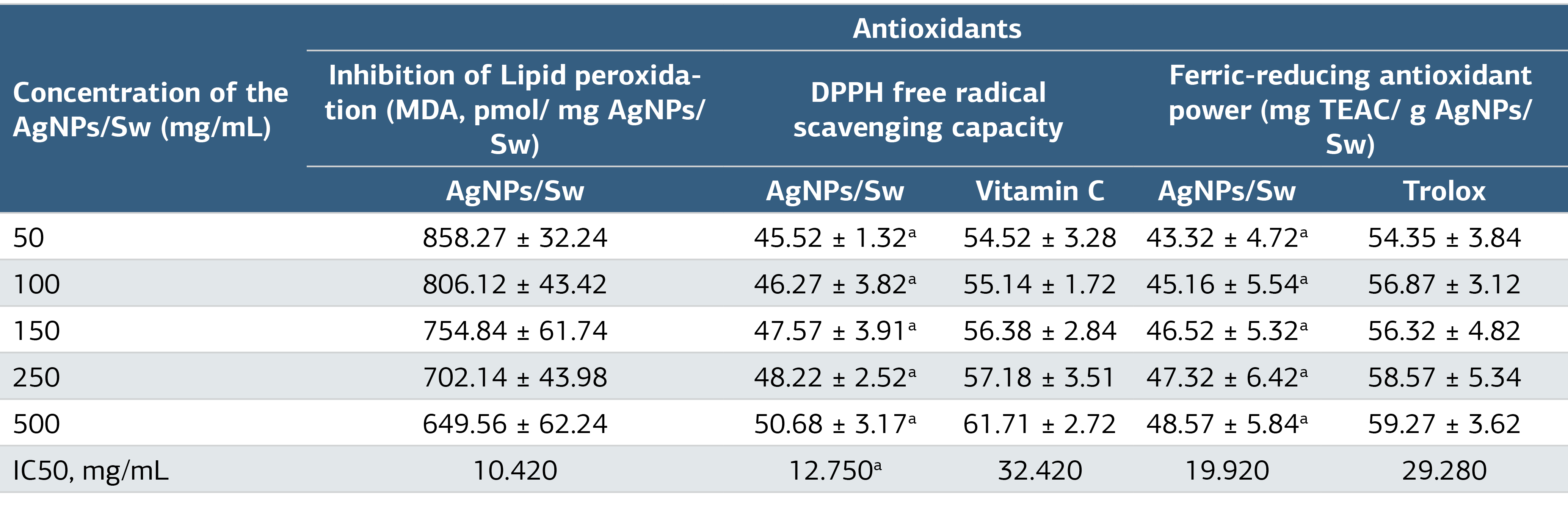
*Values are expressed as x ± standard deviation. a The significant group is compared to the standard group. A P value of ≤ 0.05 indicates a significant difference, as determined by one-way ANOVA followed by Tukey's test.
Table 3. In Vitro Anti-Aging/Anti-Alzheimer/Anti-Obesity/Antidiabetic Inhibitory Activities of AgNPs/Sw*
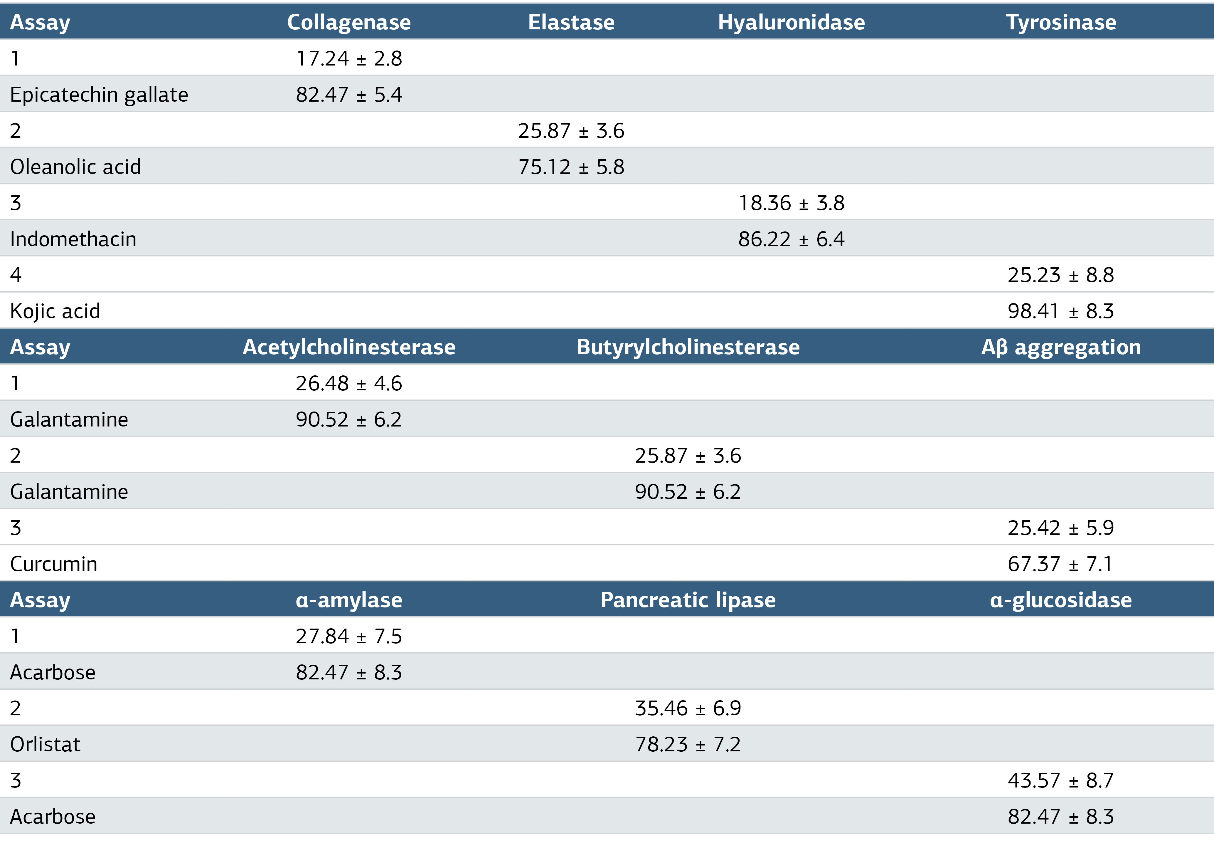
*Mean values were obtained triplicate measurement, expressed as percentages and presented as x±SD.
Figures
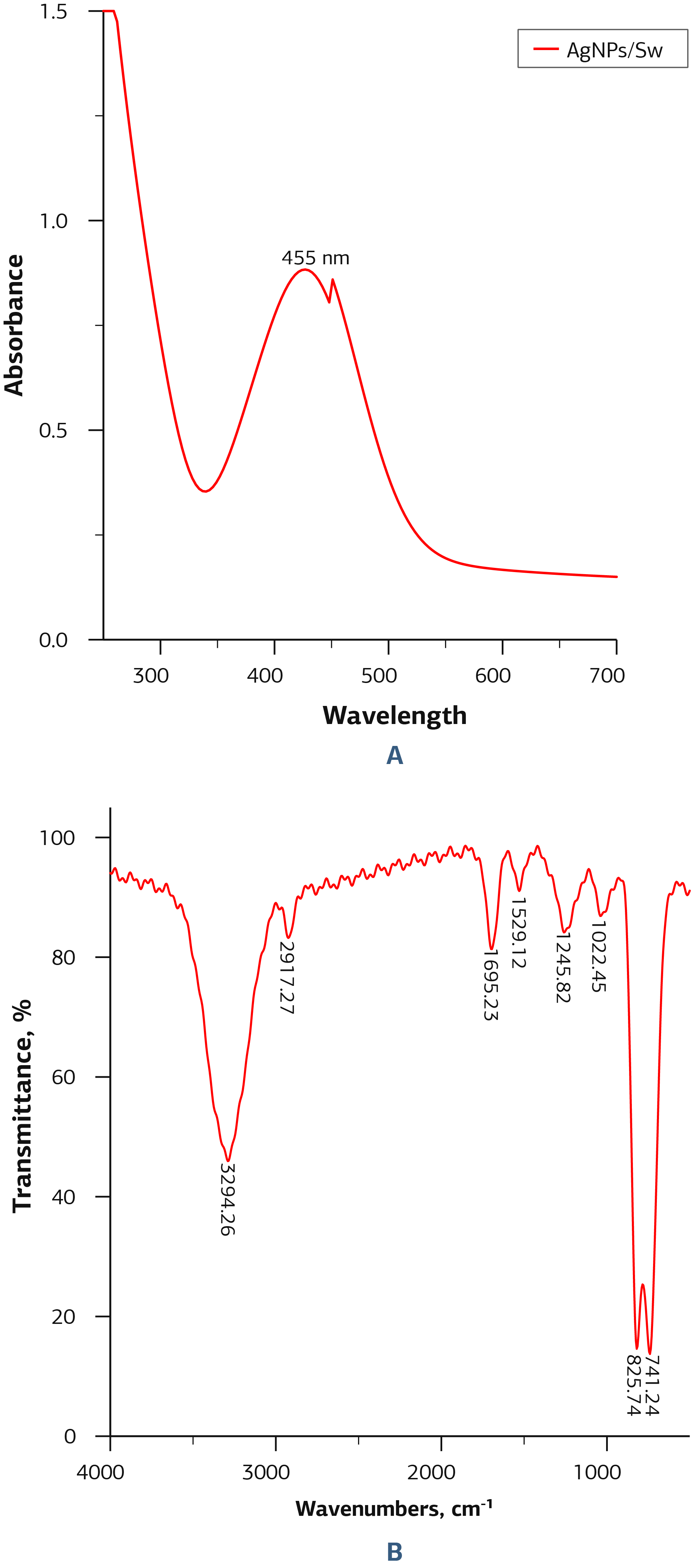
Figure 1. A, UV and B, FTIR Spectra of AgNPs/Sw
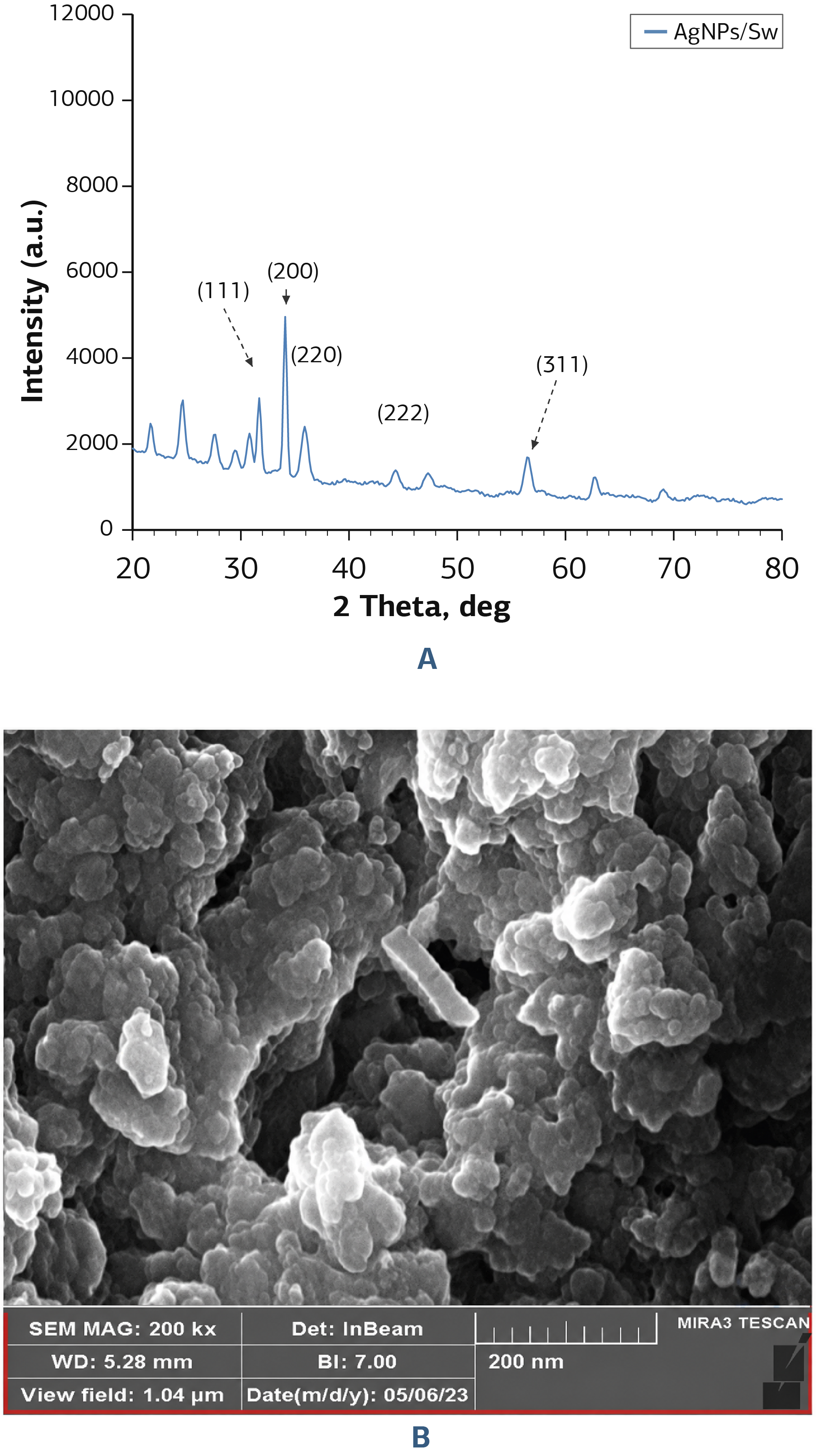
Figure 2. A, XRD Pattern and B, SEM Image of AgNPs/Sw
Additional Information
Publisher’s Note
Bayrakol MP remains neutral with regard to jurisdictional and institutional claims.
Rights and Permissions

This work is licensed under a Creative Commons Attribution-NonCommercial 4.0 International License (CC BY-NC 4.0). To view a copy of the license, visit https://creativecommons.org/licenses/by-nc/4.0/
About This Article
How to Cite This Article
Emrah Çaylak, Gökhan Nur. In vitro characterisation of antioxidant, anti-aging, anti-alzheimer, antiobesity, antidiabetic activities of silver nanoparticles synthesised from salvia willeana. Ann Clin Anal Med 2024;15(Suppl 1):1-7. doi:10.4328/ACAM.22120
Publication History
- Received:
- 18.01.2024
- Accepted:
- 15.03.2024
- Published Online:
- 19.03.2024
- Printed:
- 20.03.2024