
Abstract
AimDiagnosis of acute appendicitis is a challenging task in children presenting with abdominal pain. We conducted an analytical interpretation of the basic parameters in patients with appendicitis, according to decisive pathologic diagnosis.MethodsWe analyzed the clinical files of 12126 patients with abdominal pain and included a sum of 1066 hospitalized patients suggestive of acute abdomen. We accomplished a statistical evaluation of variables as physical examination, symptoms, laboratory findings and ultrasound screening.ResultsHistopathological analysis of 657 appendectomies revealed negative appendectomy in 12.3% and complicated appendicitis in 15% (perforated 6.7%). There was no statistical difference in terms of pyrexia and CRP between negative appendectomy and acute appendicitis. WBC count, neutrophil count, and neutrophil lymphocyte rate showed a statistically significant difference (P < .001) amongst histopathologic groups. ROC analysis revealed AUC (cut-off) values as 0.62 (≥14500), 0.769 (≥9.6), 0.689 (≥4.89) respectively. US revealed 65% sensitivity and 50% specificity rate with 90% of positive predictive value, along with 17% negative predictive value. Secondary findings as presence of peri-appendiceal fluid collection and mesenteric heterogeneity revealed 93.4% and 94.3% sensitivity consecutively.
ConclusionWe found WBC count, neutrophil count, neutrophil lymphocyte rate, presence of peri-appendiceal fluid and mesenteric heterogeneity in ultrasound
screening as highly predictive in differential diagnosis.
Keywords
Introduction
Appendicitis is one of the most common surgical causes of acute abdominal pain in pediatric patients.1,2 The clinical picture of appendicitis has long been regarded as equivalent to inevitable death historically, posing one of the greatest fears of the humankind.3 The overall lifetime risk is estimated to be around 8%, with an age peak during the teen years.4
Acute appendicitis in children may mimic atypical and non-specific abdominal symptoms, and this entity may explain the 20-50% increased incidence of perforation within the pediatric age group.5 Laboratory serological investigations are less invasive and routinely performed on most children with abdominal pain presenting to emergency.6 These tests are known to be non-specific for appendicitis but have been reported to achieve a high discriminatory power when combined with clinical history and physical examination.6 Today, contemporary research is vastly focused on physiopathology, imaging studies, digital data analysis, scoring algorithms and environmental or genetics related investigations, aiming to achieve a timely and precise diagnosis.2,7,8 However, researchers are still far from establishing a pathognomonic parameter available as a sole indicator of appendicitis amongst patients presenting with acute abdomen. Obtaining a detailed anamnesis and proper physical examination stands the test of time in diagnostic decision making.
Ultrasound (US) is a fast, and easy excess to conduct, with zero radiation. It is more useful when compared to CT or MRI. According to the American College of Radiology, ultrasound is the best method suited for initially imaging a patient with suspected acute appendicitis.9 It has, however, inherent limitations due to radiologists’ experience and factors like accumulated bowel gas and body mass index.
In our eight-year retrospective study, we evaluated the data of our patients applying with acute abdomen and analyzed the impact of numerous parameters attributable to diagnostic accuracy while treating appendicitis. We aimed to interpret and compare our results in the light of published scoring algorithms and imaging studies.
Materials and Methods
The files of 12126 children under the age of 18, attending emergency services with a complaint of abdominal pain, between 2012 and 2020 were evaluated. Our department was requested to consult a total of 3068 of these patients, out of which 1868 were diagnosed as having urinary tract infection, acute gastroenteritis, and constipation following evaluation by the pediatric surgeon. None of these patients were readmitted later with a similar clinical picture.
The remaining 1200 patients were hospitalized in the pediatric surgical ward with an initial diagnosis of acute abdomen. Amongst these, a total of 134 cases operated for reasons other than appendicitis, and four appendectomy cases without an exact histopathological confirmation were excluded from the study. Of the 1062 cases included in our study; 653 cases who underwent appendectomy were determined as “Group 1”, and 409 cases treated medically and later discharged without surgery as “Group 2”. All patients were divided into four subgroups according to their age: infants-toddlers (0-3), preschoolers (>3-6), primary school students (>6-10) and teenagers (>10-18).
The comparison was based on the clinical history, physical examination findings, laboratory tests and US results between these two groups for which data was available. The physical examination findings of the physician during the initial examination were considered in comparison to the consultant pediatric surgeon’s evaluation process. Thus, we aimed to investigate the reliability of data conducted by primary and secondary care physicians who do not have as much experience as the pediatric surgeon. Routine laboratory parameters with minimal additional cost were verified and analyzed according to histopathology results during the decision-making process of an accurate and timely diagnosis of appendicitis.Histopathological ClassificationThe exact histopathological findings of the appendix tissue verified by our Medical Pathology Laboratory were classified as; a. Negative appendectomy (NA) in cases with rich lymphoid follicles in the lamina propria under the luminal surface epithelium and not accompanied by acute and chronic inflammation. b. Acute appendicitis (AA) in cases presenting with neutrophil leukocytes infiltrating the lamina propria, muscularis mucosa, submucosa and muscularis propria starting from the crypt basal of the epithelium. c. Phlegmonous appendicitis (FA) was defined as neutrophil leukocyte infiltration in the appendiceal wall, accompanied by mucosal ulceration and crypt abscess. d. Suppurative appendicitis (SA) in the presence of extensive ulceration, transmural inflammation, and necrosis. Finally, in case of transmural inflammation, necrosis and macroscopic perforation in the lumen, it was reported as perforated appendicitis (PA).Ethical ApprovalThis study was approved by Ethics Committee of Zonguldak Bülent Ecevit University (Date: 29.04.2020, Decision No: 2020/09).Statistical AnalysisStatistical analysis of the data was performed via IBM SPSS Statistics version 22 program. Categorical variables were compared according to Pearson Chi-Square and Fisher’s Exact tests. Continuous variables were compared with Mann-Whitney U test for two groups. Kruskal Wallis statistical analyzes were used for comparisons between more than two nonparametric groups. ROC analyses were used for calculating the diagnostic impact of laboratory values. P value of less than 0.05 was considered statistically significant.Reporting GuidelinesThis study was reported in accordance with the STROBE guideline.
Results
Appendectomy group encompassed 250 (38.3%) female, and 403 (61.7%) male patients (P = .002). The incidence of NA in girls (P < .001), and FA in boys (P = .018) were significantly higher when compared to other forms. The incidence of appendectomy was statistically higher amongst primary school students and teenagers (P < .001). There was no statistical difference according to histopathological classification between the age groups (p˃0.05). We did not observe any patients with perforated appendicitis under the age of three. Amongst 653 children operated with suspected appendicitis, 80 (12.3%) were diagnosed as NA, 395 (60.5%) as AA, 80 (12.3%) as FA, 54 (8.3%) as SA and 44 (6.7%) as PA. A number of appendectomies performed during the summer were statistically lesser when compared to other seasons (P = .005).
Nausea–vomiting (76.8% vs 50.5%, P < .001), fever (17.9% vs 8.1%, P < .001), and spontaneous passage of stool (77.1% vs 57.5%, P < .001) were significantly higher in Group 1. Observation of abdominal tenderness and normal physical examination were surprisingly significantly higher in Group 2 patients (P < .001). Most of these patients were found to have normal abdominal physical examinations following pediatric surgical consultation. Rebound pain and abdominal guarding, on the other hand, were significantly higher in Group 1. The study demonstrates more common right lower abdominal quadrant tenderness finding in NA and less common rebound pain in AA cases.
White blood cell (WBC) count, C-reactive protein (CRP), neutrophile count, percent of neutrophile, neutrophile lymphocyte ratio (N/L), platelet lymphocyte ratio, and creatinine values were significantly higher in Group 1 (P < .001). Lymphocyte (P < .001), platelet count (P < .003) and urea (P < .002) values were significantly higher in Group 2. All histopathological groups were statistically different from NA in terms of WBC count, neutrophile count and N/L. In addition, FA, SA, and PA groups were statistically different from AA (p≤0.001) (Table 1). There was no significant difference between CRP levels of NA and AA patients (P > .05).
The diagnostic values of WBC count, neutrophile and N/L were further investigated using ROC curves (Figure 1).
An US screening was accomplished amongst 800 patients where an appendix was clearly visualized in 50.8%. The sensitivity, specificity, positive predictive value (PPV) and negative predictive value (NPV) for US were defined as 65%, 50%, 90%, and 17% consecutively (Table 2 and Table 3).
Discussion
Abdominal discomfort or pain is quite common in the pediatric age and mostly difficult to decide whether it is surgical in nature or not. Diagnostic accuracy of complicated appendicitis on the contrary is mostly dependable. Exclusion of a probable surgical cause is important since delay in diagnosis may result in catastrophic outcome.10-12 Main aim must be to obtain a decrease in diagnostic failure and prompt interference to avoid consequences of further clinical complications.
In contrast to numerous clinical causes which may provoke abdominal pain in girls, inflammation of the Appendix vermiformis is more common in boys.13-16 Our retrospective study revealed a similar outcome with published international data, associating a higher incidence of negative appendicitis (NA) in girls (P < .001). The reported rate of NA differs widely according to the literature. The reported range is between 1% to 40% in the English, and 1% to 17% in the Turkish literature.17 With regards to Maloney et al.’s18 study, this variation is preferable to differences in the definition of appendicitis and NA as reported by the pathology institutions. An extended analysis of our 80 patients diagnosed as NA revealed added the presence of an intraluminal faecaloid in 28, local peritonitis neighboring appendix in two and Enterobius vermicularis in one, as reported by the pathologist. Amongst these 80 patients, 33 were operated within 24 hours of admission. An extended time of observation had no impact in the outcome in majority of these patients. Even though our NA rate is 12.3%, which is well within the expected range, we may speculate that if these specific patients diagnosed as NA were discharged unoperated, may well have presented later with similar clinical picture, due to reported silent intraluminal pathologies. One of the patients was later diagnosed with Chron’s disease and another with Familial Mediterranean Fever during follow-up.
Changes in climate and environmental factors specifically related to summer period are accused of exerting impact on appendicitis by some of the researchers; however, the literature mostly declares a lack of evidence of a specific correlation.16,17,19-22 The incidence of appendicitis is low during summer according to our study.
The encountered peak incidence of AA is between six to 12 years of age according to Zhang et al.21 In our study, AA is the most confronted histopathologic subtype regardless of age. On the other hand, FA in boys (P = .018) is significantly higher when compared to other forms. In Zani et al.’s study group, 32.2% is diagnosed as complicated appendicitis.6 Aneiros et al. mention presence of an inverse correlation between age and perforated appendicitis (PA) in their report.1 The incidence of PA under the age of five is reported as 56.5% by Yıldız et al.19 Contradictory to published reports we did not encounter PA under the age of three. Owing to an increased awareness due to the higher probability of a grave prognosis and motivation for a preemptive approach, our NA ratio was found as 25% for this age group. Twenty-nine patients out of 44 diagnosed as PA, disclosed late presentation due to parental ignorance. Fourteen referred by remote hospitals were delayed due to misdiagnosis and finally remaining one because of our mismanagement.
During initial examination symptoms such as anorexia, nausea, vomiting, fever, and spontaneous passage of stool are commonly questioned. According to our emergency department’s documentation, the mentioned symptoms were significantly prominent in our appendectomized patients. Anorexia did not pose a significant difference amongst our groups. However, due to a lack of sufficient data regarding most of our cases, we think that an accurate assessment of the situation is unavailable. Patients suffering from habitual constipation quite often present with abdominal pain and must be sought for during differential diagnosis of acute abdomen. A significant difference was found between the two groups with regards to stool output within the last 24 hours and 57.5% of the cases reported absence of passage of stool in Group 2. An extended investigation showed that fever was within normal levels in 82.1% of the appendectomized cases, and AA cases did not have fever. As opposed to PAS and Alvarado scorings, anorexia complaints and fever findings were not statistically significant in our study.
During medical education, the practice of detailed anamnesis followed by specific and careful physical examination is emphasized in patients presenting with abdominal pain. Observation of abdominal tenderness and normal physical examination were surprisingly significantly higher in Group 2 patients (P < .001). In the meantime, we failed to obtain a significant difference amongst groups 1 and 2 with regards to right lower abdominal quadrant tenderness (Table 1). Most of these patients were found to have normal abdominal physical examinations following pediatric surgical consultation. Data obtained from general practitioners and pediatricians during the initial examination, revealed 15.5% rebound pain and 12.9% abdominal guarding in patients later confirmed as NA. 83.3% of the patients histopathologically confirmed as appendicitis, presented with right lower abdominal quadrant tenderness according to their initial physical examination (Table 2). Rebound pain and abdominal guarding, on the other hand, were significantly higher in Group 1 as conducted by pediatric surgeons (Table 1).
Physical examination indicating “general abdominal stiff tenderness” is envisioned as a reflection of perforation and referred to as “abdominal guarding” in our study. The review of physical findings recorded by general practitioners and pediatricians, however, proved their evaluation to be wrong owing to patients who were later followed without an operation. On the other hand, a histopathologically confirmed appendicitis was present in only 30.8% of these patients following physicians’ examination. Our study emphasizes the importance of performing a systematic physical examination, aiming to exclude diseases that do not require surgical treatment, whilst obtaining proper communication with the child and avoiding initial physical contact from where the pain is originating.
An increase in WBC count is an early marker of inflammation of the appendix, CRP on the other hand, is superior in manifesting possible perforation.23 Only 1.9% of the patients presenting normal WBC count and CRP levels are diagnosed as AA, according to Monsalve et al.’s23 study. In our study, an ROC analysis supported a correlation between increased WBC, neutrophile count and N/L values with regard to the clinical picture. Zani et al.6 confirmed comparable correlation between WBC count, CRP values and clinical picture, but failed to support their results in terms of neutrophile count. According to their study, children with CRP levels lower than 40 mg/L reflected an 80% probability of uncomplicated appendicitis. Mentioned data roughly reflects that such parameters when combined with clinical history and detailed physical examination, significantly increase discrimination in differential diagnosis.
US is preferred as the leading non-invasive, cost-effective, bedside diagnostic tool for acute abdomen, further saving patients from hazardous radiation. Due to Löfvenberg and Salö’s study, US performers’ visualization rates of the appendix illustrated no significant differences.24 Pediatric radiologists, however, demonstrated a significantly higher sensitivity (88%) in US performance compared to general radiologists (71%). Their US performance disclosed a scale of 47% visualization rate of the appendix, displaying a rate of 82% sensitivity, 97% specificity, 92% PPV and 93% NPV. US screening is mostly accomplished by the registrars in our center, and their performance rate is comparable, displaying a 50.8% rate matching with the literature.24 The appendix was clearly visualized in 63.5% of the appendectomized patients and 24.6% of those followed conservatively. However, their rate of NPV as low as 17%, demonstrated the limitation of US as a diagnostic tool in excluding appendicitis. This low ratio either reflects the inadequacy of the technique or performers in distinguishing patients within the range of normal appendiceal parameters. We advocate pediatric surgeons’ collaboration with the radiologist in case of diagnostic indecision. Recent studies indicate the importance of evaluating secondary findings.25 The impact of the mentioned findings is utilized to decrease the need for further advanced visualization methods in patients presenting with low visualization ratios with respect to NPV in otherwise possible presence of appendicitis.25
Scoring systems and algorithms that are in use such as Alvarado and PAS are accepted as relatively accurate diagnostic tools applicable in patients with abdominal pain. Our study reflected correlative results with respect to PAS concerning vomiting, right lower quadrant tenderness, abdominal tenderness, rebound pain, abdominal guarding, WBC count≥14500/mm3 and neutrophile count≥9.6 mcL. According to our results, loss of appetite, constipation, and laboratory cut off values concerning lymphocyte count<1.8µL, neutrophil percentage≥74%, and N/L≥4.7, and secondary US findings as peri-appendiceal mesenteric echogenic disturbance (94.3% sensitivity), presence of free fluid or collection (93.4% sensitivity) may additionally facilitate decisive diagnosis.
Such algorithms are user-dependent and rely on subjective data calculated by the provider. The system inherits unexpected possible misinterpretation by the user as mentioned above in our data, thus the ability of the scoring systems depends on the clinical judgment of a seniorsurgeon. Recently, the rapid development of innovative technologies and integration of artificial intelligence, machine learning, and big data, and their wide use in screening, and near-precise diagnostic procedures in diseases are becoming useful tools in various fields of research.12 They are expected to facilitate quantitative assessment of complex medical cases and help doctors in achieving accurate diagnosis.
Limitations
This study has several limitations, including its retrospective design and incomplete clinical data for some patients. In addition, ultrasonographic findings may have been influenced by operator experience.
Conclusion
Finally, it is imperative that a modest communication followed by appropriate age-specific gentle examination in a child may help reveal most of the underlying cause of abdominal pain. We believe that continuous periodical training sessions of physicians will facilitate cognitive perception of the mentioned scoring algorithm systems.
Declarations
Animal and Human Rights Statement
All procedures performed in this study were in accordance with the ethical standards of the institutional and/or national research committee and with the 1964 Helsinki Declaration and its later amendments or comparable ethical standards.
Informed Consent
Informed consent was waived due to the retrospective nature of the study.
Data Availability
The datasets used and/or analyzed during the current study are not publicly available due to patient privacy reasons but are available from the corresponding author on reasonable request.
Conflict of Interest
The authors declare that there is no conflict of interest.
Funding
None.
Abbreviations
AUC: Area under the curve
CRP: C-reactive protein
N/L: Neutrophil-to-lymphocyte ratio
NPV: Negative predictive value
PAS: Pediatric appendicitis score
PPV: Positive predictive value
ROC: Receiver operating characteristic
US: Ultrasound
WBC: White blood cell
References
- Aneiros B, Cano I, García A, Yuste P, Ferrero E, Gómez A. Pediatric appendicitis: age does make a difference. Rev Paul Pediatr. 2019;37(3):318-324. doi:10.1590/1984-0462/;2019;37;3;00019
- Di Saverio S, Podda M, De Simone B, et al. Diagnosis and treatment of acute appendicitis: 2020 update of the WSES Jerusalem guidelines. World J Emerg Surg. 2020;15(1):27. doi:10.1186/s13017-020-00306-3
- Dunn JCY. Appendicitis. In: Coran AG, Caldamone A, Adzick NS, Krummel TM, Laberge JM, Shamberger RC, eds. Pediatric Surgery. 7th ed. Elsevier Saunders; 2012:1255-1263. doi:10.1016/b978-0-323-07255-7.00100-8
- St Peter SD, Wester T. Appendicitis. In: Holcomb GW, Murphy JP, St Peter SD, eds. Holcomb and Ashcraft’s Pediatric Surgery. 7th ed. Elsevier; 2019:664-678.
- Mekhail P, Naguib N, Yanni F, Izzidien A. Appendicitis in paediatric age group: correlation between preoperative inflammatory markers and postoperative histological diagnosis. Afr J Paediatr Surg. 2011;8(3):309-312. doi:10.4103/0189-6725.91676
- Zani A, Teague WJ, Clarke SA, et al. Can common serum biomarkers predict complicated appendicitis in children? Pediatr Surg Int. 2017;33(7):799-805. doi:10.1007/s00383-017-4088-1
- Thompson G. Clinical scoring systems in the management of suspected appendicitis in children. In: Lander A, ed. Appendicitis: A Collection of Essays From Around the World. IntechOpen; 2012. doi:10.5772/25485
- Doria AS, Moineddin R, Kellenberger CJ, et al. US or CT for diagnosis of appendicitis in children and adults? A meta-analysis. Radiology. 2006;241(1):83-94. doi:10.1148/radiol.2411050913
- Khan U, Kitar M, Krichen I, et al. To determine validity of ultrasound in predicting acute appendicitis among children keeping histopathology as gold standard. Ann Med Surg (Lond). 2018;38:22-27.
- Burjonrappa S, Rachel D. Pediatric appendectomy: optimal surgical timing and risk assessment. Am Surg. 2014;80(5):496-499. doi:10.1177/000313481408000522
- Taşar S, Taşar MA, Ayyıldız NK, Güder L, Arıkan Fİ, Dallar YB. The importance of the pediatric appendicitis score and ultrasonographic findings for the diagnosis of acute appendicitis in the pediatric emergency department. Turk J Pediatr Dis. 2015;9(3):184-188.
- Aydin E, Türkmen İU, Namli G, et al. A novel and simple machine learning algorithm for preoperative diagnosis of acute appendicitis in children. Pediatr Surg Int. 2020;36(6):735-742. doi:10.1007/s00383-020-04655-7
- Salö M, Ohlsson B, Arnbjörnsson E, Stenström P. Appendicitis in children from a gender perspective. Pediatr Surg Int. 2015;31(9):845-853. doi:10.1007/s00383-015-3729-5
- Wray CJ, Kao LS, Millas SG, Tsao K, Ko TC. Acute appendicitis: controversies in diagnosis and management. Curr Probl Surg. 2013;50(2):54-86. doi:10.1067/j.cpsurg.2012.10.001
- Andersson RE. Short- and long-term mortality after appendectomy in Sweden, 1987 to 2006: influence of appendectomy diagnosis, sex, age, co-morbidity, surgical method, hospital volume, and time period. World J Surg. 2013;37(5):974-981. doi:10.1007/s00268-012-1856-x
- Ahmed W, Akhtar MS, Khan S. Seasonal variation of acute appendicitis. Pak J Med Sci. 2018;34(3):564-567. doi:10.12669/pjms.343.14793
- Sander S. Türkiye çocuk cerrahisi merkezlerinin apandisitle ilgili çalışmalarına toplu bir bakış ve çocuk apandisit dizini ön çalışma. Çocuk Cerrahisi Derg. 2016;30(2):61-88.
- Maloney C, Edelman MC, Bolognese AC, Lipskar AM, Rich BS. The impact of pathological criteria on pediatric negative appendectomy rate. J Pediatr Surg. 2019;54(9):1794-1799. doi:10.1016/j.jpedsurg.2018.10.106
- Yıldız T, Bozdağ Z, Erkorkmaz Ü, Emre A, Turgut T, İlçe Z. Çocuk apandisitlerinde risk faktörlerinin analizi. Ulus Travma Acil Cerrahi Derg. 2013;19(6):554-558.
- Reinisch A, Heil J, Woeste G, Bechstein W, Liese J. The meteorological influence on seasonal alterations in the course of acute appendicitis. J Surg Res. 2017;217:137-143. doi:10.1016/j.jss.2017.05.015
- Zhang Y, Lyu F, Kang Q, et al. Association of meteorological factors with pediatric acute appendicitis in China: a 7-year retrospective analysis. Medicine (Baltimore). 2018;97(42). doi:10.1097/md.0000000000012913
- Zangbar B, Rhee P, Pandit V, et al. Seasonal variation in emergency general surgery. Ann Surg. 2016;263(1):76-81. doi:10.1097/sla.0000000000001238
- Monsalve S, Ellwanger A, Montedonico S. White blood cell count and C-reactive protein together remain useful for diagnosis and staging of acute appendicitis in children. S Afr Med J. 2017;107(9):773-776. doi:10.7196/samj.2017.v107i9.12206
- Löfvenberg F, Salö M. Ultrasound for appendicitis: performance and integration with clinical parameters. Biomed Res Int. 2016;2016:5697692. doi:10.1155/2016/5697692
- Reddan T, Corness J, Mengersen K, Harden F. Ultrasound of paediatric appendicitis and its secondary sonographic signs: providing a more meaningful finding. J Med Radiat Sci. 2016;63(1):59-66. doi:10.1002/jmrs.154
Tables
Table 1. Correlation of the Histopathological Groups According to Laboratory Results
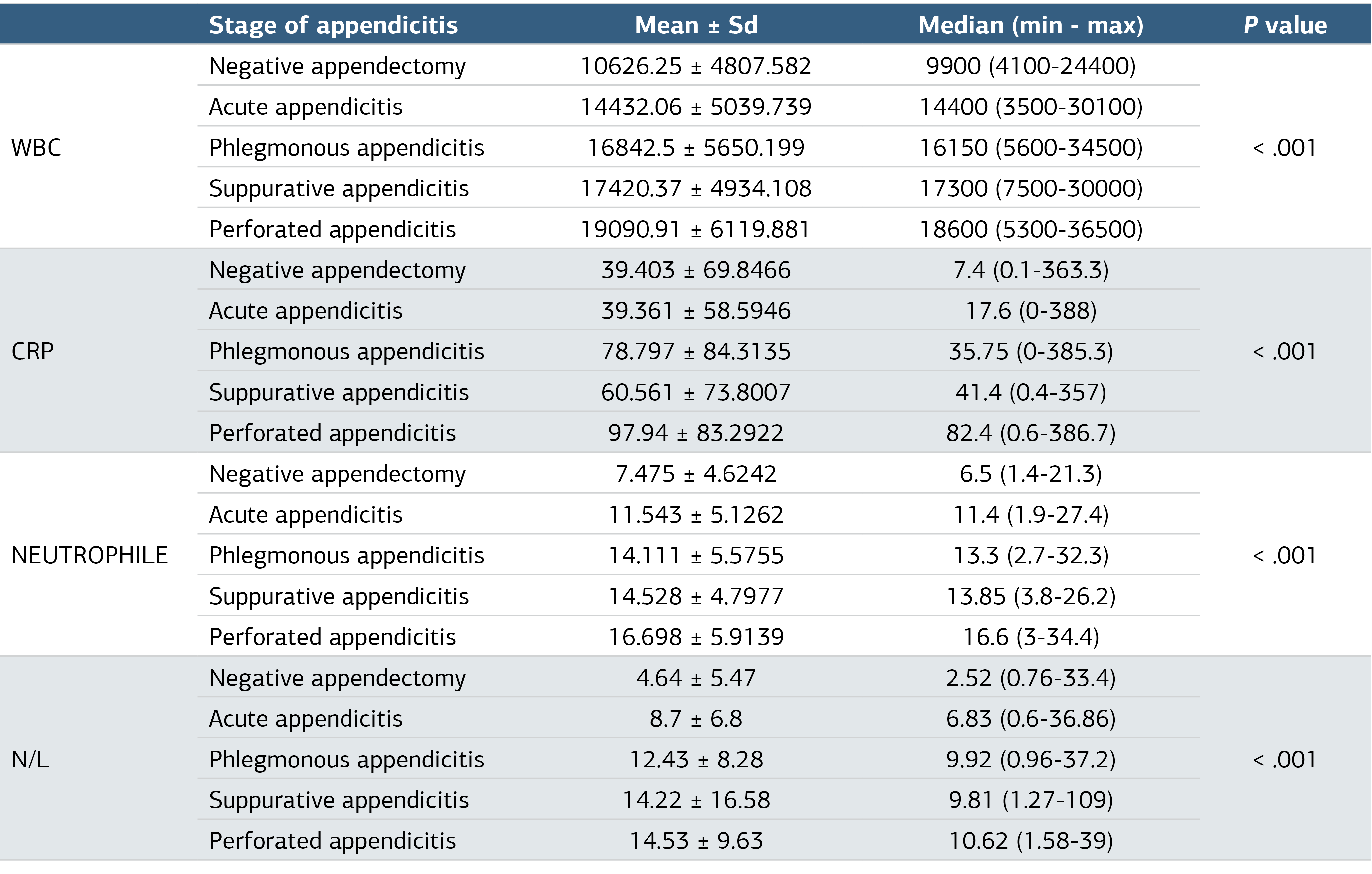
P values are based on Kruskal-Wallis test.
Table 2. Comparison of Visualization of Appendix in US Between Groups
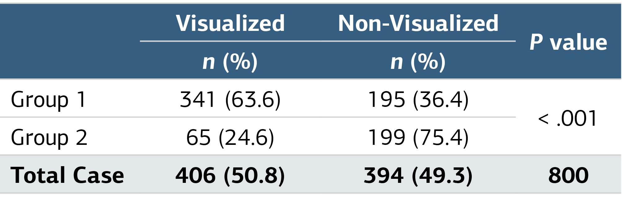
P values are based on Chi-squared test.
Table 3. Comparison of Visualization of Appendix in US Between the Histopathological Groups
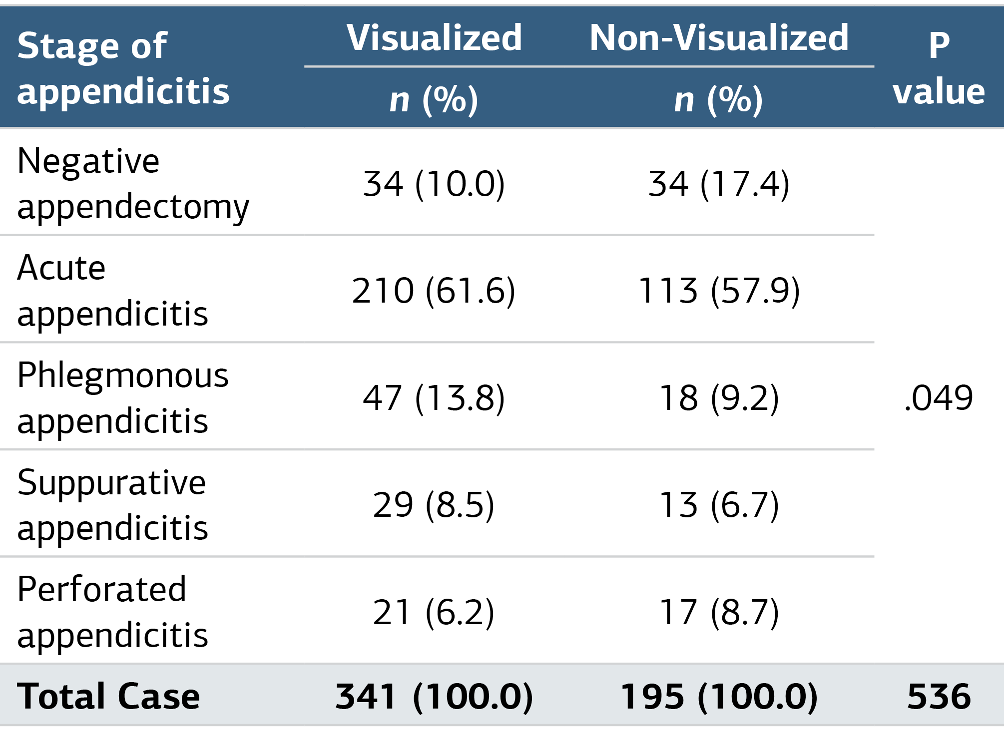
P values are based on Chi-squared test.
Figures
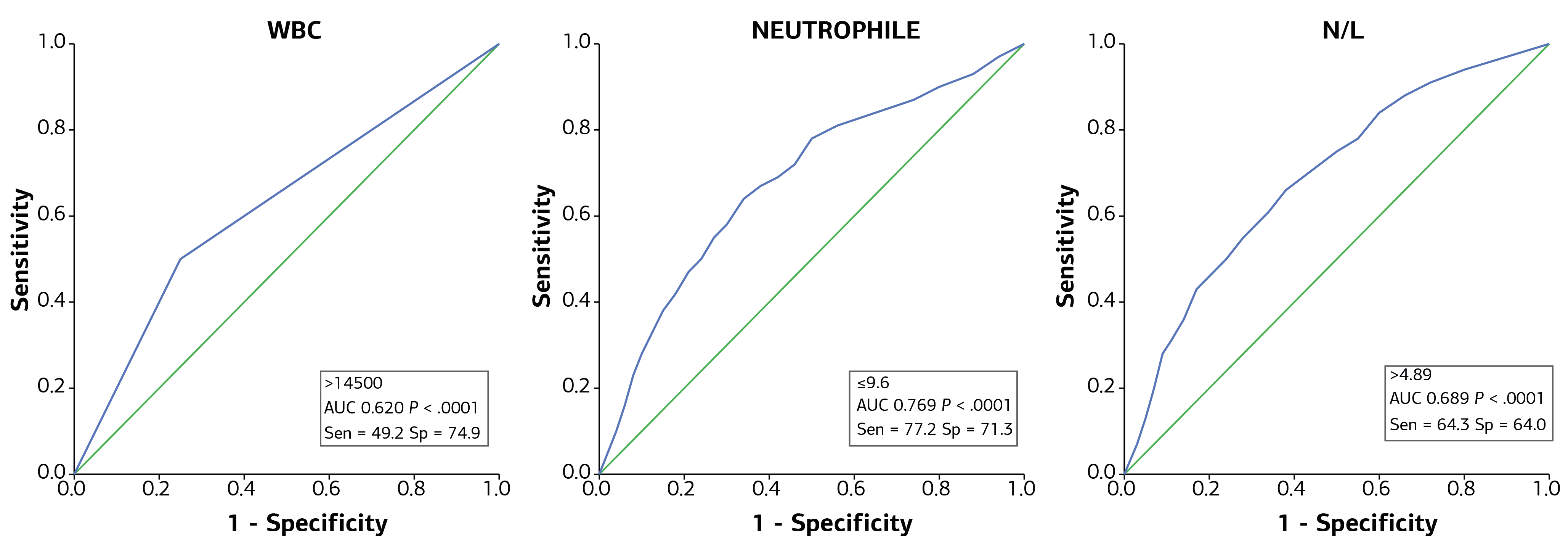
Figure 1. ROC Analysis and AUC (cut-off) Values for Accurate Diagnosis of Appendicitis
Additional Information
Publisher’s Note
Bayrakol MP remains neutral with regard to jurisdictional and institutional claims.
Rights and Permissions

This work is licensed under a Creative Commons Attribution-NonCommercial 4.0 International License (CC BY-NC 4.0). To view a copy of the license, visit https://creativecommons.org/licenses/by-nc/4.0/
About This Article
How to Cite This Article
Yusuf Doruk Bilgili, Duygu Tatlı Uçarcı, B. Haluk Güvenç. Diagnostic parameters for acute appendicitis in pediatric patients withabdominal pain: an analytical interpretation. Ann Clin Anal Med 2024;15(4):228-233. doi:10.4328/ACAM.21949
Publication History
- Received:
- 23.09.2023
- Accepted:
- 08.02.2024
- Published Online:
- 24.02.2024
- Printed:
- 01.04.2024