
Abstract
AimThe aim of this study was to compare plate positions in Lapidus arthrodesis in terms of displacement and plate stress.MethodsIn this study, the Lapidus arthrodesis was performed in a computerised environment using a finite element foot model with differentially positioned plates. The arthrodesis (Lapidus arthrodesis) was applied to the first metatarsocuneiform joint with 2 proximal 2 distal 3.5 mm screws using a 4-hole plate in three different directions: medial, dorsal and plantar.ResultsDisplacement in the arthrodesis area and stress on the fixation material, the plate, were evaluated. After loading, the plantar plate arthrodesis model produced more total displacement (1, 97121 mm) and stress (2242 MPa) than other arthrodesis models. On the other hand, the dorsal plate arthrodesis model has less total displacement (1, 20479 mm) and stress (1253 MPa) than other arthrodesis models. In the volar, dorsal and medial finite element models, it was concluded that the least displacement and plate stress were in the dorsal plate application.ConclusionIn the finite element study, complications can be seen more frequently in Lapidus arthrodesis performed with plantar and medial plates compared to Lapidus arthrodesis performed with dorsal plates. Therefore, Lapidus arthrodesis performed with a dorsal plate may be more stable.
Keywords
Introduction
Lapidus arthrodesis is a widely preferred and established procedure for the correction of various foot surgical problems, as this surgical technique allows the alignment of the first toe in all three axes. Lapidus arthrodesis is recommended as a first step in the treatment of hallux limitus, hallux valgus, hallux varus, hindfoot deformity, hypermobility of the first toe and metatarsal adductus.1
Although the Lapidus arthrodesis was first advocated by Albrecht in 1911, it was Lapidus who popularized the method.2,3 The described arthrodesis treatment has survived to this day under the name Lapidus.4
The Lapidus procedure is a form of surgical arthrodesis performed on the first tarsometatarsal joint. Although the results of Lapidus arthrodesis are very good, non-union or malunion is seen in 5-15% of patients. To minimize the risk of complications in Lapidus arthrodesis, the arthrodesis should be as stable as possible.5 This shows the importance of stability for the arthrodesis.
Lapidus arthrodesis is one of the surgical options for the treatment of hallux valgus.1-3 Lapidus arthrodesis used for the treatment of hallux valgus may be associated with various complications such as non-union, malunion and implant failure.6 While Myerson et al.7 indicated that the incidence of non-union in tarsometatarsal arthrodesis was 4.5%, Sangeorzan et al.8 emphasized that this rate was up to 13%. In a meta-analysis on lapidus arthrodesis,9 non-union was reported in 5.4% of patients after arthrodesis and false union in 6.1%.
Although there are different fixation methods in Lapidus arthrodesis, plate fixation is particularly recommended to minimize the risk of non-union, allow early weight bearing and rapid return to reference range of motion.10,11 However, there is no complete consensus on the correct location of the plate, and it is emphasized that there is a need for studies on the location and design of the plate system and the effectiveness of the results of plate detection for the treatment of hallux valgus.1,12-14
The aim of this study was to determine the stability of the arthrodesis and the plate stress after loading in a computer environment of 3 different Lapidus arthrodesis fixed with volar, dorsal, and medial 4-hole plates and compression screw modelled with finite elements. It is therefore desirable to demonstrate the importance of plate location in complications such as non-union, malunion and implant failure that may occur in Lapidus surgery.
Materials and Methods
In this study, the stress in the plate and the displacement in the arthrodesis were evaluated using finite element analysis according to the volar, dorsal and medial locations of the plate used for Lapidus arthrodesis.
The effect of the plate placed in different positions used in the foot first toe tarsometatarsal arthrodesis (Lapidus arthrodesis) created by finite element modelling on the stability in the arthrodesis and the tension in the plate used was analysed using Ansys Workbench software.
The relationship between plate fixation, which is one of the fixation methods, and such complications was demonstrated using modelling. For this purpose, a three-dimensional (3D) finite element bone model was constructed.
To create a 3D foot first tarsometatarsal joint arthrodesis model, CT images of the right ankle in 90° flexion were digitized to create solid bone models. These models were segmented into cortical and cancellous bone in the tibia, fibula, and foot bone models.
To analyse the arthrodesis of the first tarsometatarsal joint of the foot, the articular surface of the first tarsometatarsal joint of the foot was removed and a bony interface connection was created. The arthrodesis line of the first metatarsophalangeal joint was horizontal with respect to the joint plane and its direction was parallel to the x-axis of the global coordinates. The models were assembled in Ansys/SpaceClaim software according to the surgical procedure with the solid part of the implants. Volar, dorsal, and medial 4-hole plate and screw (PS) fixation methods were used (Figure 1).
A method called plate and screw fixation has been used in tarsometatarsal joint arthrodesis of the first toe. According to this surgical procedure, the incision is made at the level of the tarsometatarsal joint of the first toe and the chondral surface of the joint is removed and fixed.10,11
In the finite element model we prepared, as described in this surgical technique, 3 different models of volar, medial, and dorsal 4-hole plates were placed for fixation of the first metatarsophalangeal and medial cuneiform joint arthrodesis. The plates (Parcus Medical, Sarasota, FL, USA) were secured with 2 screws distal to the arthrodesis line and 2 screws proximal to the medial cuneiform of the first metatarsal. The plate used to connect the first finger tarsometatarsal joint arthrodesis is made of titanium alloy material. Linear elastic isotropic material models were used for the plate, bone and joint. The resulting solid models were converted into finite element models using Ansys Workbench software. The material parameters used for the plate and bone are given in Table 1.15
The finite element models are constrained so that they cannot make any translations or rotations from the lower boundary of the model. Loads were applied to the upper boundary of the model, the tibia, with a force of 600 N applied vertically downwards.16,17
The boundary conditions and loads were used. To simulate the real state of the fibula and tibia in the tissue, horizontal movement from the top of the model was prevented. The elements used for the connection are fixed to the foot, similar to the real conditions. Considering the arthrodesis interface as a frictional contact, the coefficient of friction was set at 0.2.
In order to obtain the best results in the finite element analysis, mesh optimization was performed, and the best mesh density was determined. With the best mesh density, the finite element models of the 3D models were also analysed using Ansys Workbench 2020R2 Finite Element software under the given loads. Then, the foot, fixed with a plate screw under load, was fixed in 3 different ways for arthrodesis in the first toe Lapidus arthrodesis modelling (Figure 1).
Two criteria were chosen to present the results of the numerical analysis (FEA – Finite Element Analysis). One is the von Mises stress criterion on the metal component of the fixation methods. The von Mises stress criterion determines where a given material will yield. The other criterion is the displacement and rotation of the arthrodesis space. The displacement and rotation measured after the application of physical stress is defined as the movement between the components. In particular, interfragmentary movement in the axial direction plays an important role in the stability of the arthrodesis, and this movement prevents bone healing and causes non-union.18
Points A and B were placed on the arthrodesis surface at the maximum and minimum effective points, respectively. The relative distance measurements of these points were calculated in three axes (X-mediolateral direction, Y-anteroposterior, Z-superoinferior).Ethical ApprovalNot applicable.Statistical AnalysisNot applicable.Reporting GuidelinesNot applicable.
Results
The material parameters used for Lapidus arthrodesis models are given in Table 1. As shown in Figure 1, in Lapidus arthrodesis models, which were fixed with plate screws in 3 different ways under load, first the displacement in the arthrodesis line and then the stress on the plate were evaluated. It was determined that the post-loading displacements of the plantar plate in the x, y and z directions were 0.58 mm, 0.71272 mm and 0.67849 mm, respectively. The total displacement of the plantar plate was 1.97121 mm.
Displacements of the dorsal plate in x, y and z directions were 0.40327 mm, 0.126 mm, and 0.67552 mm, respectively. The total displacement of the dorsal plate was also 1.20479 mm.
For the medial plate, the displacements in x, y and z directions were 0.4577, 0.7486 and 0.57177 mm, respectively (Table 2) (Figure 2, 3).
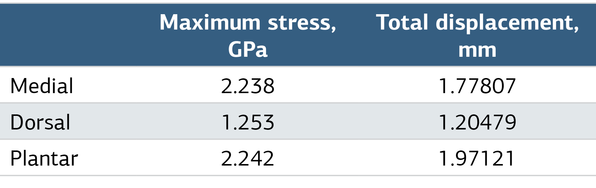
The maximum stress values in the plates after the analyses are such that for the plantar plate it is 2242 MPa, for the dorsal plate 1253 MPa and for the medial plate 2238 MPa (Table 3). If we compare them, we can see that the minimum stress is on the dorsal plate.
The dorsal plate was the most suitable combination in terms of displacement and minimum stress when three different plate placements were compared. On the other hand, the largest total displacement and stress were found on the plantar plate (Figure 3).
In terms of inter-particle displacement, the lowest displacement was found in the dorsal plate fixation method and the highest displacement was found in the medial plate fixation method.
Discussion
According to our study results, the reason why the dorsal plate placement is more suitable than others is that the dorsal plate is more effective than the plantar and medial plates. This is because the stress distribution on the dorsal plate under load is more homogeneous than others. It causes the load to spread and causes the stresses on the dorsal plate to decrease. Others carry the load mainly with the center of the plate, but the dorsal plate carries the load with the whole body.
Our results are similar to those reported in a previous biomechanical study. In this study by Ray et al,19 it was emphasized that the use of a plate increased the stability of the first metatarsocuneiform arthrodesis. In addition, fixation of the arthrodesis with 2 proximal and 2 distal 3.5 mm screws using a 4-hole dorsal plate was recommended to protect the fusion site from loading. The dorsal plate is more effective than the plantar and medial plates. These results are consistent with our work in the computer environment using the FEA technique.
On the other hand, a biomechanical study suggests the superiority of the plantar plate position in detection. Plantar and medial plate positions are superior to displacement in arthrodesis. In particular, arthrodesis performed with the plate positioned medially is superior in terms of less displacement and resistance to loading forces. Dorsal plate placement was not superior for any of the outcomes measured. It was emphasized that plantar and medial plates offer biomechanical advantages. It is emphasized that clinical trials with similarly matched constructs are needed to show whether these findings translate into improved clinical outcomes.20 When these results are evaluated, they differ from the results of our study. In our study, the dorsal plate was found to be superior to the plantar and medial plates in terms of loading and displacement. These results need to be evaluated in long-term clinical trials.
Another study compared plate fixation for arthrodesis with intramedullary fixation. In this study, Roth et al.13 presented metatarsocuneiform (MTC) fusion (Lapidus arthrodesis) as a treatment option for hallux valgus. They emphasized that plantar plate fixation creates a stronger and more rigid structure than IM fixation. They concluded that plate fixation provides a more stable fixation and reduces the risk of non-union and the duration of weight-bearing disability. They state that plate fixation is appropriate in Lapidus arthrodesis.13 However, there is no suggestion in the article for different placements of the plate.
A review of the efficacy of the Lapidus plate system in 2022 states that there are insufficient studies on the efficacy of different types of Lapidus plate systems in foot surgery. In addition, it is emphasised that there is a need to increase knowledge about the fixation level, system type and placement of the plate in Lapidus surgery.1
In a study of dorsal-medial locking plate and screw fixation in Lapidus arthrodesis, the dorsal-medial plate showed better preservation of intermetatarsal angle (IMA) correction compared to screw fixation at mid-term follow-up.21
A study evaluating the soft tissue effect of Lapidus arthrodesis found that Lapidus arthrodesis with a plantar plate resulted in high patient satisfaction and significant deformity improvement at mid-term follow-up. On the other hand, it was reported that more than half of the patients had tendinopathy symptoms on postoperative magnetic resonance imaging (MRI). It is emphasised that these findings should be taken into account when choosing the method of fixation and implant placement in Lapidus arthrodesis.22
Another study also highlights that the superficial peroneal nerve (SPN) is at risk of iatrogenic injury due to incisions made along the medial column, including the medial branch of the medial dorsal cutaneous nerve (MDCN) branch, dorsal longitudinal incision for Lapidus fusion. The study concluded that the surgeon must be careful to avoid nerve injuries during dissection.23
A study of 3-plane first tarsometatarsal arthrodesis using a combination of dorsal and medial plates reported statistically significant improvements in radiographic correction, low deformity recurrence and early return to activity with low complication rates up to 24 months post-operatively. In addition, statistical improvements in patients’ health-related quality of life are reported up to 24 months.24 Although the fixation results of double plate application are good, it has disadvantages such as additional incision, soft tissue dissection and cost.
In a study comparing the biomechanical stability of Lapidus arthrodesis performed with the plantar Lapidus plate in patients with severe symptomatic hallux valgus with proximal chevron osteotomy performed with plate fixation, it is emphasised that the angular correction potential and clinical value of modified Lapidus arthrodesis performed with the plantar plate is better than chevron osteotomy.25
In a study on the use of different types of the Lapidus Plate System, it is stated that “the use of screwed or screwless plates, proper levelling and system placement are related to existing foot discomfort”.1 It is emphasized that there is a need for studies on the level of fixation and placement in foot surgery and studies to increase knowledge of the plate system and plate outcomes in foot surgery. For this purpose, in our study, we addressed this controversial issue from a different perspective with a finite element study.
Limitations
This study was limited to finite element modeling and did not include clinical validation. Therefore, the findings may not fully reflect in vivo biomechanical conditions.
Conclusion
Although the Lapidus arthrodesis is a recommended surgical technique for the treatment of hallux valgus, various studies show a symptomatic non-union rate of up to 12% and an overall failure rate of up to 20%. In our study, in finite element modeling of dorsal, medial and plantar plate screw fixation, it was determined that the dorsal plate fixation method provided better stability in Lapidus arthrodesis than other plate locations. We believe that these rates may be further reduced with new studies on the design and placement of the proposed plate system in Lapidus arthrodesis. For this purpose, we tried to show the most appropriate plate location in Lapidus arthrodesis in terms of arthrodesis displacement and plate loading using finite element modelling.
Declarations
Animal and Human Rights Statement
All procedures performed in this study were in accordance with the ethical standards of the institutional and/or national research committee and with the 1964 Helsinki Declaration and its later amendments or comparable ethical standards.
Informed Consent
Not applicable.
Data Availability
The datasets used and/or analyzed during the current study are not publicly available due to patient privacy reasons but are available from the corresponding author on reasonable request.
Conflict of Interest
The authors declare that there is no conflict of interest.
Funding
None.
Abbreviations
CT: Computed tomography
FEA: Finite element analysis
MDCN: Medial dorsal cutaneous nerve
MRI: Magnetic resonance imaging
MTC: Metatarsocuneiform
PS: Plate and screw
SPN: Superficial peroneal nerve
References
- López-López D, Larrainzar-Garijo R, De-Bengoa-Vallejo RB, Losa-Iglesias ME, Bayod-López J. Effectiveness of the Lapidus plate system in foot surgery: a PRISMA-compliant systematic review. Int Wound J. 2022;19(3):507-514.
- Hecht PJ, Lin TJ. Hallux valgus. Med Clin North Am. 2014;98(2):227-232. doi:10.1016/j.mcna.2013.10.007
- Baravarian B, Briskin GB, Burns P. Lapidus bunionectomy: arthrodesis of the first metatarsocuneiform joint. Clin Podiatr Med Surg. 2004;21(1):97-vi. doi:10.1016/s0891-8422(03)00112-5
- Symeonidis PD, Anderson JG. Original and modified Lapidus procedures: proposals for a new terminology. J Bone Joint Surg Am. 2021;103(4). doi:10.2106/jbjs.20.01010
- Cohen DA, Parks BG, Schon LC. Screw fixation compared to H-locking plate fixation for first metatarsocuneiform arthrodesis: a biomechanical study. Foot Ankle Int. 2005;26(11):984-989. doi:10.1177/107110070502601114
- Willegger M, Holinka J, Ristl R, Wanivenhaus AH, Windhager R, Schuh R. Correction power and complications of first tarsometatarsal joint arthrodesis for hallux valgus deformity. Int Orthop. 2015;39(3):467-476. doi:10.1007/s00264-014-2601-x
- Klos K, Simons P, Schopp P, et al. Biomechanical comparison of medio-plantar and plantar plate fixation for first tarsometatarsal joint arthrodesis. J Clin Med. 2023;12(12):3896. doi:10.3390/jcm12123896
- Roukis TS. Nonunion after arthrodesis of the first metatarsal-phalangeal joint: a systematic review. J Foot Ankle Surg. 2011;50(6):710-713. doi:10.1053/j.jfas.2011.06.012
- Baravarian B, Ben-Ad R. Contemporary approaches and advancements to the Lapidus procedure. Clin Podiatr Med Surg. 2014;31(2):299-308. doi:10.1016/j.cpm.2014.01.001
- Sorensen MD, Hyer CF, Berlet GC. Results of Lapidus arthrodesis and locked plating with early weight bearing. Foot Ankle Spec. 2009;2(5):227-233. doi:10.1177/1938640009348389
- Cottom JM, Baker JS. Comparison of locking plate with interfragmentary screw versus plantarly applied anatomic locking plate for Lapidus arthrodesis: a biomechanical cadaveric study. Foot Ankle Spec. 2017;10(3):227-231. doi:10.1177/1938640016676341
- Roth KE, Peters J, Schmidtmann I, Maus U, Stephan D, Augat P. Intraosseous fixation compared to plantar plate fixation for first metatarsocuneiform arthrodesis: a cadaveric biomechanical analysis. Foot Ankle Int. 2014;35(11):1209-1216. doi:10.1177/1071100714547082
- Dayton P, Santrock R, Kauwe M, et al. Progression of healing on serial radiographs following first ray arthrodesis in the foot using a biplanar plating technique without compression. J Foot Ankle Surg. 2019;58(3):427-433. doi:10.1053/j.jfas.2018.09.001
- Hess F, Sommer C. Minimally invasive plate osteosynthesis of the distal fibula with the locking compression plate: first experience of 20 cases. J Orthop Trauma. 2011;25(2):110-115. doi:10.1097/bot.0b013e3181d9e875
- Öner K, Durusoy S, Özer A. Is the fracture morphology in the sagittal plane important in determining the ideal placement of the lag screw in intertrochanteric femoral fractures? Injury. 2021;52(3):562-568. doi:10.1016/j.injury.2020.10.023
- Zhao Z, Yan T, Guo W, Yang R, Tang X. Is double-strut fibula ankle arthrodesis a reliable reconstruction for bone defect after distal tibia tumor resection? A finite element study based on promising clinical outcomes. J Orthop Surg Res. 2021;16(1):230. doi:10.1186/s13018-021-02362-0
- Li J, Wei Y, Wei M. Finite element analysis of the effect of talar osteochondral defects of different depths on ankle joint stability. Med Sci Monit. 2020;26. doi:10.12659/msm.921823
- Claes LE, Heigele CA, Neidlinger-Wilke C, et al. Effects of mechanical factors on the fracture healing process. Clin Orthop Relat Res. 1998;(355 Suppl). doi:10.1097/00003086-199810001-00015
- Ray RG, Ching RP, Christensen JC, Hansen ST Jr. Biomechanical analysis of the first metatarsocuneiform arthrodesis. J Foot Ankle Surg. 1998;37(5):376-385. doi:10.1016/s1067-2516(98)80045-8
- Drummond D, Motley T, Kosmopoulos V, Ernst J. Stability of locking plate and compression screws for Lapidus arthrodesis: a biomechanical comparison of plate position. J Foot Ankle Surg. 2018;57(3):466-470. doi:10.1053/j.jfas.2017.10.025
- Abben KW, Sakow CF, Sorensen T, Chang HC, Boffeli TJ. First tarsometatarsal joint fusion for hallux valgus deformity: a retrospective comparison of two fixation constructs regarding initial maintenance of correction and complications: traditional crossing screw fixation versus dorsomedial locking plate and intercuneiform compression screw. J Foot Ankle Surg. 2023;62(2):347-354. doi:10.1053/j.jfas.2022.09.002
- Niehaus R, Hodel S, Eid K, Bensler S, Lenz CG. Plantar plating in the modified Lapidus procedure: evaluation of function and impairment of the tibialis anterior tendon. J Foot Ankle Surg. 2022;61(6):1203-1208. doi:10.1053/j.jfas.2022.02.003
- Boffeli TJ, Gervais SJ. Accuracy of palpation to identify the medial branch of the medial dorsal cutaneous nerve in medial column surgery: correlation of preoperative nerve marking with intraoperative findings during Lapidus fusion. J Foot Ankle Surg. 2021;60(1):2-5. doi:10.1053/j.jfas.2019.07.010
- Liu GT, Chhabra A, Dayton MJ, et al. One- and two-year analysis of a five-year prospective multicenter study assessing radiographic and patient-reported outcomes following triplanar first tarsometatarsal arthrodesis with early weightbearing for symptomatic hallux valgus. J Foot Ankle Surg. 2022;61(6):1308-1316. doi:10.1053/j.jfas.2022.04.008
- Kasparek MF, Benca E, Hirtler L, et al. Biomechanical evaluation of the proximal chevron osteotomy in comparison to the Lapidus arthrodesis for the correction of hallux valgus deformities. Int Orthop. 2022;46(10):2257-2264. doi:10.1007/s00264-022-05514-x
Tables
![Table 1. Material Properties of Bone and Plate[18]](https://acamedicine.org/archive/vol15/issue4/10.4328-ACAM.22077/table-1.png)
Table 2. Displacements of Plates in the X, Y, and Z Directions
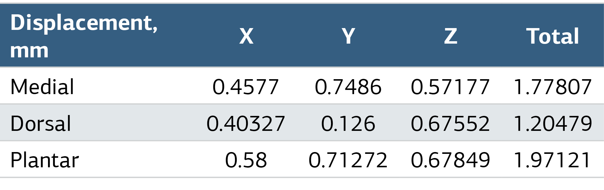
Table 3. Max Stress and Total Displacement Values in Plates After Analyses
Figures
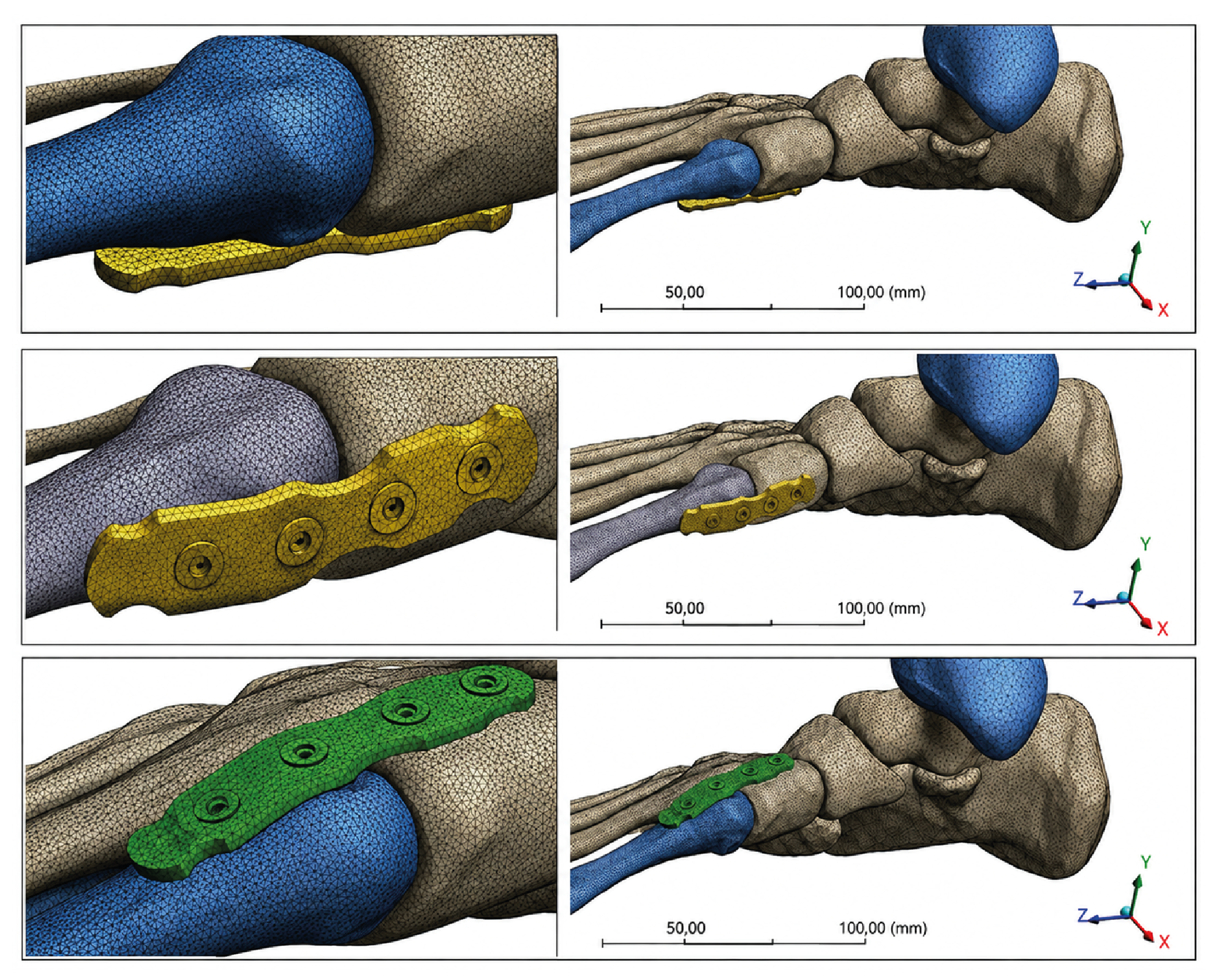
Figure 1. The Finite Element Model of Volar, Medial, and Dorsal Plates in Lapidus Arthrodesis
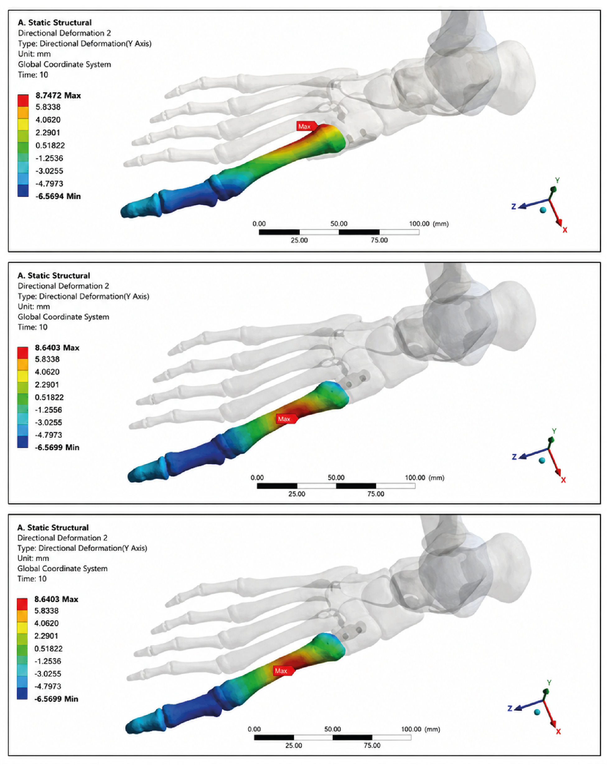
Figure 2. Displacements in Plates After Analyses, Medial (Upper), Dorsal (Middle), and Plantar (Bottom)
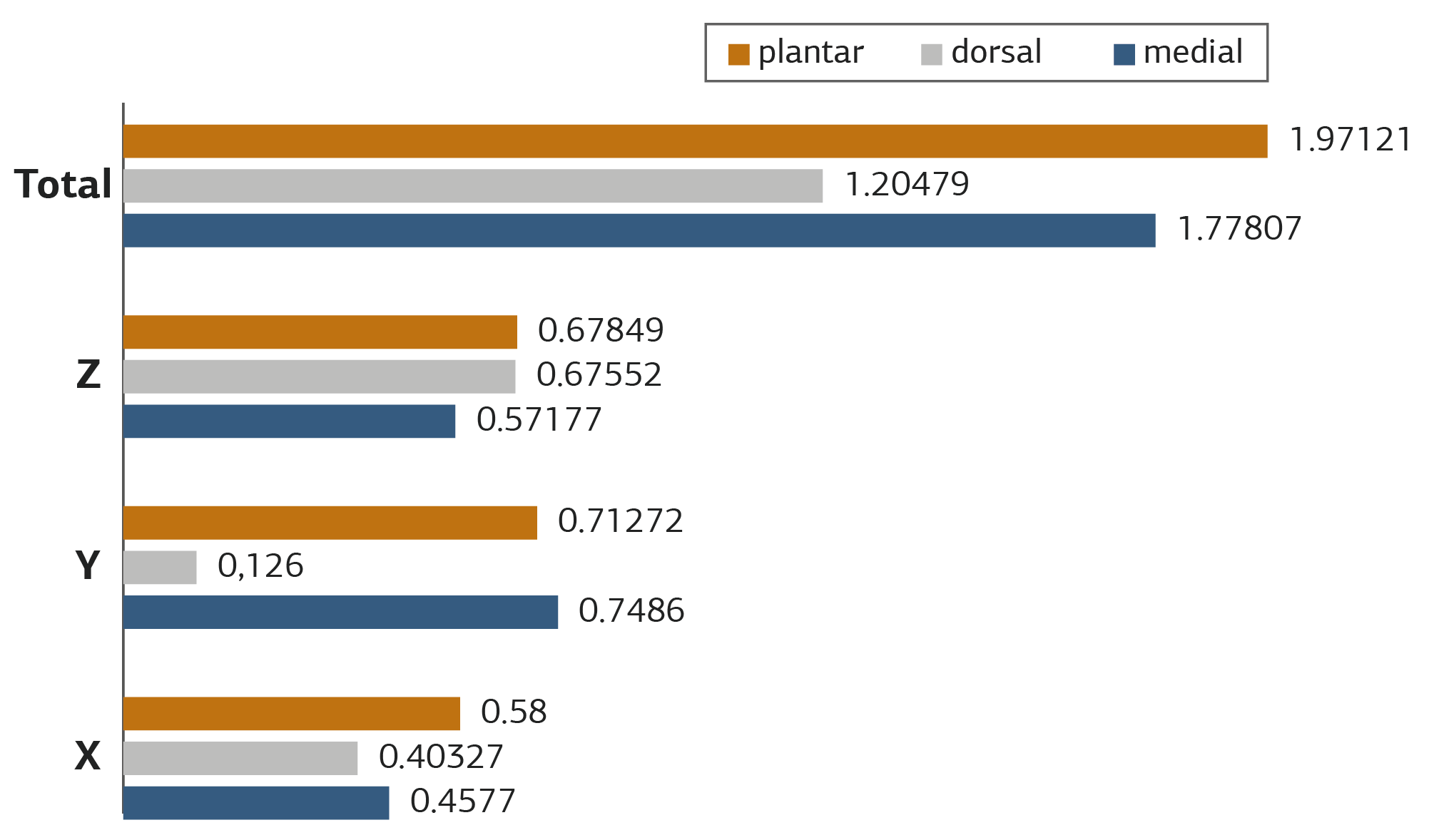
Figure 3. Displacements of Plates in the X, Y, and Z Directions
Additional Information
Publisher’s Note
Bayrakol MP remains neutral with regard to jurisdictional and institutional claims.
Rights and Permissions

This work is licensed under a Creative Commons Attribution-NonCommercial 4.0 International License (CC BY-NC 4.0). To view a copy of the license, visit https://creativecommons.org/licenses/by-nc/4.0/
About This Article
How to Cite This Article
Hacı Ali Olçar, Alaettin Özer, Halil Burak Mutu, Tolgahan Kuru, Göker Yurdakul, Davut Aydın, Murat Korkmaz. The biomechanical analysis of first metatarsocuneiform arthrodesis usingthe finite element method: a comparison of plate osteosynthesis in differentpositions. Ann Clin Anal Med 2024;15(4):255-259. doi:10.4328/ACAM.22077
Publication History
- Received:
- 20.12.2023
- Accepted:
- 22.02.2024
- Published Online:
- 09.03.2024
- Printed:
- 01.04.2024