
Abstract
AimThis study focused on determining the exit variations and branching pattern of the sciatic nerve (SN), obtaining morphometric data of the lower extremity and correlating these data with nerve blockade procedures.MethodsThe lower extremities of twenty human cadavers were prospectively dissected, and the distances between superficial and deep reference points, the thickness of the SN and its branches were measured. The exit of the SN to the gluteal region, its course at the back of the thigh, and its terminal branching level were examined.ResultsThe literature review determined that at least 5 and at most 13 different sciatic nerve types were defined according to their relationship with the piriformis. During the dissections, it was observed that 95% of the SN passed under the piriformis as a single root and bifurcated in all lower extremities. The bifurcation level of the SN in the upper, middle and lower thighs was close to each other (35%, 35%, 30%, respectively). Statistically significant (P < .05) correlations were detected between distances using the greater trochanter as a reference and thigh length. ConclusionThe obtained correlations support the use of the greater trochanter in determining the point of trans-gluteal SN blockade. If sciatic nerve blockade in the thigh cannot be performed with imaging methods, performing it at the level above the popliteal fossa as much as possible, considering the person’s thigh length, will increase success.
Keywords
Introduction
The sciatic nerve (SN), a continuation of the sacral plexus, is the body’s thickest and longest peripheral nerve. The SN, whose initial part is located in the pelvis, leaves the pelvis through the infrapiriform foramen and exits to the gluteal region. It usually extends from the lower border of the piriformis in the gluteal region to the upper level of the popliteal fossa, where it bifurcates into its terminal branches (tibial and common peroneal nerves). A common sheath surrounds it up to the bifurcation (BFR), while the tibial nerve (TN) and common peroneal nerve (CPN) are surrounded by different sheaths.1-3 Many studies have investigated the relationship between SN and piriformis, and some have classified this relationship.3-9
The incidence of damage to the SN is high due to its thickness and long course. In addition, piriformis syndrome, intervertebral disc, hip joint and pelvis pathologies, gluteal intramuscular injection, nerve inflammation, or pressure during labor may cause sciatica, motor and sensory loss. Although variations in the SN and its branches, other than those related to the piriformis, do not cause symptoms, undesirable results may occur in interventions in this region.6,8,10
This study, which includes a literature review on SN anatomy, aims to contribute to nerve blockade applications by additionally performing gluteal and posterior thigh dissections.
Materials and Methods
The study was performed prospectively in the Anatomy Laboratory on 20 lower extremities of 10 adult human cadavers, aged between 40 and 80 years, fixed with 10% formalin. Variations and nerve branching were recorded with a digital camera and schematized. The same person repeated the length and distance measurements three times using a tape measure and the thickness measurements using a caliper.
Measurements and dissections were made in the prone position. Before dissection, palpable bone formations were marked as reference points. Then, thigh length (distance between the greater trochanter and lateral condyle of femur, Thigh length (TL)), lower extremity length (distance between anterior superior iliac spine and medial malleolus, Lower extremity length (LEL)), the distance between the greater trochanter and the posterior superior iliac spine (GT-PSIS), and the distance between the greater trochanter and the sacral hiatus (GT-SH) were measured. The superficial GT-SH distance was marked by dividing it into three parts (medial, middle and lateral) to determine the position of the dissected SN. The point where the line drawn perpendicularly from the midpoint of GT-PSIS intersects with GT-SH was defined as the superficial blocking point of SN.11
Following superficial measurements, gluteus maximus, connective and fatty tissue dissection was performed and piriformis and SN were exposed. Hamstring muscles and later parts of SN at the back of the thigh were dissected. The passage of the SN to the subgluteal region was evaluated according to the relationship of the nerve with the piriformis.1-5 The branching pattern (bifurcation, trifurcation, etc.) and level (distal 1/3, middle 1/3, proximal 1/3) of the SN were determined. The connecting branches between TN and CPN were evaluated according to the Testut classification.9
The exit point of the SN was determined as medial, middle and lateral according to its location on the superficially defined GT-SH line. Then, the following measurements were made in the subgluteal and posterior femoral regions;
– Distance (cm) of the SN exit point (EP) to the greater trochanter (GT-EP) and sacral hiatus (SH-EP).
– Distance of nerve blocking point to the greater trochanter (GT-BP, cm)
– Distance between SN exit point and BFR level (EP-BFR, cm)
– Distance between the lower border of gluteus maximus and BFR (GM-BFR, cm)
– SN exit point diameter (EPd, mm) and SN blocking point’s projection diameter determined on the superficial plane (BPd, mm)
– TN diameter (Td, mm) and CPN diameter (CPd, mm) at BFR level.Ethical ApprovalThis study was approved by the Ethics Committee of Necmettin Erbakan University Non-Pharmaceutical and Non-Medical Device Research (Date: 05.02.2021, Decision No: 2021/3091).Statistical AnalysisData obtained from lower limbs were analyzed by using SPSS 21.0 (IBM-Statistics software, Chicago Illinois) statistical program. Descriptive statistics (mean, standard deviation) were calculated for all parameters. The relationship between the parameters was determined by Pearson Correlation test. The significance level of statistical analyses was P < .01 and P < .05.
Reporting GuidelinesNot applicable.
Results
In 19 of 20 lower extremities (95%), it was observed that the SN passed from the lower border of the piriformis to the gluteal region as a single root, and in 1 (5%) it branched before reaching the muscle. In this lower extremity, the CPN had pierced the piriformis and the TN was passing through the lower border of the piriformis and advancing towards the gluteal region (Figure 1). The SN was bifurcated in all (%100) of the lower extremities. The BFR level, determined according to thigh lengths, was detected in the distal 1/3 of the thigh in 7 extremities (35%), in the middle 1/3 of the thigh in 7 extremities (35%), and in the proximal 1/3 of the thigh in 6 extremities (30%)(Figure 2A-C). After BFR, connections between SN branches were detected in only two lower extremities (Figure 3A-B). The average measurement data of length, distance and thickness of 19 lower extremities were as follows:
Length measurements: LEL, 81.38 ± 5.2cm; TL, 36.48 ± 3cm.
Distances measurements: GT-SH, 18.47 ± 1.52cm; GT-PSIS, 17.62 ± 0.93cm; GT-EP, 10.02 ± 1.35cm; GT-BP, 8.30 ± 1.29cm; SH,-EP, 8.45 ± 1.32cm; IT-EP, 5.41 ± 1.36 cm; EP-BFR, 22.30 ± 9.67cm; GM-BFR, 9.54 ± 9.87cm; EP-FB, 8.35 ± 3.99cm; EP-LB, 14.66 ± 4.66cm.
Diameter measurements: EPd, 16.41 ± 3.61mm; BPd, 12.56 ± 3.11mm; Td, 5.06 ± 0.99mm; CPd, 3.33 ± 0.85mm.
The origin of the SN was medial to the midpoint of GT-SH in 16 cases (0.5-4.15cm), over the middle in one case, and lateral to the midpoint in three cases (0.5cm). The blocking point determined according to the classical SN blocking technique (back) was approximately 2cm closer to the greater trochanter than the exit point. The SN was, on average, 4mm thinner at the site of the blocking point than at the exit point, and the thicker terminal branch was TN in all cases.
GT-PSIS and GT-BP correlated with each other (r = 0.567, P = .011). GT-PSIS with TL (r = 0.641, P = .003) and GT-BP with TL (r = 0.703, P = .001) were found to have a statistically significant correlation. A positive and significant correlation was observed between the diameter of the SN at its exit point and its diameter at the blocking point (r = 0.649, P = .003). No significant relationship was detected between TL and total LEL of the TN and CPN.
Discussion
The SN is significant clinically because of its long course (gluteal, subgluteal, and posterior femoral region) and wide innervation area. In addition, the relationship of the nerve with the piriformis, as well as injuries that occur during invasive procedures (superficial and deep injections, abscess drainages, hip joint surgery, etc.) applied to the area, may cause sensory and motor disorders.6,8,12,13
The first classification based on the relationship between the piriformis and SN and referenced in many studies belongs to Beaton and Anson.1,5,8,13,14 Barbosa et al.1 made the most comprehensive classification in a systematic review that included 12 studies. Beaton and Anson.5 defined 7 types (1 normal, Type a; 6 variations, Type b-g) for SN and accepted that SN occurs in all cases. Barbosa et al.1 described 13 types. In 10 of these types, the SN was formed before the piriformis (in 3 types, arising as a single root; in 7 types, early branched before the piriformis and branches not fused distally); in 2, formed distally and in one, not formed (Table 1).
The incidence of SN with a standard course was reported as 71.87% – 98.3% in cadaver and imaging studies.1,2,4,7,9,14-19 The second most common (1.7-22.5%) muscle-nerve relationship is the emergence of the peroneal root from the inside of the piriformis and the tibial root from the infrapiriform foramen. However, in these cases, Beaton and Anson.5 stated that these roots combined to form SN (Type b), while Barbosa et al.1 mentioned that the roots continue as separate nerves (Type 3) (Table 1). Although it is argued in the literature that SN variations have a strong relationship with piriformis syndrome, Barbosa et al.1 stated that there may be variations in asymptomatic patients, and on the contrary, these variations may not be found in symptomatic patients.
Most researchers stated that the SN and its branches emerge from the superior, inferior, or inside of the piriformis.1,5,7,10,13-17 Natsis et al.8 reported that the piriformis had two (superior, inferior) or three bellies (superficial, medium, deep), while Jacomo et al.20 mentioned accessory piriformis. Huang et al.21 identified a connecting branch from the TN passing through the infrapiriform foramen to the CPN passing through the inside of the piriformis in a lower extremity during routine cadaveric dissection. They described this case as the reoccurrence of a split of SN. In our study, we observed that in 19 lower extremities (95%), the SN emerged as a single root from the infrapiriform foramen, while in one extremity (5%), the SN bifurcated early into terminal branches, the CPN emerged from the inside of piriformis, and the TN arose from the infrapiriform foramen, and these branches did not join. This type is suitable for Type b, according to Beaton and Anson.5 And Type 3 because the SN bifurcated early, according to Barbosa et al.1 The frequency of this variation detected in our study is close to the lower limit (4.1-22.5%) of incidence rates in adults reported in the literature.
Although it has been reported that the SN divides into two terminal branches by bifurcation.4,7,18,22,23 Berihu and Debeb.2 Mentioned 5.36% trifurcation (TN, CPN and accessory nerve). It was determined that the SN was bifurcated into two branches in all lower extremities included in our study (100%). Cadaveric studies report differences in the level of bifurcation of the SN, but there is no standard classification.4,7,16,18,22 (Table 2). In three of these studies describing the BFR level, the level was stated in the lower 1/3 of the thigh, while in the others it was frequently stated in the popliteal fossa (Table 2). Güvençer et al.7 identified BFR in the popliteal fossa (52%) and the pelvis (48%). Ogeng’o et al.16 frequently (67.1%) found BFR in the popliteal fossa. Barbosa et al.4 defined BFR level according to the popliteal fossa and most frequently detected it below the apex of the popliteal fossa (67.96%) (Table 2). In our cadaveric study with a relatively small sample size, BFR levels detected in the upper 1/3, middle 1/3, and lower 1/3 of the thigh were close to each other (35%, 35%, 30%, respectively). In an imaging study for popliteal SN block, the BFR level was reported to be approximately 6±2.7cm (0-11.5cm) above the popliteal fossa fold. However, literature knowledge and our study data show that this level may be higher. We suggest that it would be useful to consider the TL along with the popliteal fossa in estimating the location of SN blockade in the region of the back of the thigh above the BFR level. According to our results, we recommend knowing different BFR levels for invasive interventions to the region, including nerve block, intervening with imaging methods, and using the thigh length as well as the popliteal fossa as a reference in cases where imaging is not possible (blindly). Between the terminal branches of the SN, connecting branches can be found along their course.9,16 Testut classified these connection branches in his book “Tratado de Anatomia Humana (1902)”.9 Tubbs et al.9 detected various connections between the terminal branches of the SN in 30 of 40 lower extremities of 20 cadavers and evaluated them according to the Testut classification. They reported that the neural connections detected in the majority of sides (75%) were always located within 20 cm of the greater sciatic notch and the presence of up to 3 communications in one connection pattern. Ogeng’o et al.16 reported but did not classify connecting branches in 3.7% of 164 lower extremities. In our study, neuronal connections between terminal branches were detected in two left extremities (10%). There was a connection from CPN to TN (Type A according to the Tesut classification) in one of these extremities. In the other, there was a connection between the proximal and distal parts of the TN. In the second case, which was similar to Type G (the connection between proximal and distal portions of the nerve) according to the Testut classification, there was an additional joint branch arising from the connecting branch. Since the number of studies on connection branches in the literature is limited, a comprehensive comparison could not be made.
Success in hip arthroscopy is closely related to the location of the opened ports.12,13 Especially in the posterolateral approach, the SN should be protected due to its proximity to the portal opened adjacent to the posterior and upper part of the apex of the greater trochanter.13 In three of our cases, the origin of the SN was lateral to the midpoint of the GT-SH, and in the others it was medial. In surgical intervention or nerve block where ultrasonography cannot be used, the superficial projection of the SN from the exit site is determined, and its course is estimated. There are a limited number of studies conducted on adults, including lower extremity lengths, distances between some reference structures (posterior superior iliac spine, greater trochanter, ischial tuberosity, sacral hiatus) and SN thickness.15,17,19 (Table 3).
Compared to other studies evaluating similar parameters in samples with almost the same limb lengths, the GT-PSIS distance was longer and the bifurcation level of the SN was higher in this study (Table 3). Additionally, in our study, the relationship between the bifurcation level of the SN and the lower border of the gluteus maximus was evaluated; It was observed below the lower border (1.19-23cm) in 13 cases, above the lower border [(-3.7)-(-13)cm] in four cases, and at the lower border (0cm) in three cases. This study found significant correlations between the distance of the SN blocking point determined by the trans gluteal SN block technique to the greater trochanter, the lower extremity length, and the GT-PSIS. This relationship supports the use of the greater trochanter in determining the SN blocking point. However, there was no significant relationship between nerve thickness and lower extremity lengths. This result was incompatible with the expectation that the nerve thickness would be higher in tall people.
Limitations
Since it became very difficult to obtain cadavers during the COVID-19 epidemic, the low number of samples limited our study. On the other hand, the strength of our research is the comparison of classifications found in the literature and obtaining morphometric data.
Conclusion
The literature review observed that the relationship variations between SN and piriformis muscle, which have been defined since long ago, have recently been added to new ones. It is concluded that not all individual differences have yet been defined, so current and extended variation classifications may change. While we support using the greater trochanter as a reference in determining the point of SN blockade, we suggest that it would be useful to consider the TL together with the popliteal fossa in estimating the location of SN blockade on the posterior thigh.
Declarations
Animal and Human Rights Statement
All procedures performed in this study were in accordance with the ethical standards of the institutional and/or national research committee and with the 1964 Helsinki Declaration and its later amendments or comparable ethical standards.
Informed Consent
Not applicable.
Data Availability
The datasets used and/or analyzed during the current study are not publicly available due to patient privacy reasons but are available from the corresponding author on reasonable request.
Conflict of Interest
The authors declare that there is no conflict of interest.
Funding
None.
Acknowledgements
The authors sincerely thank those who donated their bodies to science so that anatomical research could be performed.
Abbreviations
BFR: Bifurcation
CPN: Common peroneal nerve
EP: Exit point
GT: Greater trochanter
GT-BP: Distance between the greater trochanter and blocking point
GT-EP: Distance between the greater trochanter and sciatic nerve exit point
GT-PSIS: Distance between the greater trochanter and posterior superior iliac spine
GT-SH: Distance between the greater trochanter and sacral hiatus
LEL: Lower extremity length
SN: Sciatic nerve
TL: Thigh length
TN: Tibial nerve
References
- Barbosa ABM, Santos PV, Targino VA, et al. Sciatic nerve and its variations: is it possible to associate them with piriformis syndrome? Arq Neuropsiquiatr. 2019;77(9):646-653. doi:10.1590/0004-282x20190093
- Berihu BA, Debeb YG. Anatomical variation in bifurcation and trifurcations of sciatic nerve and its clinical implications: in selected university in Ethiopia. BMC Res Notes. 2015;8:633. doi:10.1186/s13104-015-1626-6
- Lewis S, Jurak J, Lee C, Lewia R, Gest T. Anatomical variations of the sciatic nerve, in relation to the piriformis muscle. Transl Res Anat. 2016;5:15-19. doi:10.1016/j.tria.2016.11.001
- Barbosa FT, Barbosa TRBW, Cunha RM da, Rodrigues AKB, Ramos FW da S, Sousa-Rodrigues CF de. Anatomical basis for sciatic nerve block at the knee level. Braz J Anesthesiol. 2015;65(3):177-179. doi:10.1016/j.bjane.2014.03.010
- Beaton LE, Anson BJ. The relation of the sciatic nerve and of its subdivisions to the piriformis muscle. Anat Rec. 1937;70(1):1-5. doi:10.1002/ar.1090700102
- Bretonnier M, Lemée J-M, Berton J-E, Morandi X, Haegelen C. Selective neurotomy of the sciatic nerve branches to the hamstring muscles: an anatomical study. Orthop Traumatol Surg Res. 2019;105(7):1413-1418. doi:10.1016/j.otsr.2019.07.019
- Güvençer M, Iyem C, Akyer P, Tetik S, Naderi S. Variations in the high division of the sciatic nerve and relationship between the sciatic nerve and the piriformis. Turk Neurosurg. 2009;19(2):139-144.
- Natsis K, Totlis T, Konstantinidis GA, et al. Anatomical variations between the sciatic nerve and the piriformis muscle: a contribution to surgical anatomy in piriformis syndrome. Surg Radiol Anat. 2014;36:273-280. doi:10.1007/s00276-013-1180-7
- Tubbs RS, Collin PG, D’Antoni AV, Loukas M, Oskouian RJ, Spinner RJ. Sciatic nerve intercommunications: new finding. World Neurosurg. 2017;98:176-181. doi:10.1016/j.wneu.2016.10.118
- Marco C, Miguel-Perez M, Perez-Bellmunt A, et al. Anatomical causes of compression of the sciatic nerve in the pelvis: piriformis syndrome. Rev Esp Cir Ortop Traumatol. 2019;63(6):424-430.
- Enneking KF, Chan V, Greger J, Hadžić A, Lang SA, Horlocker TT. Lower-extremity peripheral nerve blockade: essentials of our current understanding. Reg Anesth Pain Med. 2005;30(1):4-35. doi:10.1016/j.rapm.2004.10.002
- Ciloğlu O, Karaali E, Görgülü FF, Toğrul E. Evaluation of the relationship between stiffness and thickness of the sciatic nerve and clinical outcomes after total hip arthroplasty: a prospective case-controlled study. Acta Orthop Traumatol Turc. 2021;55(6):500-507. doi:10.5152/j.aott.2021.20324
- Gomes BA, Ramos MRF, Fiorelli RKA, Almeida CR de, Fiorelli SKA. Topographic anatomical study of the sciatic nerve relationship to the posterior portal in hip arthroscopy. Rev Col Bras Cir. 2014;41:440-444. doi:10.1590/0100-69912014006010
- Smoll NR. Variations of the piriformis and sciatic nerve with clinical consequence: a review. Clin Anat. 2010;23(1):8-17. doi:10.1002/ca.20893
- Mbaka G, Osinubi A. Morphometric study of sciatic nerve and its topographic anatomical variations in relation to landmark structures around pelvis: a Nigerian population study. Folia Morphol (Warsz). 2022;81(1):44-51. doi:10.5603/fm.a2020.0144
- Ogeng’o JA, El-Busaidy H, Mwika PM, Khanbhai MM, Munguti J. Variant anatomy of sciatic nerve in a black Kenyan population. Folia Morphol (Warsz). 2011;70(3):175-179.
- Wan-Ae-Loh P, Huanmanop T, Agthong S, Chentanez V. Evaluation of the sciatic nerve location regarding its relationship to the piriformis muscle. Folia Morphol (Warsz). 2020;79(4):681-689. doi:10.5603/fm.a2019.0140
- Prakash, Bhardwaj AK, Devi MN, Sridevi NS, Rao PK, Singh G. Sciatic nerve division: a cadaver study in the Indian population and review of the literature. Singapore Med J. 2010;51:721-723.
- Haladaj R, Pingot M, Polguj M, Wysiadecki G, Topol M. Anthropometric study of the piriformis muscle and sciatic nerve: a morphological analysis in a Polish population. Med Sci Monit. 2015;21:3760-3768.
- Jacomo AL, Martínez CAR, Saleh SO, Andrade M, Akamatsu FE. Unusual relationship between the piriform muscle and sciatic, inferior gluteal and posterior femoral cutaneous nerves. Int J Morphol. 2014;32(2):432-434. doi:10.4067/s0717-95022014000200008
- Huang B, Decater T, Iwanaga J, Dumont AS, Tubbs RS. Reunification of a split sciatic nerve. Anat Cell Biol. 2020;53(3):369-371. doi:10.5115/acb.20.096
- Javia MD, Vikani SK. Cadaveric study on variations in the level of bifurcation of sciatic nerve and its clinical implications. Int J Anat Radiol Surg. 2019;8:29-32.
- Sawant SP. The study of division of sciatic nerve in 100 specimens with its clinical significance. Int J Sci Res. 2013;2:306-309.
Tables
Table 1. Classification of Variations According to the Relationship of the Sciatic Nerve (SN) and Its Roots with the Piriformis
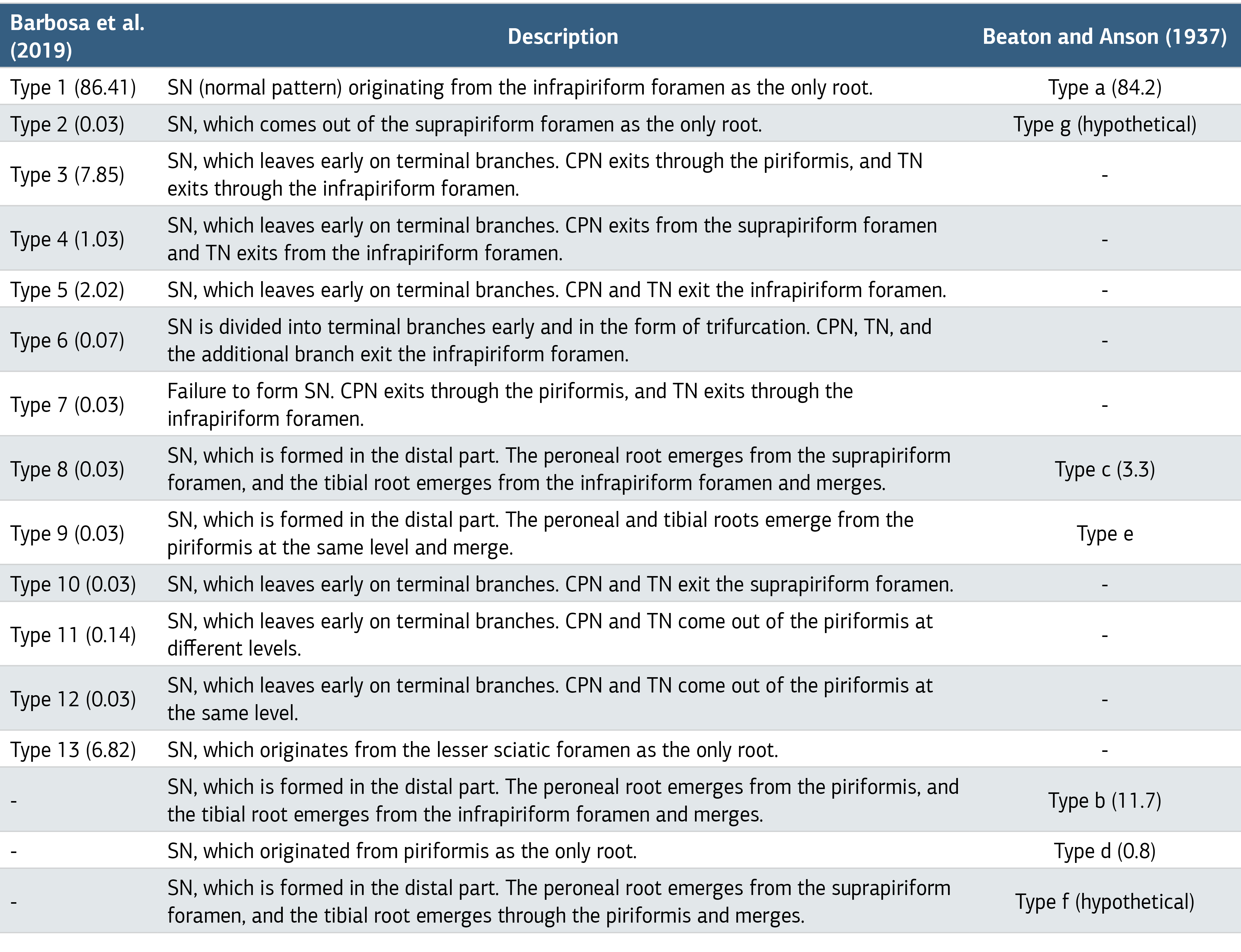
CPN, common peroneal nerve; TN, tibial nerve.
Table 2. Comparison of Cadaver Studies Investigating the Level of Sciatic Nerve Bifurcation (%)
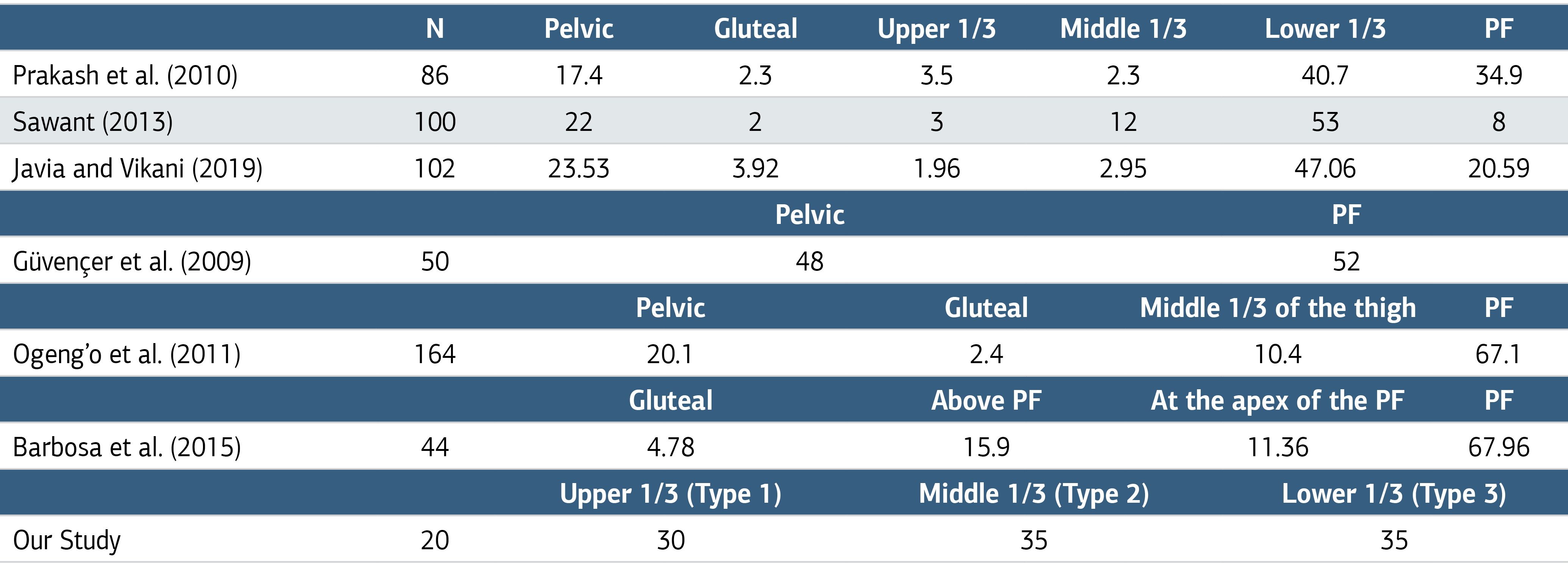
N, Number of extremities; PF, popliteal fossa.
Figures
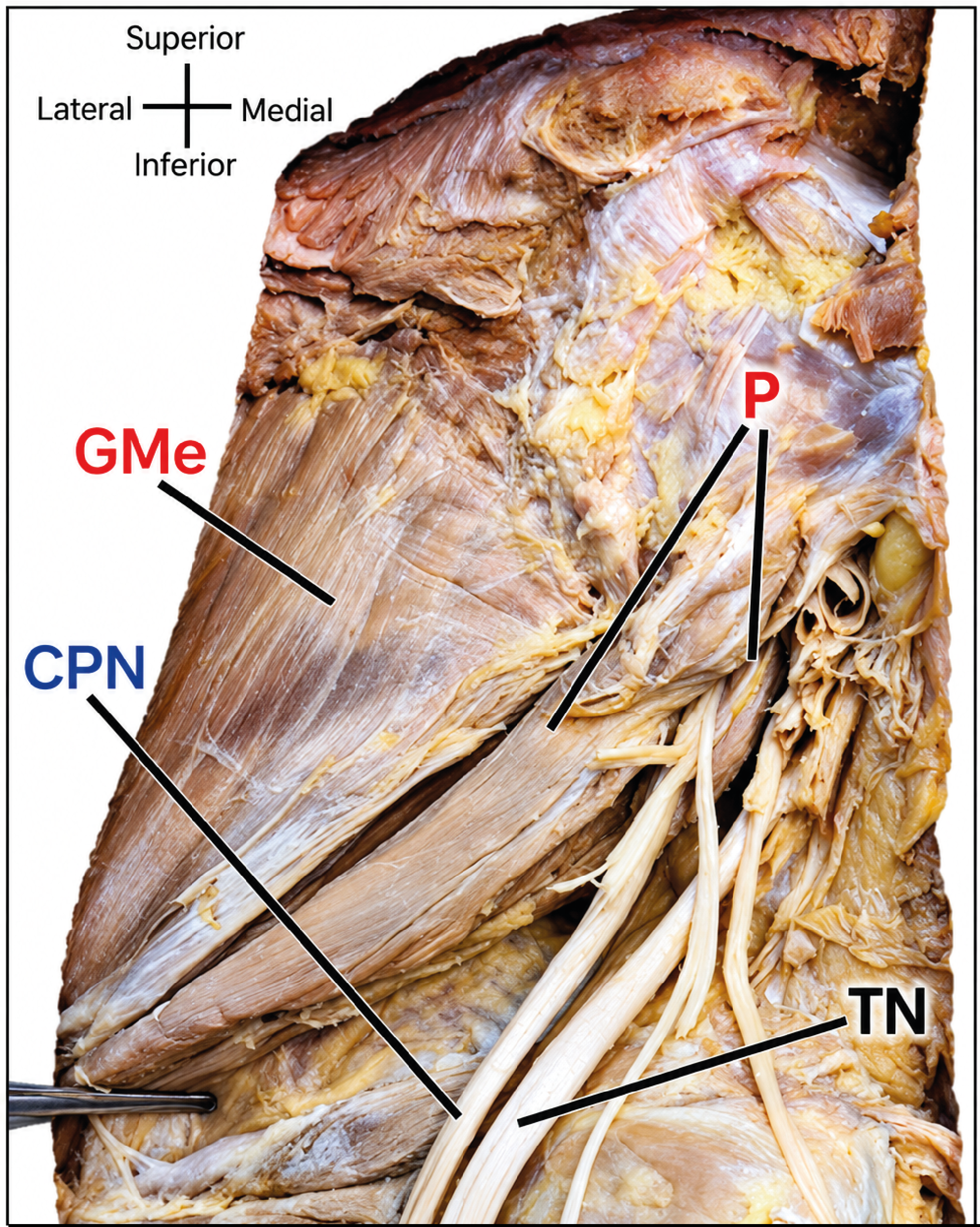
Figure 1. The Relationship of the Terminal Branches of the Sciatic Nerve in A Left Lower Limb with the Piriformis
GMe, gluteus medius; P, piriformis; CPN, common peroneal nerve; TN, tibial nerve.
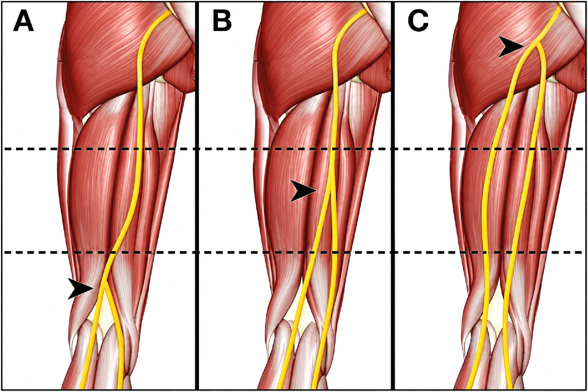
Figure 2. Sciatic Nerve Bifurcation Levels
A, distal; B, middle; C, proximal.
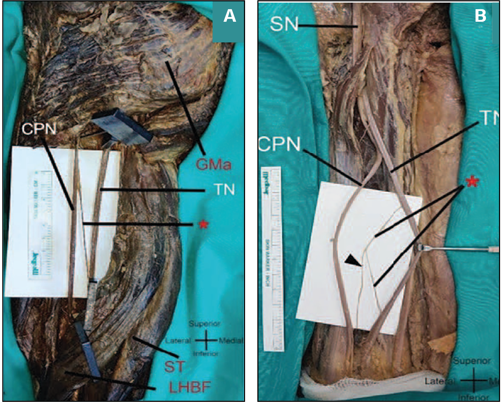
Figure 3. Connections (*) between the Terminal Branches of the Sciatic Nerve (SN)
(A) from the common peroneal nerve (CPN) to the tibial nerve (TN) (B) from TN to itself and an articular branch (arrowhead) GMa, gluteus maximus; ST, semitendinosus; LHBF, long head of biceps femoris.
Additional Information
Publisher’s Note
Bayrakol MP remains neutral with regard to jurisdictional and institutional claims.
Rights and Permissions

This work is licensed under a Creative Commons Attribution-NonCommercial 4.0 International License (CC BY-NC 4.0). To view a copy of the license, visit https://creativecommons.org/licenses/by-nc/4.0/
About This Article
How to Cite This Article
Alperen Doğuhan Oğuz, İsmihan İlknur Uysal, Betül Diğilli Ayas, Ahmet Safa Göksan. Overview of clinical anatomy of the sciatic nerve: re-evaluation with a cadaveric study. Ann Clin Anal Med 2024;15(11):753-758. doi:10.4328/ACAM.22184
Publication History
- Received:
- 20.03.2024
- Accepted:
- 06.05.2024
- Published Online:
- 24.08.2024
- Printed:
- 01.11.2024