
Abstract
AimThis study aims to examine the effects of subchorionic hematoma (SCH) on maternal serum alpha-fetoprotein (MSAFP) levels and the outcomes of second-trimester triple screening tests in patients who were diagnosed with threatened abortion during the first trimester.MethodsA retrospective study involved 922 patients who were diagnosed with threatened abortion between August 2013 and August 2015. After excluding cases of abortion, lack of follow-up, and those that did not meet inclusion criteria, 435 patients were included. Out of these, 102 had SCH (Group 1) while 248 did not (Group 2). Data collected comprised demographic characteristics, ultrasound findings, and triple screening test results. Statistical analyses included the use of student’s t-test, chi-square test, and correlation analysis. Additionally, ROC analysis was conducted to evaluate the diagnostic value of SCH as a predictor for elevated MSAFP levels.ResultsThe patients had an average age of 27.5 ± 5.6 years, and the mean gestational age at the time of diagnosis was 10.8 ± 3.1 weeks. Patients with SCH showed significantly higher MSAFP levels compared to those without SCH (413 ± 178 ng/mL vs. 393 ± 255 ng/mL, P = .036). However, no significant differences in MoM values or NTD risk were detected between the groups. Correlation analysis revealed a low yet significant correlation between AFP levels and hematoma size (r = 0.231, P = .020). ROC analysis revealed that while SCH is a significant factor for elevated MSAFP levels, it exhibits low sensitivity and specificity (AUC: 0.571, 95% CI: 0.507-0.636, P = .033).ConclusionSCH is linked to elevated MSAFP levels in patients experiencing threatened abortion in the first trimester. However, SCH does not have a significant impact on MoM values or NTD risk in second trimester screening tests. Clinicians should take SCH into account when interpreting elevated MSAFP levels and monitor patients with larger hematomas more closely. Further research is required to better understanding the clinical implications and improve the management of pregnancies complicated by SCH.
Keywords
Introduction
Threatened abortion, also known as imminent miscarriage, is defined by vaginal bleeding and abdominal pain in the first trimester, with a closed cervix and a viable fetus. It affects about 20–25% of pregnancies, and among these, 20–25% are found to have a subchorionic hematoma (SCH) on ultrasound. SCH results from partial detachment of the chorionic membranes from the uterine wall, causing blood accumulation between the wall and the chorion.1,2
The second‑trimester screening test typically includes maternal serum human chorionic gonadotropin (MShCG), maternal serum alpha‑fetoprotein (MSAFP), and maternal serum unconjugated estriol (MSuE3). Elevated MSAFP is a significant marker for neural tube defects (NTDs), severe congenital anomalies of the central nervous system.3 Elevated MSAFP may also indicate fetal, placental, or maternal complications, with fetomaternal hemorrhage being the most common cause aside from NTDs.4
Alpha‑fetoprotein (AFP), produced by the fetal liver and yolk sac, enters maternal circulation via transplacental diffusion and amniotic fluid. In non‑pregnant women, serum AFP is around 1–2 ng/mL. During pregnancy, MSAFP rises by ~15% per week from weeks 12–32, reaching 500–550 ng/mL at 32 weeks, then gradually decreases until term.4,5
Previous studies have linked SCH in threatened abortion with increased MSAFP. Seppala and Ruoslahti reported that 83% of pregnancies with threatened abortion at 13 weeks had elevated MSAFP, attributing this to fetomaternal hemorrhage.5 Elevated MSAFP has also been observed after invasive procedures such as chorionic villus sampling (CVS) and amniocentesis due to secondary fetomaternal hemorrhage.6-9
Given the potential impact of SCH on MSAFP levels and the risks involved, it is important to evaluate how SCH affects second‑trimester screening outcomes. This study aims to determine whether SCH in threatened abortion during the first trimester influences MSAFP levels and the results of triple screening tests, with a particular focus on NTD risk assessment.
Materials and Methods
Study Design and Patient SelectionThis retrospective study was conducted at the Early Pregnancy Clinic of Etlik Zübeyde Hanım Women’s Health Education and Research Hospital. The medical records of 922 patients who presented with vaginal bleeding and were diagnosed with threatened abortion between August 2013 and August 2015 were reviewed.
Inclusion criteria: single viable pregnancy between 6 and 18 weeks, no systemic diseases or bleeding disorders.
Exclusion criteria: miscarriage (n = 80), no follow‑up (n = 315), not meeting inclusion criteria (n = 92).
Ultimately, 435 patients were eligible: 102 with subchorionic hematoma (Group 1) and 248 without (Group 2).
Data collected: age, BMI, gestational age at diagnosis, gravidity, parity, ultrasound findings. Ultrasonographic evaluations were performed with a GE Logiq P5 machine (transabdominal and transvaginal probes) by two obstetricians. The size and location of SCH were recorded. Triple screening test results (AFP levels, corrected MoM values, NTD risk) were retrieved from the hospital information system.Ethical ApprovalThis study was approved by the Ethics Committee of Etlik Zübeyde Hanım Women’s Health Education and Research Hospital (Date: 25.02.2016, Decision No: 205).Statistical AnalysisData analysis was performed using SPSS for Windows version 11.5. Descriptive statistics for demographic and clinical characteristics
Student’s t‑test for normally distributed continuous variables
Chi‑square test (Pearson, Yates correction, Fisher’s exact test) for categorical variables
Correlation analysis for continuous variables
ROC analysis for cut‑off values
A P < .05 was considered statistically significant.
Reporting GuidelinesThis study was reported in accordance with the STROBE guideline.
Results
Table 1 displays the demographic characteristics of the 350 patients included in the study. The average age was 27.5 ( ± 5.6) years, with a mean gestational age of 10.8 ( ± 3.1) weeks at diagnosis. Comparing SCH‑positive and SCH‑negative groups, mean age was similar (27.5 ± 5.5 vs. 27.5 ± 5.7 years, P = .748). BMI was significantly higher in the SCH‑negative group (25.4 ± 4.5 vs. 24.2 ± 3.9, P = .011).
Gravidity and parity were similar (gravidity: 2.2 ± 1.2 vs. 2.3 ± 1.4, P = .787; parity: 0.78 ± 0.86 vs. 0.76 ± 0.91, P = .647). Gestational age at diagnosis did not differ significantly (10.7 ± 3.0 vs. 10.8 ± 3.1 weeks, P = .920) (Table 1). No significant differences were found in age, gravidity, or parity.Previous Pregnancy Outcomes and Smoking StatusNo significant differences were observed between groups in abortion, ectopic pregnancy, voluntary curettage, or smoking habits.Triple Screening Test ResultsTable 2 shows triple screening results. AFP levels differed significantly (413 ± 178 ng/mL vs. 393 ± 255 ng/mL, P = .036). MoM values were comparable (1.1 ± 0.4 vs. 1.0 ± 0.7, P = .689). Gestational age at screening did not differ (17.1 ± 0.7 vs. 16.9 ± 0.8 weeks, P = .071). NTD risk was similar (3% vs. 4.4%, P = .765).
Correlation analysis: AFP correlated weakly but significantly with hematoma size (r = 0.231, P = .020). No significant correlation with hematoma location (r = 0.450, P = .081), maternal age, gravida, parity, or gestational age.
Table 3 summarizes NTD risk by MSAFP cut‑off. Patients with MSAFP <2.0 MoM: 96.2% normal risk, 3.8% increased risk. Patients with MSAFP ≥2.0 MoM: 62.5% normal risk, 37.5% increased risk. This difference was statistically significant (P < .001).
ROC Curve AnalysisROC analysis evaluated SCH as a predictor of elevated MSAFP. AUC = 0.571 (95% CI: 0.507–0.636, P = .033), indicating SCH is a significant but low‑accuracy predictor. Sensitivity was 62.7%, specificity 51.4% (Figure 1).
Discussion
This study investigated the impact of subchorionic hematoma (SCH) on MSAFP levels and triple screening test outcomes in patients diagnosed with threatened abortion during the first trimester. Our findings indicate that the presence of SCH is associated with elevated MSAFP levels. However, there were no significant differences in MoM values or NTD risk between patients with and without SCH.
The observed rise in MSAFP levels in patients with SCH aligns with previous studies that have associated elevated MSAFP levels with fetomaternal hemorrhage.10,11 For instance, Seppala and Ruoslahti found elevated MSAFP levels in 83% of pregnancies with threatened abortion at 13 weeks, attributing this to fetomaternal hemorrhage.4 Similarly, Christmas et al. examined the effect of fetomaternal hemorrhage on adverse pregnancy outcomes in patients with elevated second‑trimester maternal serum AFP levels and found a significant correlation.11 In this regard, Lachman et al. reported that the detection and measurement of fetomaternal hemorrhage using serum alpha‑fetoprotein and the Kleihauer technique confirmed these observations.7
Notwithstanding, elevated MSAFP levels have been observed following invasive procedures like chorionic villus sampling (CVS) and amniocentesis due to secondary fetomaternal hemorrhage. Katiyar and colleagues detected fetomaternal hemorrhage following CVS, which was associated with a rise in maternal serum alpha‑fetoprotein levels.6 Moreover, Fuhrmann et al. reported that maternal serum AFP levels increased following CVS due to fetomaternal hemorrhage.8 Conversely, Makrydimas et al. investigated fetomaternal hemorrhage following coelocentesis and found no significant impact on AFP levels.12
Despite the elevated MSAFP levels in patients with SCH, our study did not find significant differences in MoM values or NTD risk between the groups. This suggests that while SCH can elevate MSAFP levels, it may not substantially impact the accuracy of NTD risk assessment using triple screening tests. This finding is crucial for clinical practice as it indicates that SCH should be considered when interpreting elevated MSAFP levels, but it does not necessarily indicate a higher risk of NTDs. This aligns with the work of Maso et al., who observed that the presence of SCH did not significantly alter pregnancy outcomes.9
The absence of a significant difference in MoM values and NTD risk between SCH‑positive and SCH‑negative groups could be attributed to several factors. First, the size of the hematoma may play a role. In our study, correlation analysis showed a low but significant correlation between AFP levels and hematoma size (r = 0.231, P = .020), indicating that larger hematomas are associated with higher AFP levels. However, the overall impact of hematoma size on MSAFP levels and subsequent NTD risk assessment may be limited. This finding is consistent with previous studies that have shown variable impacts of hematoma size on pregnancy outcomes. Similarly, Tuuli et al. conducted a meta‑analysis and found that while SCH was associated with adverse pregnancy outcomes, the size of the hematoma was a critical factor in determining the level of risk.13
Second, the timing of the hematoma’s formation and its resolution could influence MSAFP levels. Hematomas that resolve earlier in pregnancy may have less impact on second‑trimester screening results compared to those that persist. Regan et al. noted that the timing and resolution of hematomas significantly affected pregnancy outcomes, suggesting that early resolution might mitigate some risks.14 Early resolution of SCH can lead to normalization of MSAFP levels, thereby reducing the potential for false‑positive NTD screening results.
Another important consideration is the clinical management of patients with SCH. In our study, the lack of significant differences in adverse pregnancy outcomes such as abortion history, ectopic pregnancy, voluntary curettage, and smoking status between SCH‑positive and SCH‑negative groups suggests that the presence of SCH alone may not be a decisive factor in determining pregnancy outcomes. This aligns with previous studies that have shown variable impacts of SCH on pregnancy outcomes, with some studies reporting increased risks of adverse outcomes and others finding no significant associations. Stephenson et al. reported similar findings in a study involving 197 couples, where the presence of SCH did not consistently predict adverse outcomes.15
Additionally, SCH may have a transient effect on MSAFP levels. Wilcox et al. demonstrated that early pregnancy losses are common and often associated with transient elevations in serum markers, including MSAFP.16 This transient nature might explain why some studies, including ours, do not find long‑term impacts of SCH on pregnancy outcomes despite initial elevations in MSAFP levels.
The findings of this study carry several clinical implications. First, the presence of SCH should be taken into account when evaluating elevated MSAFP levels during the second trimester. While elevated MSAFP levels may indicate an increased risk of NTDs, SCH can also contribute to these elevated levels. Therefore, clinicians should take into account the presence of SCH and potentially use additional diagnostic tools such as detailed ultrasound evaluations to accurately assess NTD risk. Alberman emphasized the importance of considering other diagnostic tools in conjunction with MSAFP levels to provide a comprehensive assessment.17 Second, the correlation between hematoma size and AFP levels suggests that patients with larger SCH should be monitored more closely. These patients may require more frequent follow‑up visits and additional diagnostic tests to ensure the well‑being of the fetus and to manage any potential complications. Kurki and Ylikorkala suggested that close monitoring and follow‑up are crucial in managing pregnancies complicated by SCH.18
Furthermore, the results of this study underscore the importance of considering other potential factors that could influence MSAFP levels. For example, maternal weight, diabetes, and smoking status are known to affect MSAFP levels and should be taken into account when interpreting screening results. Blumenfeld and Brenner noted that various maternal factors, including thrombophilia and lifestyle choices, significantly influence MSAFP levels.19 Additional research is necessary to investigate the influence of these factors on MSAFP levels in patients with SCH and to create more comprehensive risk assessment models that include these variables.
Limitations
This study has several limitations. The retrospective design may introduce selection bias, and the sample size, particularly for patients with SCH, was relatively small. Additionally, the study did not consider other potential factors that might affect MSAFP levels, such as maternal weight, diabetes, and smoking status. Future research should focus on addressing these limitations by conducting larger prospective studies that account for these confounding factors. Additionally, investigating the longitudinal effects of SCH on pregnancy outcomes and MSAFP levels throughout pregnancy could provide valuable insights into the temporal dynamics of these associations. Harlap and Shiono emphasized the importance of prospective studies to gain a deeper understanding of long‑term impacts of SCH on pregnancy outcomes.20 Furthermore, the clinical significance of the low correlation between AFP levels and hematoma size warrants further investigation. Understanding the mechanisms underlying this correlation could provide valuable insights into the pathophysiology of SCH and its impact on pregnancy outcomes. For instance, examining the role of placental vascularization and the degree of fetomaternal hemorrhage in relation to hematoma size could help elucidate the biological processes driving the observed associations. Blumenfeld and Brenner noted the importance of understanding the pathophysiological mechanisms to improve clinical management and outcomes.19
Conclusion
In conclusion, this study demonstrates that subchorionic hematoma is associated with elevated MSAFP levels in patients with threatened abortion during the first trimester. However, the presence of SCH does not significantly impact MoM values or NTD risk in second trimester screening tests. Clinicians should consider SCH when interpreting elevated MSAFP levels and monitor patients with larger hematomas more closely. Further research is needed to explore the clinical implications of these findings and to improve the management of pregnancies complicated by SCH.
Declarations
Animal and Human Rights Statement
All procedures performed in this study were in accordance with the ethical standards of the institutional and/or national research committee and with the 1964 Helsinki Declaration and its later amendments or comparable ethical standards.
Informed Consent
Informed consent was waived due to the retrospective nature of the study.
Data Availability
The datasets used and/or analyzed during the current study are not publicly available due to patient privacy reasons but are available from the corresponding author on reasonable request.
Conflict of Interest
The authors declare that there is no conflict of interest.
Funding
None.
Abbreviations
AFP: Alpha-fetoprotein
BMI: Body mass index
CVS: Chorionic villus sampling
MSAFP: Maternal serum alpha-fetoprotein
NTD: Neural tube defect
ROC: Receiver operating characteristic
SCH: Subchorionic hematoma
SPSS: Statistical Package for the Social Sciences
References
- Dolk H, Loane M, Garne E. The prevalence of congenital anomalies in Europe. In: De La Paz MP, Groft SC, eds. Rare Diseases Epidemiology. Springer; 2010:349-364. doi:10.1007/978-90-481-9485-8_20
- Graves JC, Miller KE, Sellers AD. Maternal serum triple analyte screening in pregnancy. Am Fam Physician. 2002;65(5):915-921.
- Kim GJ, Seong JS, Oh JA. Prenatal screening for neural tube defects: from maternal serum alpha-fetoprotein to ultrasonography. Obstet Gynecol Sci. 2023;66(1):1-10. doi:10.5468/ogs.22263
- Seppala M, Rouslahti E. Alpha-fetoprotein in normal and pregnancy sera. Lancet. 1972;299(7746):375-376. doi:10.1136/bmj.4.5843.769
- Seppälä M, Ruoslahti E. Alpha-fetoprotein in abortion. Br Med J. 1972;4(5843):769-771.
- Katiyar R, Kriplani A, Agarwal N, Bhatla N, Kabra M. Detection of fetomaternal hemorrhage following chorionic villus sampling by Kleihauer Betke test and rise in maternal serum alpha feto protein. Prenat Diagn. 2007;27(2):139-142. doi:10.1002/pd.1632
- Lachman E, Hingley SM, Bates G, Ward AM, Stewart C, Duncan S. Detection and measurement of fetomaternal haemorrhage: serum alpha-fetoprotein and the Kleihauer technique. Br Med J. 1977;1(6073):1377-1379. doi:10.1136/bmj.1.6073.1377
- Fuhrmann W, Altland K, Köhler A, et al. Feto-maternal transfusion after chorionic villus sampling. In: Human Genetics. Springer; 1988:83-85. doi:10.1007/bf00291241
- Maso G, D’Ottavio G, De Seta F, Sartore A, Piccoli M, Mandruzzato G. First-trimester intrauterine hematoma and outcome of pregnancy. Obstet Gynecol. 2005;105(2):339-344. doi:10.1097/01.aog.0000152000.71369.bd
- Kumbak B, Sahin L. Elevated maternal serum alpha-fetoprotein levels in patients with subchorionic hematoma. J Matern Fetal Neonatal Med. 2010;23(7):717-719. doi:10.3109/14767050903366093
- Christmas JT, Vanner LV, Daniels RM, Bodurtha JN, Hays PM, Redwine FO. The effect of fetomaternal bleeding on the risk of adverse pregnancy outcome in patients with elevated second-trimester maternal serum α-fetoprotein levels. Am J Obstet Gynecol. 1994;171(2):315-320. doi:10.1016/s0002-9378(94)70029-x
- Makrydimas G, Lolis D, Georgiou I, Navrozoglou I, Nicolaides KH. Feto-maternal bleeding following coelocentesis. Hum Reprod. 1997;12(4):845-846. doi:10.1093/humrep/12.4.845
- Tuuli MG, Norman SM, Odibo AO, Macones GA, Cahill AG. Perinatal outcomes in women with subchorionic hematoma: a systematic review and meta-analysis. Obstet Gynecol. 2011;117(5):1205-1212. doi:10.1097/aog.0b013e31821568de
- Regan L, Braude PR, Trembath PL. Influence of past reproductive performance on risk of spontaneous abortion. BMJ. 1989;299(6698):541-545. doi:10.1136/bmj.299.6698.541
- Stephenson M. Frequency of factors associated with habitual abortion in 197 couples. Fertil Steril. 1996;66(1):24-29.
- Wilcox AJ, Weinberg CR, O’Connor JF, et al. Incidence of early loss of pregnancy. N Engl J Med. 1988;319(4):189-194. doi:10.1056/nejm198807283190401
- Alberman E. The epidemiology of repeated abortion. In: Early Pregnancy Loss. Springer; 1988:9-17. doi:10.1007/978-1-4471-1658-5_2
- Kurki T, Ylikorkala O. Coitus during pregnancy is not related to bacterial vaginosis or preterm birth. Am J Obstet Gynecol. 1993;169(5):1130-1134. doi:10.1016/0002-9378(93)90268-n
- Blumenfeld Z, Brenner B. Thrombophilia-associated pregnancy wastage. Fertil Steril. 1999;72(5):765-774. doi:10.1016/s0015-0282(99)00360-x
- Harlap S, Shiono P. Alcohol smoking and incidence of spontaneous abortions in the first and second trimester. Lancet. 1980;316(8187):173-176. doi:10.1016/s0140-6736(80)90061-6
Tables
Table 1. Demographic Characteristics of the Study Population and Comparison Between SCH-Positive and SCH-Negative Groups
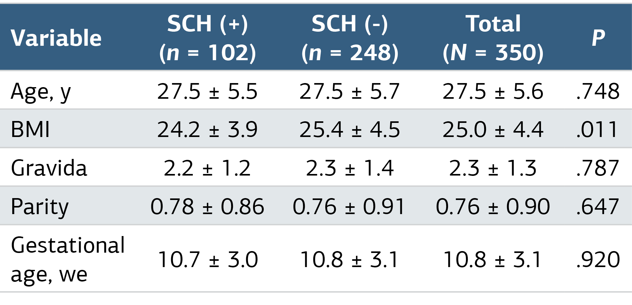
BMI, Body Mass Index; (kg/m²); SCH, subchorionic hematoma.
Table 2. Triple Screening Test Results
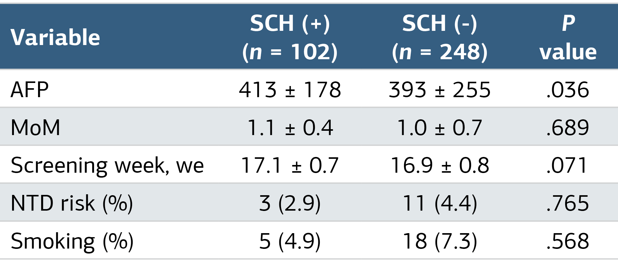
SCH, subchorionic hematoma; AFP, Alpha fetoprotein; (ng/mL); MoM, Multiple of Median; NTD, Neural Tube Defect.
Table 3. Comparison of NTD Risk with MSAFP Cut-off Levels in Study Participants
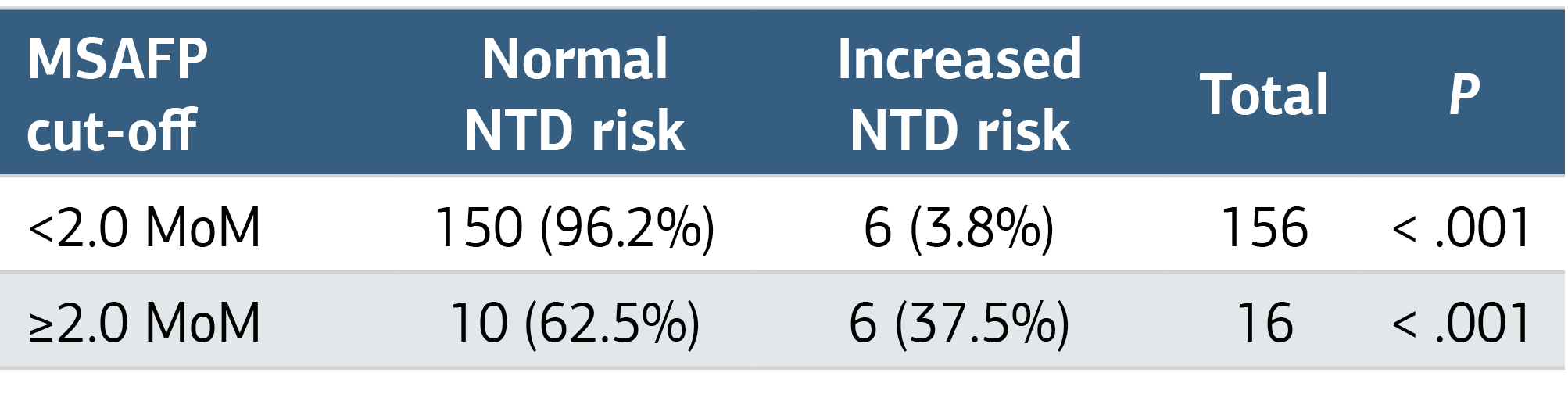
MoM, Multiple of Median; AFP, Alpha fetoprotein; (ng/mL); NTD, Neural Tube Defect.
Figures
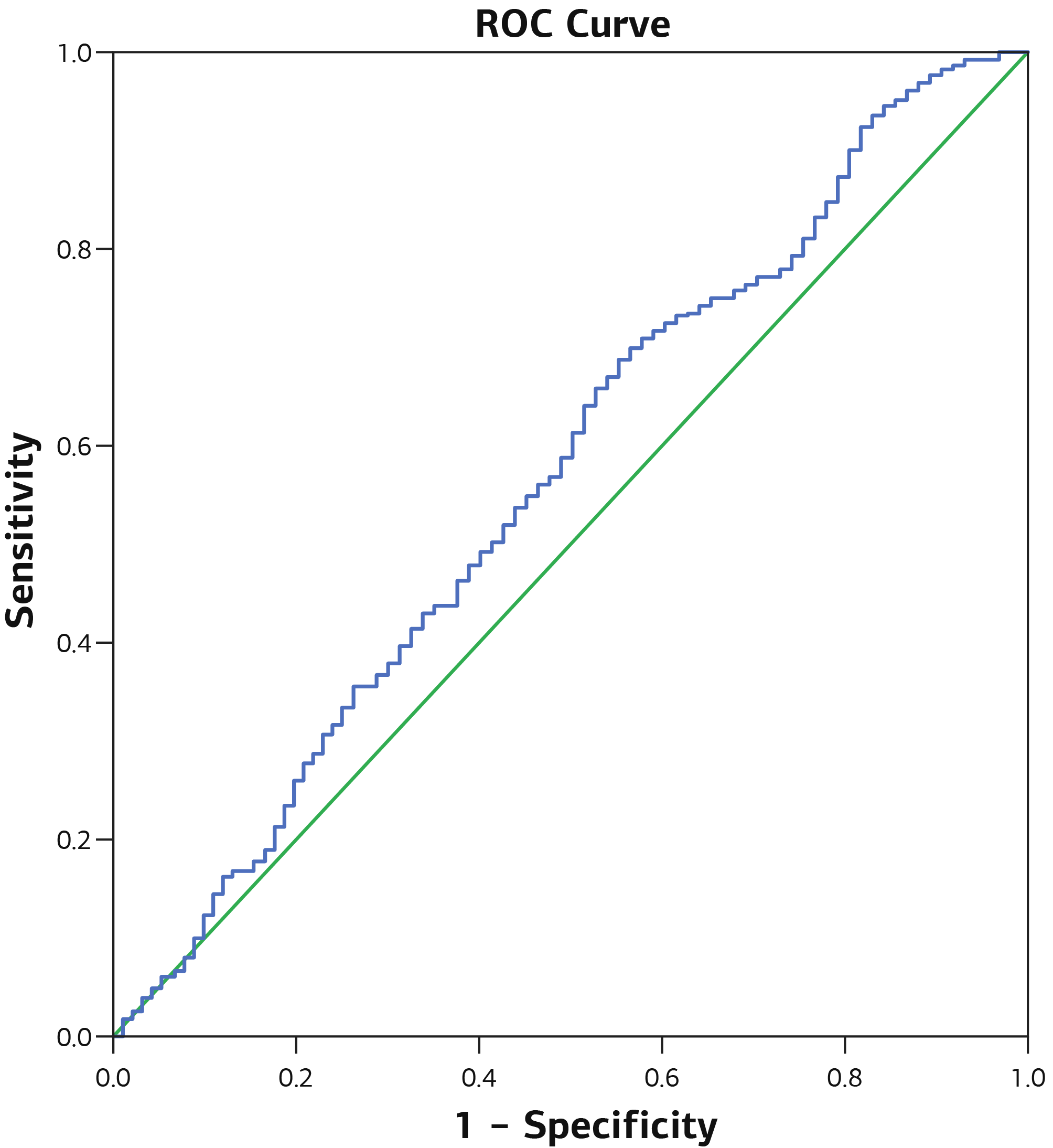
Figure 1. ROC Curve of Subchorionic Hematoma as a Variable for Elevated MSAFP Levels
Additional Information
Publisher’s Note
Bayrakol MP remains neutral with regard to jurisdictional and institutional claims.
Rights and Permissions

This work is licensed under a Creative Commons Attribution-NonCommercial 4.0 International License (CC BY-NC 4.0). To view a copy of the license, visit https://creativecommons.org/licenses/by-nc/4.0/
About This Article
How to Cite This Article
Tuğrul Başar, Abdurrahman Alp Tokalıoğlu, Rıza Dur, Tufan Arslanca, Metin Altay. Impact of subchorionic hematoma on maternal serum alpha-fetoproteinlevels and second trimester screening outcomes in threatened abortion cases. Ann Clin Anal Med 2024;15(11):803-807. doi:10.4328/ACAM.22347
Publication History
- Received:
- 31.07.2024
- Accepted:
- 02.09.2024
- Published Online:
- 18.09.2024
- Printed:
- 01.11.2024