
Abstract
AimAcute lymphoblastic leukemia is the most common cancer in children and adolescents. The application of chemotherapy by risk group and genetic modification have significantly improved the survival rate of childhood acute lymphoblastic leukemia. In this study, we aimed to determine genetic abnormalities and investigate the correlation between gene abnormalities and treatment results of childhood acute lymphoblastic leukemia.MethodsThis was a descriptive cross-sectional study on childhood acute lymphoblastic leukemia patients who were admitted to the hospital between November 2017 and May 2022.ResultsThere were 83 new patients with acute lymphoblastic leukemia. The results of multiplex PCR tests showed that 24.1% of patients had genetic abnormalities, of which, 12.1% had TEL/AML1 fusion, 4.8% had BCR/ABL1 fusion, 3.6% had E2A/PBX1 fusion, 2.4% had MLL/AF4 fusion and 1.2% had SET/NUP214 fusion. There were 12.1% of patients with a favorable prognosis, 80.7% of patients with an intermediate prognosis and 7.2% of patients with a poor prognosis. There were statistical correlations between the remission rate, the relapsed rate, the overall survival rate, the event-free survival rate and the genetic risk group. The overall survival rate and the event-free survival rate for the poor prognosis, intermediate prognosis and good prognosis risk groups after 2 years were 50.0 ± 25.0%, 0%; 86.7 ± 4.9%, 77.9 ± 6.1% and 100.0 ± 0.0%, 100.0 ± 0.0%.ConclusionGenetic abnormalities help in classification, prognosis and treatment of childhood acute leukemia.
Keywords
Introduction
Acute lymphoblastic leukemia (ALL) is the most common cancer in children and adolescents, accounting for 20% of all new cancers diagnosed in children under 15 years of age.1 It is a disease of the hematopoietic system caused by the uncontrolled proliferation of one or more malignant immature cell lines. The incidence of acute lymphoblastic leukemia is about 2–5 cases per 100,000 children and ALL is the most common in children aged 2–5 years.2 Diagnosis and classification of acute lymphoblastic leukemia is based on clinical features combined with morphological malignancies, cytochemical staining, immunocytochemical markers, and genetic tests. Regarding the treatment of childhood acute lymphoblastic leukemia, the application of chemotherapy by risk group and genetic modification has significantly improved the survival rate of childhood acute lymphoblastic leukemia. In developed countries, the overall survival rate for acute lymphoblastic leukemia has risen from 10% in the 1960s to more than 90% in recent years.1,2 Therefore, I carried out this study to determine genetic abnormalities and investigate the correlation between gene abnormalities and the treatment results of childhood acute lymphoblastic leukemia.
Materials and Methods
PatientsThe study included 83 children diagnosed with acute lymphoblastic leukemia treated at Pediatric Oncology – Hematology – Bone Marrow Transplant Department, Pediatric Center – Hue Central Hospital, from 11/2017 to 5/2022.Inclusion Criteria• The patient was diagnosed with acute lymphoblastic leukemia
• Age <16
• All patients underwent multiplex RT-PCR genetic analysis, with the Hemavision 28N kit, 28 basic genetic mutations in acute lymphoblastic leukemia were detected.
Criteria for diagnosis of acute lymphoblastic leukemia: Clinical: Systemic symptoms: fever, fatigue, poor appetite. Anemia, bleeding under the skin or mucous membranes. Symptoms of extramedullary infiltrates: liver, spleen, lymphadenopathy, gingival hypertrophy, subcutaneous papules, central nervous system infiltrates, mediastinal infiltrates, or testicular infiltrates.
Peripheral blood count: There is usually a decrease in hemoglobin (Hb), the white blood cell count may be elevated, normal or decreased, but often there is a severe decrease in neutrophils, and peripheral bleeding may or may not be visible. The platelet count is usually low.
More than 20% of leukemic blasts were found in the bone marrow. According to the results of flow cytometry, acute lymphoblastic leukemia was diagnosed.
Exclusion criteria
• Patients were diagnosed ALL, L3
• Pediatric patients with secondary or relapsed acute lymphoblastic leukemia
• The child and the representative did not agree to participate in the study.MethodThis was a descriptive, prospective longitudinal study. The longitudinal follow-up time to the end of the study was June 1, 2022.
Data were analyzed according to genetic tests, the remission rate, the relapsed rate, overall survival rate.Ethical ApprovalAll ethical regulations were followed, and this study was approved by the Hue Central Hospital Ethics Committee ( Date: 20.10.2017, Decision No: 18/NCKH-BVH) .Statistical AnalysisAll statistical analysis was performed using SPSS v.18.0 (IBM Corp, Armonk, NY).Reporting GuidelinesThis study was reported in accordance with the STROBE guideline.
Results
A total of 83 new patients with ALL were identified from 11/2017 to 5/2022 who met eligible criteria. Among these patients, 48 were males and 35 were females, the male-to-female ratio was 1.37:1. The mean age was 5.2 ± 3.5 years. 61.4% of patients were standard risk, and 38.6% were high-risk patients. The percentages of B-ALL and T-ALL were 84.3% and 15.7%, respectively.
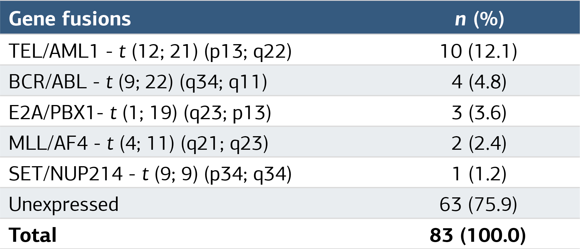
The results of the analysis with multiplex PCR showed that 24.1% of patients had genetic abnormalities in the following order: TEL/AML1 (12.1%), BCR/ABL1 (4.8%), E2A/PBX1 (3.6%), MLL/AF4 (2.4%) and SET/NUP214 (1.2%) (Table 1).
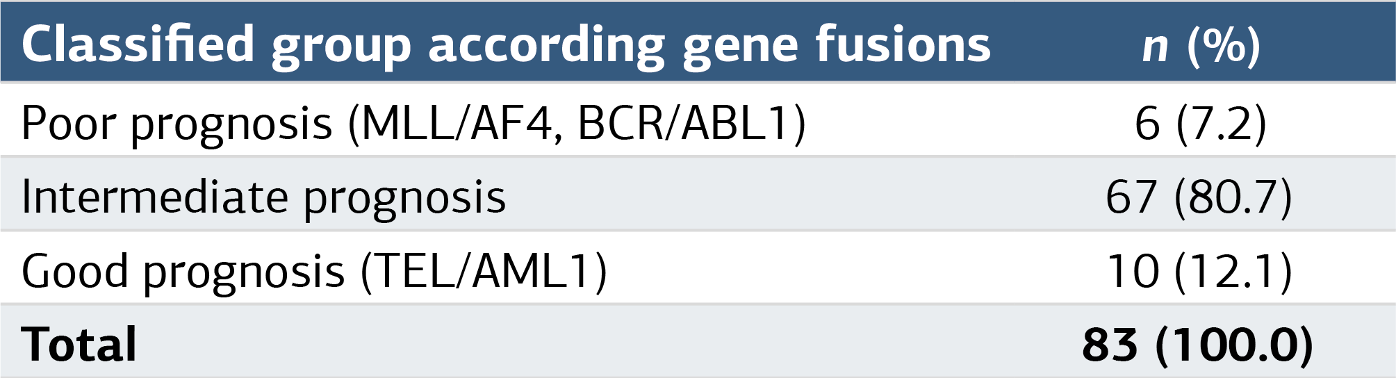
Prognostic grouping according to genetic mutation: good prognosis (12.1%), intermediate prognosis (80.7%), poor prognosis (7.2%) (Table 2). The difference in the distribution rate of gene fusion between the two groups of standard and high risk, or between the two groups B-ALL and T-ALL was not statistically significant (P > .05).
Results showed that 91.6% of ALL patients recovered based on blast cell count <5% after the induction phase. The remission rate for the standard-risk group was 98.0%, while the rate for the high-risk group was 81.7%. The difference was statistically significant (P < .05). Regarding the correlation between genetic abnormalities and the rate of remission in childhood acute lymphoblastic leukemia, it was shown that remission rates varied between groups, in which the poor prognosis group accounted for the lowest (66.6%), 94.0% for the intermediate prognosis group, and 90.0% for the good prognosis group. The difference was statistically significant (P < .05).
In the good and intermediate prognosis groups, MRD<10⁻⁴ after induction therapy was 88.9% and 92.2%, respectively, while this rate was only 50% in the group with poor prognosis. There was a statistically significant difference between the three groups (P < .02).
When doing the evaluation of ALL relapse rates based on genetic risk groups, the results showed that there was a difference in the relapse rates between the genetic risk groups, in which this rate of the poor prognosis group was highest (66.7%), followed by the intermediate prognosis group (11.1%), while the group with good prognosis did not show any recurrence. The difference was statistically significant (P < .05).
Regarding the evaluation of the overall survival rate according to genetic risk group, the result showed that at the end of the study, out of a total of 83 patients with ALL, 13 patients died, of which 3/6 patients belonged to the poor prognosis group, 10/67 patients belonged to the intermediate prognosis group and no patient in the good prognosis group died. The number of surviving patients was 70, accounting for 84.3%. At 3 years, overall survival rates for groups with good, intermediate, and poor prognosis were 100.0 ± 0.0%, 76.3 ± 7.1%, and 25.0 ± 21.7%, respectively. The difference was statistically significant (P < .05) (Figure 1). Regarding the event-free survival rate, in the 2nd year, the event-free survival rate in the groups with good, intermediate and poor prognoses were: 100 ± 0.0%, 77.9 ± 6.1%, and 0%, respectively (Figure 2). The difference was statistically significant (P < .05).
At the end of the study, out of a total of 83 patients with ALL, 13 patients died, of which 3/6 patients belonged to the poor prognosis group, 10/67 patients belonged to the intermediate prognosis group and no patient in the good prognosis group died. The number of surviving patients was 70 patients, accounting for 84.3%. At 3 years, overall survival for the good, intermediate, and poor prognosis groups was 100.0 ± 0.0%, 76.3 ± 7.1%, and 25.0 ± 21.7%, respectively. The difference was statistically significant (P < .05).
Discussion
The male/female ratio was 1.37:1 and the mean age was 5.2 ± 3.5 years, similar to some studies by Fadoo, Al-Sudairy, Jovanovska and Yasmeen.3-6 Regarding cell lineages, B-cell lineage accounted for a high percentage (84.3%), and T-cell lineage accounted for 15.7%. The result was similar to the results of studies in developing countries. According to Fadoo, there were 78.5% of B-cell patients and 17.5% of T-cell patients.3 According to Al-Sudairy, there were 89.5% of pre-B cell patients and 10.5% of T-cell patients.4
The results of the analysis with multiplex PCR showed that there were 24.1% of patients with genetic abnormalities, including 12.1% of patients with TEL/AML1 fusion, 4.8% of patients with BCR/ABL1 fusion, 3.6% of patients with E2A/PBX1 fusion, 2.4% of patients with MLL/AF4 fusion and 1.2% of patients with SET/NUP214 fusion. Our results were similar to those of other authors, such as Veerman and Aldrich.7,8 With the multiplex-PCR, our result found a new fusion compared with other previous results in Vietnam. It was SET/NUP214 fusion, which is a rare fusion gene and often occurs in T-ALL at 10–11 years of age.9,10 A patient with SET/NUP214 will have a high percentage of relapse.10 Until now, SET/NUP214 is still poorly understood.9
Based on genetic abnormalities, some authors such as Inaba, Pui classify patients into three risk genetic groups: favorable prognosis, intermediate prognosis and poor prognosis.11,12 In our result, 12.1% of patients had a favorable prognosis, 80.7% of patients had an intermediate prognosis and 7.2% had a poor prognosis.
The rate of remission after the induction phase for ALL was 91.6%, which was similar to that of other researchers all over the world, the rate achieved 90–98%. The result showed that the percentage of remission differed among the three risk genetic groups. The rate of remission for the poor prognosis group was 66.6%, the favorable prognosis group achieved 90.0% and in the intermediate prognosis group, it was 94.0%. The difference was statistically significant (P < .05). Our results are similar to some studies by Pui, Lin, Toksvang and Cimino.12-15 Thus, genetic abnormalities made a tremendous impact on the result of treatment. The good prognosis group had 88.9% MRD <10⁻⁴ after induction phase and this percentage was only 50% for the poor prognosis group and 92.2% for the intermediate prognosis group. The difference was statistically significant (P < .05). Our results are similar to the study by Borowitz.16 The percentage of MRD <10⁻⁴ after the induction phase in patients with TEL/AML1 fusion gene was 87.9%, however, that percentage was only 70.7% for patients with MLL rearrangement.
The poor prognosis group had the highest relapsed percentage, 66.7%, the intermediate prognosis group had 11.1% relapse and the good prognosis group had no relapse. The difference was statistically significant (P < .05). Our results are similar to the study by Sanchez, the poor prognosis has the highest relapse rate. 23% of patients with BCR/ABL1 relapsed after achieving remission, in which, there was 10% of CNS relapse or CNS and bone marrow relapse.17
Overall survival rates for the poor prognosis, intermediate prognosis and good prognosis risk groups after 3 years were 25.0 ± 21.7%, 76.3 ± 7.1% and 100.0 ± 0.0%. The difference was statistically significant (P < .05). Our results are similar to the study by Pui, the overall survival rate for patients with BCR/ABL1 fusion gene was 48.0 ± 2.0%, and the patients without BCR/ABL1 fusion genes was 94.5 ± 1.8%.12 Regarding event-free survival, event-free survival rates for the poor prognosis, intermediate prognosis and favorable prognosis risk groups after 2 years were 0%, 77.9 ± 6.1% and 100.0 ± 0.0%. The difference was statistically significant (P < .05). Our results were similar to the study by Jeha, Pui.12,18
Limitations
The main limitations of this study are the relatively small sample size and the single-center design. In addition, the follow-up period was limited to June 1, 2022, which may restrict the assessment of long-term survival and relapse outcomes.
Conclusion
Genetic abnormalities help classify, predict and treat childhood acute lymphoblastic leukemia. There were statistical correlations between remission rate, relapse rate, overall survival, event-free survival and the genetic risk group.
Declarations
Animal and Human Rights Statement
All procedures performed in this study were in accordance with the ethical standards of the institutional and/or national research committee and with the 1964 Helsinki Declaration and its later amendments or comparable ethical standards.
Informed Consent
Informed consent was obtained from all participants in this study.
Data Availability
The datasets used and/or analyzed during the current study are not publicly available due to patient privacy reasons but are available from the corresponding author on reasonable request.
Conflict of Interest
The authors declare that there is no conflict of interest.
Funding
None.
Abbreviations
ALL: Acute lymphoblastic leukemia
B-ALL: B-cell acute lymphoblastic leukemia
CNS: Central nervous system
MRD: Minimal residual disease
PCR: Polymerase chain reaction
RT-PCR: Reverse transcription polymerase chain reaction
SPSS: Statistical package for the social sciences
T-ALL: T-cell acute lymphoblastic leukemia
References
- Hunger SP, Mullighan CG. Acute lymphoblastic leukemia in children. N Engl J Med. 2015;373(16):1541-1552. doi:10.1056/nejmra1400972
- Freireich EJ, Wiernik PH, Steensma DP. The leukemias: a half-century of discovery. J Clin Oncol. 2014;32(31):3463-3469. doi:10.1200/jco.2014.57.1034
- Fadoo Z, Nisar I, Yousuf F, et al. Clinical features and induction outcome of childhood acute lymphoblastic leukemia in a lower/middle income population: a multi-institutional report from Pakistan. Pediatr Blood Cancer. 2015;62(10):1700-1708. doi:10.1002/pbc.25583
- Al-Sudairy R, Al-Nasser A, Alsultan A, et al. Clinical characteristics and treatment outcome of childhood acute lymphoblastic leukemia in Saudi Arabia: a multi-institutional retrospective national collaborative study. Pediatr Blood Cancer. 2014;61(1):74-80.
- Jovanovska A, Martinova K, Kocheva S, et al. Clinical significance of minimal residual disease at the end of remission induction therapy in childhood acute lymphoblastic leukemia. Open Access Maced J Med Sci. 2019;7(17):2818-2823. doi:10.3889/oamjms.2019.752
- Yasmeen N, Ashraf S. Childhood acute lymphoblastic leukaemia: epidemiology and clinicopathological features. J Pak Med Assoc. 2009;59(3):150-153.
- Veerman AJ, Kamps WA, van den Berg H, et al. Dexamethasone-based therapy for childhood acute lymphoblastic leukaemia: results of the prospective Dutch Childhood Oncology Group protocol ALL-9 (1997-2004). Lancet Oncol. 2009;10(10):957-966. doi:10.1016/s1470-2045(09)70228-1
- Aldrich MC, Zhang L, Wiemels JL, et al. Cytogenetics of Hispanic and White children with acute lymphoblastic leukemia in California. Cancer Epidemiol Biomarkers Prev. 2006;15(3):578-581. doi:10.1158/1055-9965.epi-05-0833
- Gao MG, Hong Y, Qin YZ, et al. Prognostic significance of SET-NUP214 fusion gene in acute leukemia after allogeneic hematopoietic stem cell transplantation. Medicine (Baltimore). 2020;99(50). doi:10.1097/md.0000000000023569
- Xu X, Zhai Q, Jin H, et al. SET-NUP214 fusion gene involved early T-cell precursor acute lymphoblastic leukemia in adult with B marker expression. Int J Gen Med. 2021;14:659-664. doi:10.2147/ijgm.s294715
- Inaba H, Pui CH. Advances in the diagnosis and treatment of pediatric acute lymphoblastic leukemia. J Clin Med. 2021;10(9):1926. doi:10.3390/jcm10091926
- Pui CH, Carroll WL, Meshinchi S, Arceci RJ. Biology, risk stratification, and therapy of pediatric acute leukemias: an update. J Clin Oncol. 2011;29(5):551-565. doi:10.1200/jco.2010.30.7405
- Lin PC, Chang TT, Lin SR, Chiou SS, Jang RC, Sheen JM. TEL/AML1 fusion gene in childhood acute lymphoblastic leukemia in southern Taiwan. Kaohsiung J Med Sci. 2008;24(6):289-296. doi:10.1016/s1607-551x(08)70155-4
- Toksvang LN, Lee SHR, Yang JJ, Schmiegelow K. Maintenance therapy for acute lymphoblastic leukemia: basic science and clinical translations. Leukemia. 2022;36(7):1749-1758. doi:10.1038/s41375-022-01591-4
- Cimino G, Cenfra N, Elia L, et al. The therapeutic response and clinical outcome of adults with ALL1(MLL)/AF4 fusion-positive acute lymphoblastic leukemia according to the GIMEMA experience. Haematologica. 2010;95(5):837-840. doi:10.3324/haematol.2009.009035
- Borowitz MJ, Devidas M, Hunger SP, et al. Clinical significance of minimal residual disease in childhood acute lymphoblastic leukemia and its relationship to other prognostic factors: a Children’s Oncology Group study. Blood. 2008;111(12):5477-5485. doi:10.1182/blood-2008-01-132837
- Sanchez R, Ayala R, Alonso RA, et al. Clinical characteristics of patients with central nervous system relapse in BCR-ABL1-positive acute lymphoblastic leukemia: the importance of characterizing ABL1 mutations in cerebrospinal fluid. Ann Hematol. 2017;96(7):1069-1075. doi:10.1007/s00277-017-3002-1
- Jeha S, Choi J, Roberts KG, et al. Clinical significance of novel subtypes of acute lymphoblastic leukemia in the context of minimal residual disease-directed therapy. Blood Cancer Discov. 2021;2(4):326-337. doi:10.1158/2643-3230.bcd-20-0229
Tables
Figures
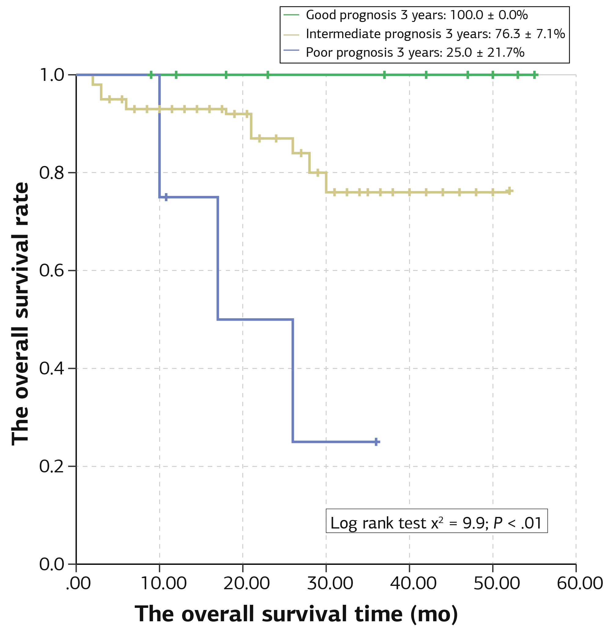
Figure 1. Overall survival curve by genetic risk group in ALL
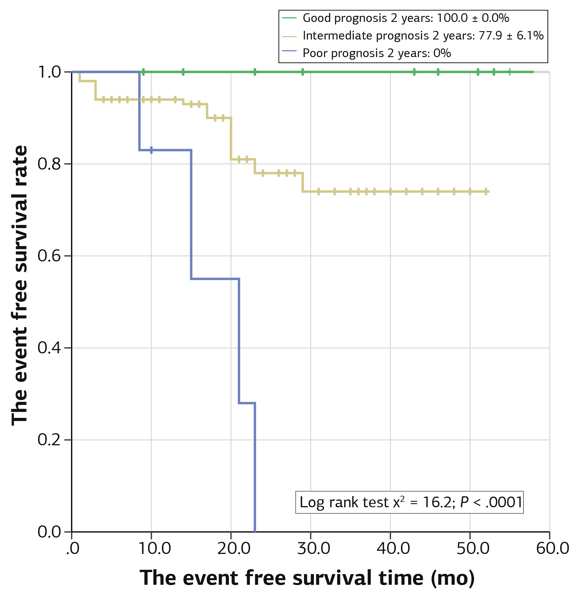
Figure 2. Event-free survival curve by genetic risk group in ALL
Additional Information
Publisher’s Note
Bayrakol MP remains neutral with regard to jurisdictional and institutional claims.
Rights and Permissions

This work is licensed under a Creative Commons Attribution-NonCommercial 4.0 International License (CC BY-NC 4.0). To view a copy of the license, visit https://creativecommons.org/licenses/by-nc/4.0/
About This Article
How to Cite This Article
Tran Kiem Hao, Nguyen Thi Kim Hoa, Phan Hung Viet. Research of genetic abnormalities in diagnosis and treatment of childhood acute lymphoblastic leukemia at hue central hospital. Ann Clin Anal Med 2024;15(1):21-24. doi:10.4328/ACAM.21847
Publication History
- Received:
- 06.08.2023
- Accepted:
- 06.11.2023
- Published Online:
- 10.11.2023
- Printed:
- 01.01.2024