
Abstract
AimThe aim of the study is to evaluate the synergistic modulatory effects of ferulic acid (FA) in combination with caffeic acid (CA) on the immune response and metabolic syndrome in male rats fed high-fat diets (HFD).MethodsForty-five adult male albino rats (Sprague-Dawley) were divided into five groups: G1, controls fed basal fat diet (BFD); G2, high-fat modified diet containing 20% fat (HFD); G3, rats fed HFD and received FA (150 mg/day) orally; G4, rats fed HFD supplemented with CA (0.8 g/100 g diet); and G5, rats fed HFD and received both FA (150 mg/day) and CA (0.8 g/100 g diet).ResultsSerum concentrations of TG, TC, LDL-c, glucose, insulin, oxidative stress biomarkers, and proinflammatory cytokines were elevated in rats fed a high-fat diet compared with the BFD group. Significant adjustments were observed after the administration of FA and CA. The maximum improvement was observed in rats administered with CA in combination with FA.ConclusionFA and CA significantly counteracted the pronounced oxidative stress effect of HFD by inhibiting lipid peroxidation, restoring antioxidant status, and improving metabolic syndrome biomarker levels. In conclusion, these findings indicate the synergistic protective effect of FA and CA on risk factors that can lead to metabolic syndrome and immune response imbalance during HFD-associated oxidative stress in rats.
Keywords
Introduction
Metabolic syndrome (MetS) is a condition where the body metabolizes proteins, fats, carbohydrates and other substances in a disorderly manner. It is characterized by a combination of risk factors, including diabetes, high cholesterol levels, high blood pressure and obesity.1 The primary culprits behind MetS are lifestyles and unhealthy eating habits like consuming high-calorie diets. It is proven that 20–30% of adults are affected by MetS.2 Therefore, it is crucial to identify strategies to combat this issue. Extensive research has demonstrated that incorporating wholegrain foods and dietary fiber into diets can have positive impacts on lowering the risk of MetS and its correlated disorders.3
Nutritional supplements frequently contain phenolic acids, like caffeic acid (CA) and ferulic acid (FA), which are characterized by antioxidant properties, hypoglycemic, hypolipidemic,4,5 anti-inflammatory and immunomodulatory roles.6 FA is a phenolic acid compound found in cereals, fruits, vegetables and other edible plants.7 It is recognized for its anti-inflammatory properties, which contribute to its ability to perform several beneficial functions in the body. These include regulating glucose tolerance and lipid metabolism, which leads to improving liver health.8
CA is a secondary metabolite derived from natural sources such as olives, berries, potatoes and carrots, and is predominantly obtained from coffee beans. It belongs to the family of hydroxycinnamic acid, which is a major constituent of the daily human diet.9 Numerous in vitro and in vivo experiments demonstrated that CA possesses anti-atherosclerotic, immunostimulant, cardioprotective and antiproliferative benefits,10 as well as its ability to improve inflammation and oxidative stress in chronic metabolic diseases.
The strategy of synergistic effects aims to enhance potency through treatment with a combination of therapeutic agents. Successful combinations involving agents and natural products can effectively attain desired outcomes while reducing toxicity levels.11 Based on this, the aim of this study is to examine the synergistic modulatory effects of FA in combination with CA on the immune response and MetS in male rats fed high-fat diets (HFD).
Materials and Methods
ChemicalsCA (≥95%) was purchased from Sigma-Aldrich (St Louis, MO, USA). FA (purity >98%) was purchased from Source Naturals Co., Miami, FL, USA. Commercial assay kits including glucose, total cholesterol (TC), triacylglycerols (TG), high-density lipoprotein-cholesterol (HDL-c), low-density lipoprotein-cholesterol (LDL-c), free fatty acid (FFA) were obtained from Siemens Healthcare Diagnostics, USA. Malondialdehyde (MDA), superoxide dismutase (SOD), and reduced glutathione (GSH) kits were purchased from BioVision Inc. Co., USA. Insulin, tumor necrosis factor-α (TNF-α), nuclear factor kappa B (NF-kB), interleukin-6 (IL-6), and interferon-γ (IFN-γ) were analyzed by enzyme-linked immunosorbent assay (ELISA) kits obtained from BioVision Inc. Co., USA. Pyruvate carboxylase (PC) assay colorimetric kits and phosphoenolpyruvate carboxykinase (PEPCK) kits were also purchased from BioVision Inc. Co., USA.Animals and Experimental DesignThe study was conducted between March and May 2023. Fifty adult male albino rats (Sprague-Dawley) weighing 127.2–134.3 g were used in this study. Animals were kept in stainless steel cages in an air-conditioned animal house at 24°C and fed a basal diet, with water allowed ad libitum throughout the experimental period (8 weeks). The rats were randomly divided into five groups (10 rats/group) as follows:
G1: Rats fed a basal fat diet (BFD).
G2: Rats fed a high-fat diet (25%) (HFD).
G3: Rats fed HFD and received FA (150 mg/day) by oral gavage (HFD+FA).
G4: Rats fed HFD supplemented with CA (0.8 g/100 g diet) (HFD+CA).
G5: Rats fed HFD supplemented with CA (0.8 g/100 g diet) and FA orally (150 mg/day) (HFD+FA+CA).Sample Collection and Biochemical AssessmentAfter eight weeks, blood was drawn from the hepatic portal vein of ether-anesthetized rats that had fasted overnight. Blood tubes were centrifuged at 4000 × g for 15 minutes at 25°C to separate the serum. Serum samples were collected in sterile plastic tubes and kept frozen at −20 ºC for later biochemical testing. The liver was separated, rinsed and washed with saline solution (NaCl 0.9%), then blotted on filter paper. The liver was rapidly freeze-clamped and stored at −20 ºC for liver enzyme analysis. Liver enzymes (PC and PEPCK) and serum biochemical parameters were determined according to analytical methods. The oral glucose tolerance test (OGTT) was performed with oral gavage of glucose (2 g/kg b.w.) after 12 hours of fasting on the first day of week eight. Blood glucose concentrations were monitored by tail blood using a glucometer with one-touch ultra test strips at 0 (before glucose infusion), 30, 60, 90 and 120 minutes (post-infusion).Ethical ApprovalThe study was approved by the Research Ethics Committee of Ain Shams University (Date: 01.03.2023; Decision No.: sci1432309001).Statistical AnalysisData were statistically evaluated. Results were presented as mean ± standard error of the mean (SEM), and one-way analysis of variance (ANOVA) was used. Differences were considered significant at P ≤ 0.01.Reporting GuidelinesThis study was reported in accordance with the STROBE guideline.
Results
Serum concentrations of TG, TC and LDL-c were elevated significantly (p≤0.01) in rats fed a high-fat diet compared with the BFD group and gradually normalized by FA and CA supplementation. The level of serum HDL-c was significantly lowered (p≤0.01) in the HFD group compared to BFD and gradually increased by adding FA and CA (G3–G5). The maximum improvement in serum lipid profile was observed in rats administered CA in combination with FA (Figure 1).
Glucose administration raised glucose levels after 30 minutes, then gradually returned to baseline levels within 90 minutes. However, when rats were fed HFD for 56 days, their glucose levels peaked at 30 minutes after receiving glucose. All groups showed normalized blood glucose levels within 60–90 minutes except for rats fed a high-fat diet (G2). Supplementation with CA alone or in combination with FA significantly normalized blood sugar levels (Figure 2). HFD rats had significantly increased serum FFA, fasting glucose and insulin levels compared with the BFD group (p≤0.01). Supplementation of FA combined with CA significantly lowered the values of FFA, glucose and insulin by 45.2%, 26.6% and 22.1%, respectively, compared with HFD rats (Table 1).
Results revealed the reduction of hepatic PC and PEPCK activities in rats fed HFD (by −28.3% for PC and −41.3% for PEPCK, respectively) compared to control rats. Supplementation with FA and CA modulated the activities of both enzymes compared to rats fed a high-fat diet. The maximum improvement in liver activities of both enzymes was observed in rats fed a diet supplemented with CA in combination with FA (by +42.1% for PC and +58.3% for PEPCK, respectively) compared with the HFD group (Table 2).
In this study, SOD activity was significantly decreased (p≤0.01) in HFD-fed rats compared to the control group. Serum levels of MDA, GSH and SOD activities were significantly (p≤0.01) restored by CA and FA supplementation compared with HFD-fed rats (Figure 3). Compared with the control group, an apparent increase in IFN-γ, TNF-α, IL-6 and NF-kB was observed in the HFD group by 3.5-, 2.7-, 2.4- and 2.2-fold, respectively. The levels of IFN-γ, TNF-α, IL-6 and NF-kB displayed a significant decrease after administration of FA separately or combined with CA. The results postulated that FA combined with CA exhibited an improving effect in HFD-caused inflammation in rats (Table 3).
Discussion
High-fat and high-energy diet is widely recognized as a factor contributing to the onset of various health issues. This study demonstrates that rats administered HFD suffered from glucose intolerance, dyslipidemia and oxidative stress in their livers with a parallel increase in serum glucose and lipid levels, including cholesterol and triglycerides.12 Supplementing FA and CA decreased serum lipid levels in the rat group fed HFD, thus avoiding glucose intolerance. Supplementation of CA combined with FA also reduced oxidative stress and lipid peroxidation in both serum and liver by repairing cellular antioxidants.
Hyperglycemia-related oxidative stress is considered one of the main reasons for metabolic and hormonal imbalances. Overnutrition may result in insulin resistance in tissues, which generally respond to insulin for glucose uptake.13 Over time this resistance leads to increased fasting glucose levels and diminished insulin-mediated glucose clearance. As a result, negative feedback signaling prompts pancreatic β-cells to produce more insulin. If this state persists without correction, it eventually causes hyperglycemia and type 2 diabetes.14
Administering CA and FA enhanced glucose utilization in the current study, as shown by OGTT results, supported by previous experiments.15 FA diminished HFD-induced glucose synthesis possibly by suppressing glucose-6-phosphatase, consequently slowing hepatic gluconeogenesis. PC and PEPCK are key enzymes regulating hepatic gluconeogenesis and glucose production. Results showed that CA and FA displayed synergistic effects on modulation of glucose metabolism by managing PEPCK and PC activities in HFD-fed rats. Son et al.16 reported that FA boosted glycogenesis by decreasing PEPCK levels. These results suggest that FA can adjust glucose homeostasis by restoring hepatic glucose metabolism disturbance. A previous study showed that FA reduced insulin resistance and diminished TG, free fatty acids, cholesterol and phospholipids in rats fed HFD. The modulatory effect of FA on lipid homeostasis was correlated to decreased lipogenic enzymes such as fatty acid synthase and acetyl-CoA carboxylase.8
CA elevated insulin sensitivity by reducing proinflammatory cytokines and increasing adiponectin under hyperglycemic states. One previous work showed that MetS diet in rats increased blood glucose, TG and LDL-c, and decreased HDL-c, while the group that received CA showed reduced serum insulin, TNF-α and IL-6. CA-supplemented rats also showed the highest liver levels of SOD, CAT and GPx antioxidant enzymes after four weeks of administration compared to FA, gallic acid and protocatechuic acid.17 This suggests that CA showed high scavenging activity and effectiveness among phenolic acids against hyperglycemic injuries.
A recent in vivo study showed that CA supplementation reduced body weight and prevented fat accumulation in HFD obese mice, improving lipid profile with increased HDL-c.18 This suggests that CA is capable of reducing FFA production, demonstrating hepatoprotective capability. Results showed that co-treatment of FA and CA reduced serum TC by 47.1% from the HFD group, higher than mono treatments (FA by 29.2% and CA by 36.3%). Similarly, FA and CA combination reduced LDL-c by 47.1% versus 31.1% by FA and 24.5% by CA. Thus, both CA and FA exhibited excellent synergistic effects on modulation of lipid profile in HFD-fed rats.
CA has shown potential antihyperglycemic effects, as well as antioxidant and anti-inflammatory properties. Its protective effects may be attributed to its ability to activate and defend cellular antioxidant enzymes by transferring hydrogen atoms and single electrons while chelating metal ions.19 Rats fed HFD had higher levels of lipid peroxidation product MDA. The decreased SOD activity and GSH observed could be related to increased oxidative stress. According to findings, MDA levels increased in HFD-fed rats, linked to reduced SOD and GSH activities, agreeing with studies suggesting compromised tissue antioxidant defenses under HFD.20
FA reduced MDA production and boosted antioxidant enzymes (SOD and GSH) activities, enhancing scavenging free radicals,21 helping to reduce lipid peroxidation and oxidative stress caused by HFD in the liver. TNF-α, NF-kB and IL-6 levels were significantly reduced after FA consumption. FA may decrease lipid peroxidation, resulting in decreased MDA and LDL-c levels.22
Inflammation develops after ingestion of HFD in peripheral tissues including liver, adipose tissue and skeletal muscles. Alterations in gut microbiota and direct effects of FFAs on intestinal cells may initiate chronic systemic inflammation. The second step may involve increased intestinal lipopolysaccharide (LPS), proinflammatory cytokines and FFA transport into systemic circulation, resulting in low-grade inflammation.23 Elevated serum FFAs and LPS can upregulate Toll-like receptors in macrophages, leading to activation and cytokine production.
Cytokines are crucial mediators in immune response. The results showed that HFD significantly increased IFN-γ, TNF-α, IL-6 and NF-kB serum levels. IFN-γ is produced by natural killer T cells during innate immune response and by CD4+ Th1 and CD8+ cytotoxic T cells in humoral immune response, essential for tumor detection, immunoregulation and inflammation.24
Our experimental results demonstrated that CA and FA showed strong synergistic antioxidant and anti-inflammatory activity. The immunomodulatory role of antioxidants is demonstrated in their capability to influence signaling pathways responsible for inflammation. NF-kB regulates expression of many genes directly related to inflammation. Recent research suggests certain polyphenols can alter NF-kB activity and lessen cellular inflammation.25 Suppression of NF-kB depends on polyphenolic compounds. Reduction of pro-inflammatory cytokine expression is one of the most prevalent ways polyphenols exert immunomodulatory effects.
Limitations
The findings are limited to this experimental rat model and require confirmation in further studies.
Conclusion
Several disease conditions primarily require combination therapy due to their complex pathophysiology and progression. The combination of CA with different compounds has been widely examined for various types of disorders. CA, owing to its free radical scavenging property, was able to increase the antioxidant efficacy of agents such as FA for better treatment outcomes. As the results of the present study showed, CA exhibited synergistic effects with FA for modulation of MetS and immune response. Through this study, we can conclude that CA and FA possess excellent promise for managing MetS through their anti-obesity, antidiabetic and hypolipidemic activities. Although there are many studies on CA and other phenolic acids, no previous research indicated that CA and FA had synergistic effects against immune disturbance and metabolic implications induced by HFD.
Declarations
Animal and Human Rights Statement
All animal procedures were performed in accordance with institutional guidelines for the care and use of laboratory animals and were approved by the Research Ethics Committee of Ain Shams University.
Informed Consent
Not applicable, as this was an experimental animal study.
Data Availability
The datasets used and/or analyzed during the current study are not publicly available due to patient privacy reasons but are available from the corresponding author on reasonable request.
Conflict of Interest
The authors declare that there is no conflict of interest.
Funding
None.
Abbreviations
BFD: Basal fat diet
CA: Caffeic acid
ELISA: Enzyme-linked immunosorbent assay
FA: Ferulic acid
FFA: Free fatty acid
GSH: Reduced glutathione
HFD: High-fat diet
IL-6: Interleukin-6
MDA: Malondialdehyde
OGTT: Oral glucose tolerance test
PC: Pyruvate carboxylase
PEPCK: Phosphoenolpyruvate carboxykinase
SEM: Standard error of the mean
SOD: Superoxide dismutase
TC: Total cholesterol
TG: Triglycerides
References
- Ye L, Hu P, Feng LP, et al. Protective effects of ferulic acid on metabolic syndrome: a comprehensive review. Molecules. 2023;28(1):281.
- Prasun P. Mitochondrial dysfunction in metabolic syndrome. Biochim Biophys Acta Mol Basis Dis. 2020;1866(10):165838. doi:10.1016/j.bbadis.2020.165838
- Khan J, Khan MZ, Ma Y, et al. Overview of the composition of whole grains’ phenolic acids and dietary fibre and their effect on chronic noncommunicable diseases. Int J Environ Res Public Health. 2022;19(5):3042. doi:10.3390/ijerph19053042
- Feldman F, Koudoufio M, Desjardins Y, Spahis S, Delvin E, Levy E. Efficacy of polyphenols in the management of dyslipidemia: a focus on clinical studies. Nutrients. 2021;13(2):672. doi:10.3390/nu13020672
- Mangrulkar S, Shah P, Navnage S, Mazumdar P, Chaple D. Phytophospholipid complex of caffeic acid: development, in vitro characterization, and in vivo investigation of antihyperlipidemic and hepatoprotective action in rats. AAPS PharmSciTech. 2021;22(1):28. doi:10.1208/s12249-020-01887-7
- Gairola K, Gururani S, Dubey SK. Polyphenols and its effect on the immune system. In: Nutraceuticals and Functional Foods in Immunomodulators. Springer Nature Singapore; 2023:121-140.
- Theodosis-Nobelos P, Papagiouvannis G, Rekka EA. Ferulic, sinapic, 3,4-dimethoxycinnamic acid and indomethacin derivatives with antioxidant, anti-inflammatory, and hypolipidemic functionality. Antioxidants (Basel). 2023;12(7):1436. doi:10.3390/antiox12071436
- Li Y, Sair AT, Zhao W, Li T, Liu RH. Ferulic acid mediates metabolic syndrome via the regulation of hepatic glucose and lipid metabolisms and the insulin/IGF-1 receptor/PI3K/AKT pathway in palmitate-treated HepG2 cells. J Agric Food Chem. 2022;70(46):14706-14717. doi:10.1021/acs.jafc.2c05676
- Ganguly R, Singh SV, Jaiswal K, Kumar R, Pandey AK. Modulatory effect of caffeic acid in alleviating diabetes and associated complications. World J Diabetes. 2023;14(2):62-75. doi:10.4239/wjd.v14.i2.62
- Mirzaei S, Gholami MH, Zabolian A, et al. Caffeic acid and its derivatives as potential modulators of oncogenic molecular pathways: new hope in the fight against cancer. Pharmacol Res. 2021;171:105759. doi:10.1016/j.phrs.2021.105759
- Das P, Delost MD, Qureshi MH, Smith DT, Njardarson JT. A survey of the structures of US FDA-approved combination drugs. J Med Chem. 2018;62(9):4265-4311.
- Zhang X, Qiu B, Wang Q, et al. Dysregulated serum lipid metabolism promotes the occurrence and development of diabetic retinopathy associated with upregulated circulating levels of VEGF-A, VEGF-D, and PlGF. Front Med (Lausanne). 2021;8:779413. doi:10.3389/fmed.2021.779413
- Kuppuswami J, Senthilkumar GP. Nutri-stress, mitochondrial dysfunction, and insulin resistance: role of heat shock proteins. Cell Stress Chaperones. 2023;28(1):35-48. doi:10.1007/s12192-022-01314-9
- Hashim KN, Chin KY, Ahmad F. The mechanism of honey in reversing metabolic syndrome. Molecules. 2021;26(4):808. doi:10.3390/molecules26040808
- Kang BB, Chiang BH. Amelioration of insulin resistance using the additive effect of ferulic acid and resveratrol on vesicle trafficking for skeletal muscle glucose metabolism. Phytother Res. 2020;34(4):808-816. doi:10.1002/ptr.6561
- Son MJ, Rico CW, Nam SH, Kang MY. Effect of oryzanol and ferulic acid on the glucose metabolism of mice fed with a high-fat diet. J Food Sci. 2011;76(1). doi:10.1111/j.1750-3841.2010.01907.x
- Muhammad Abdul Kadar NN, Ahmad F, Teoh SL, Yahaya MF. Caffeic acid on metabolic syndrome: a review. Molecules. 2021;26(18):5490. doi:10.3390/molecules26185490
- Xu W, Luo Q, Wen X, Xiao M, Mei Q. Antioxidant and antidiabetic effects of caffeic acid in a rat model of diabetes. Trop J Pharm Res. 2020;19(6):1227-1232. doi:10.4314/tjpr.v19i6.17
- Gupta A, Kumar R, Ganguly R, Singh AK, Rana HK, Pandey AK. Antioxidant, anti-inflammatory, and hepatoprotective activities of Terminalia bellirica and its bioactive component ellagic acid against diclofenac-induced oxidative stress and hepatotoxicity. Toxicol Rep. 2020;8:44-52.
- Chowdhury FI, Yasmin T, Akter R, et al. Resveratrol treatment modulates several antioxidant and anti-inflammatory genes expression and ameliorated oxidative stress-mediated fibrosis in the kidneys of high-fat diet-fed rats. Saudi Pharm J. 2022;30(10):1454-1463. doi:10.1016/j.jsps.2022.07.006
- Jing X, Zhang N, Zhao L, et al. Effect of soaked and fermented raspberry wines on the liver in mice. Food Biosci. 2022;47:101704. doi:10.1016/j.fbio.2022.101704
- Wang Y, Chen X, Huang Z, et al. Dietary ferulic acid supplementation improves antioxidant capacity and lipid metabolism in weaned piglets. Nutrients. 2020;12(12):3811. doi:10.3390/nu12123811
- Sankararaman S, Noriega K, Velayuthan S, Sferra T, Martindale R. Gut microbiome and its impact on obesity and obesity-related disorders. Curr Gastroenterol Rep. 2023;25(2):31-44. doi:10.1007/s11894-022-00859-0
- Huang LY, Chiu CJ, Hsing CH, Hsu YH. Interferon family cytokines in obesity and insulin sensitivity. Cells. 2022;11(24):4041. doi:10.3390/cells11244041
- Kanner J. Polyphenols by generating H2O2 affect cell redox signaling, inhibit PTPs, and activate Nrf2 axis for adaptation and cell surviving in vitro, in vivo, and human health. Antioxidants (Basel). 2020;9(9):797. doi:10.3390/antiox9090797
Tables
Table 1. Effect of various treatments on serum glucose, insulin, and free fatty acids (FFA)
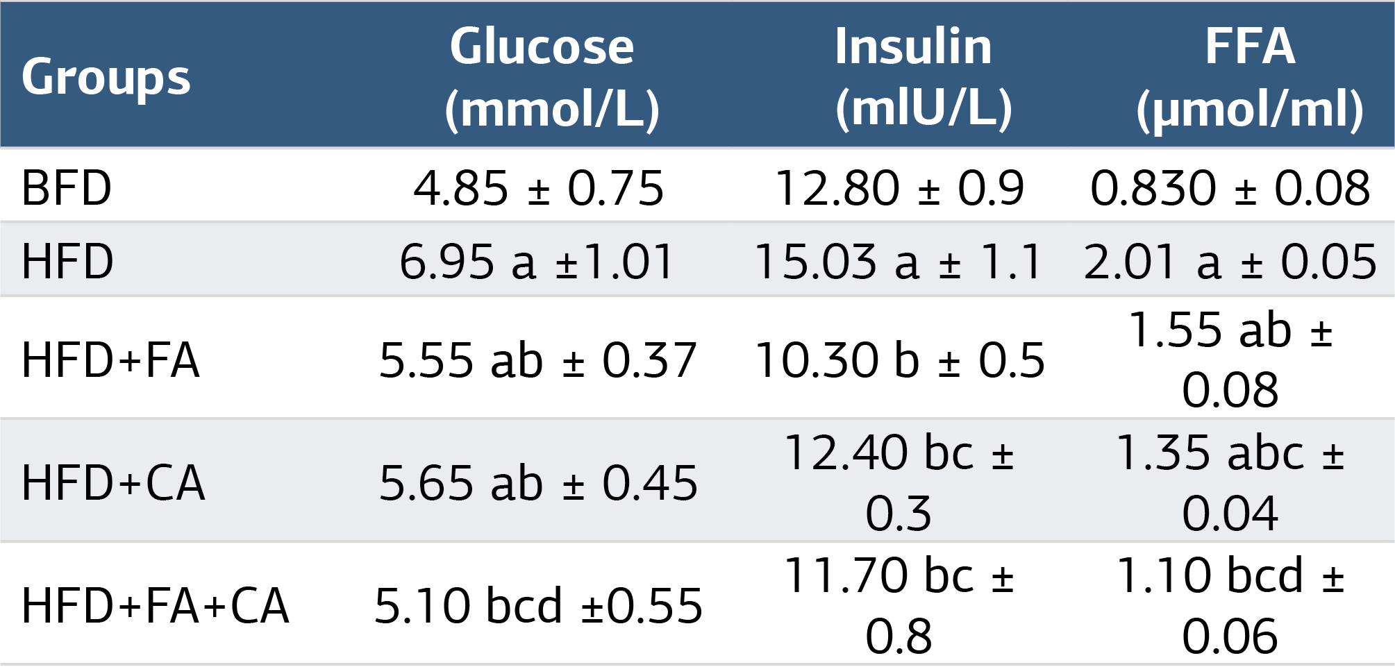
Values are expressed as means ± SD. (n = 9), a = significance against control; b= significance against HFD group; c= significance against HFD +FA group; d= significance against HFD + CA. at P ≤ 0.01
Table 2. Effect of various treatments on liver pyruvate carboxylase (PC) and phosphoenolpyruvate carboxykinase (PEPCK) activities
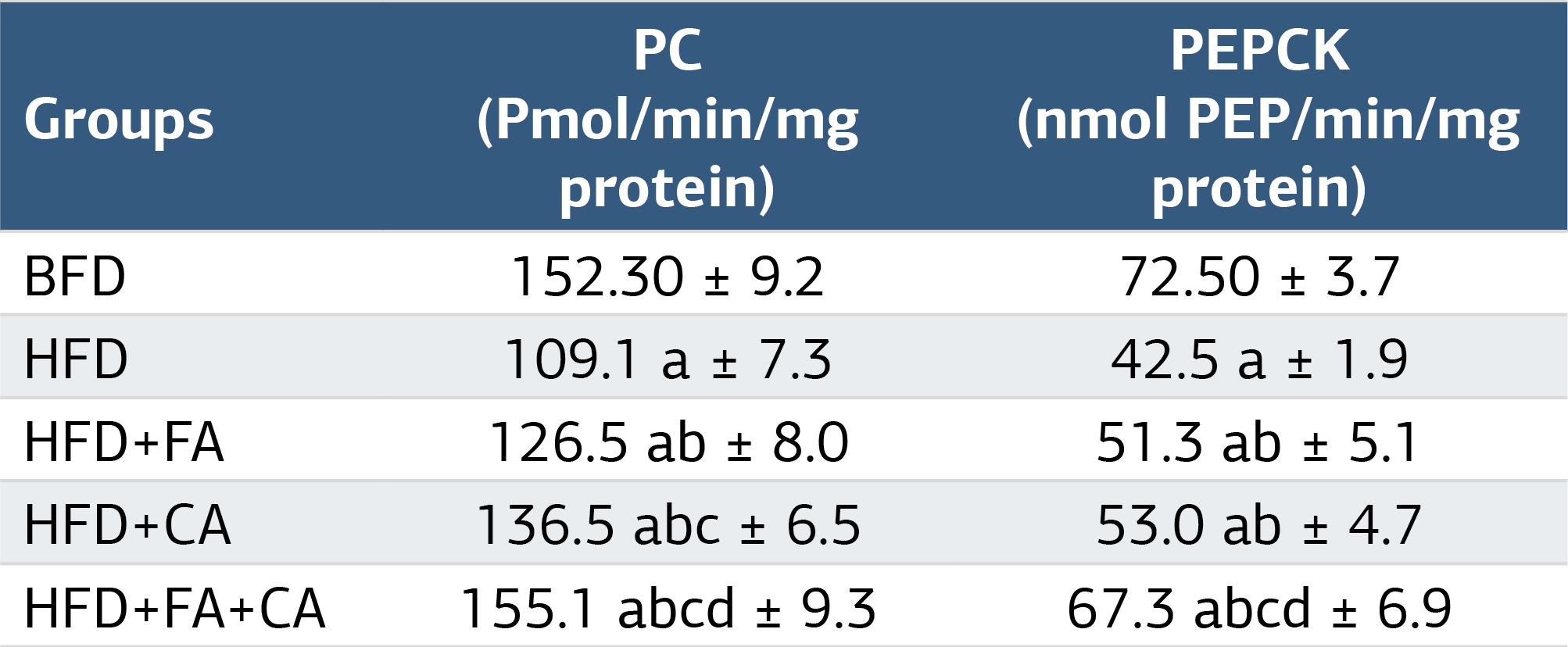
Values are expressed as mean ± SD. (n = 9), a = significance against control; b= significance against HFD group; c= significance against HFD +FA group; d= significance against HFD + CA. at P ≤ 0.01
Table 3. Effect of various treatments on serum levels of proinflammatory cytokines
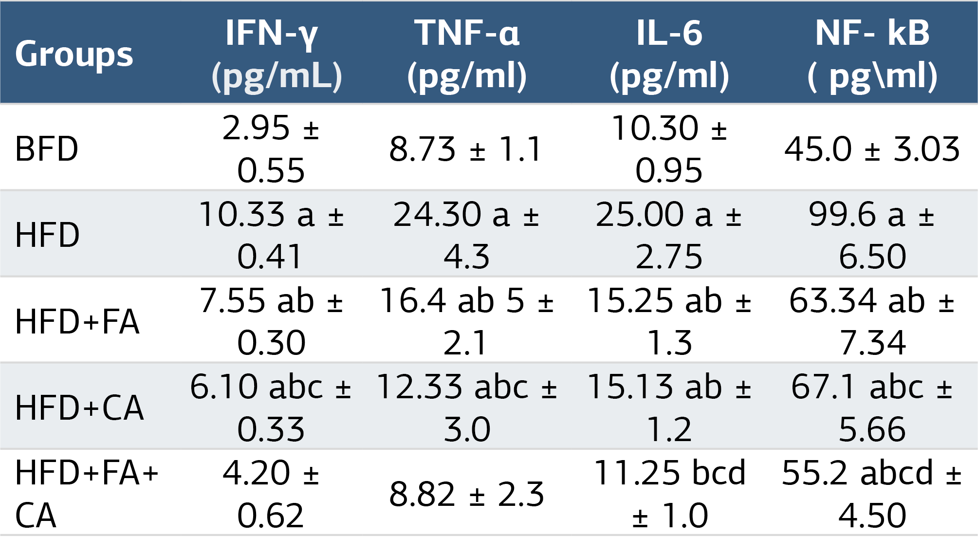
Values are expressed as mean ± SD. (n = 9), a = significance against control; b= significance against HFD group; c= significance against HFD +FA group; d= significance against HFD + CA. at P ≤ 0.01
Figures
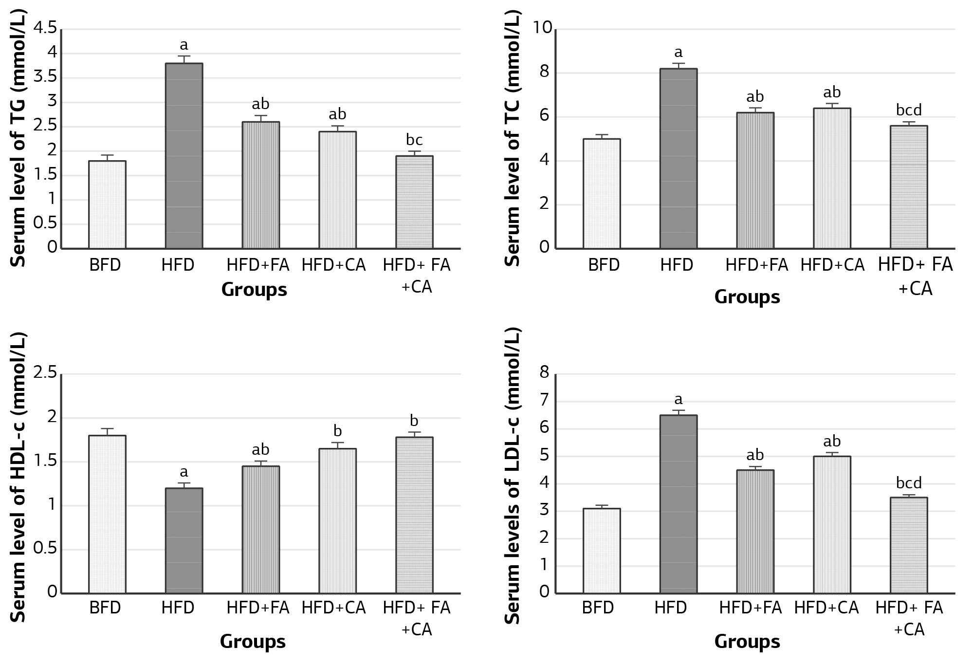
Figure 1. Effect of all treatments on serum lipids profile. BFD: basal fat diet (control); HFD: high fat diet; FA: Ferulic acid; CA: Caffeic acid. Alphabetical superscripts mean: a: significance against control; b: significance against HFD group; c: significance against HFD +FA group; d: significance against HFD + CA.
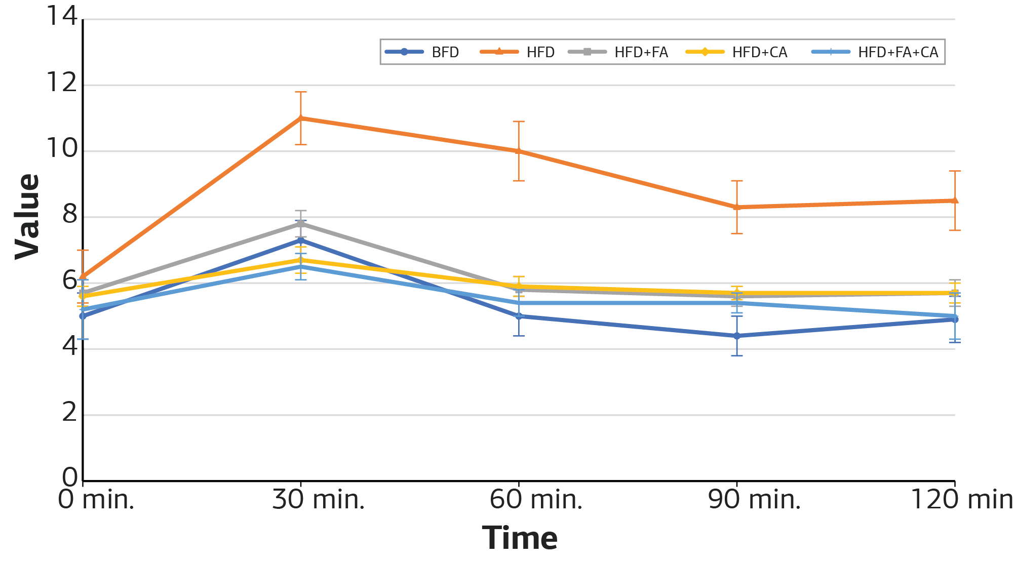
Figure 2. Oral glucose tolerance test (OGTT) after treatments. BFD: basal fat diet (control); HFD: high-fat diet; FA: Ferulic acid; CA; Caffeic acid.
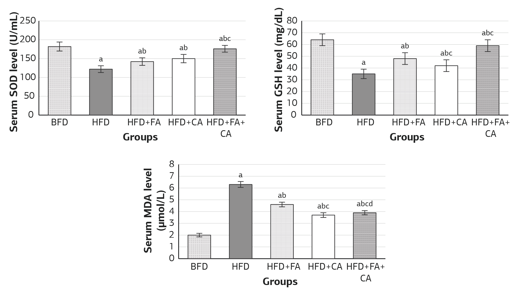
Figure 3. Effect of various treatments on serum levels of oxidative stress biomarkers. BFD: basal fat diet (control); HFD: high-fat diet; FA: Ferulic acid; CA; Caffeic acid. The alphabetical superscripts mean: a: significance against control; b: significance against HFD group; c: significance against HFD +FA group; d: significance against HFD + CA.
Additional Information
Publisher’s Note
Bayrakol MP remains neutral with regard to jurisdictional and institutional claims.
Rights and Permissions

This work is licensed under a Creative Commons Attribution-NonCommercial 4.0 International License (CC BY-NC 4.0). To view a copy of the license, visit https://creativecommons.org/licenses/by-nc/4.0/
About This Article
How to Cite This Article
Nesrin I. Tarbiah, Fares K. Khalifa, Nuha A. Alkhattabi, Abeer A. Banjabi, Reem F. Ghazali, Reem Y. Alzahri, Norah Abdu Almerabi, Ahd A. Mansour. Synergistic effect of ferulic acid and caffeic acid on metabolic syndrome and immune response in rats. Ann Clin Anal Med 2024;15(1):42-46. doi:10.4328/ACAM.22005
Publication History
- Received:
- 05.10.2023
- Accepted:
- 13.11.2023
- Published Online:
- 15.11.2023
- Printed:
- 01.01.2024