
Abstract
AimIn this study, surgical approaches were determined by making 3D anatomical modelling of paediatric cases with congenital atrial septal defect and ventricular septal defects using computed tomography data.MethodsComputed Tomography Angiography data from ten male and ten female patients with congenital atrial septal defect and ventricular septal defect were utilized in the present study. After obtaining the computed tomography images of the patients in DICOM format, 3D anatomical modelling was built by transferring them to the three-dimensional modelling software MIMICS 20.01 (The Materialise Group, Leuven, Belgium). Then, 3D printers generated a 3D-printed heart model.ResultsThe intracardiac and vascular structures of different types of congenital heart diseases, such as atrial septal defect and ventricular septal defect, were imaged through the 3D organ models. Then, conclusions were drawn about interventional or surgical procedures that can be followed for the restoration of atrial septal defect and ventricular septal defect.ConclusionThe three-dimensional modelling of the intracardiac structure, performed in this study, enables better imaging of the defects or pathologies and easier follow-up of the course of the disease and provides great support to the preoperative planning of paediatric cardiologists and cardiac surgeons. These modelling methods help to observe the risks of complicated surgeries in more detail. This allows identification of operational risks and taking surgical actions, and the duration of the operation can be shortened through patient-specific planning. When cumulatively assessed, this method reduces costs. Another significant advantage of these outcomes is their use in patient education in addition to the education of assistants and medical students. Additionally, 3D printing material systems, integrated functional applications, state-of-the-art technology products in patient-specific organ models, limitations in the field, current status and future perspectives were comprehensively discussed.
Keywords
Introduction
Today, rapid advances in the field of medical imaging have enabled the diagnosis of diseases more easily and the monitoring of their course. Computed Tomography (CT) or Magnetic Resonance Imaging (MRI) is a detailed non-invasive imaging technology for the anatomical structure of organs or tissues and allows measurement of their area and volume.1 By this means, scientists and physicians can obtain detailed information about the anatomical structures of organs or tissues.2 Advanced medical imaging tools allow carrying out key procedures such as simulation, advanced surgical planning and radiotherapy planning in addition to diagnosis. Three-dimensional (3D) printed models can be produced by utilizing MRI and CT scan data of patients in order to print custom-fit prosthesis, orthosis and implant or to perform procedures before complex high-risk surgeries. 3D printed models provide a better understanding of anatomy.3
3D printed models have been employed both preoperatively and intraoperatively to assist in planning, decision making and intraoperative guidance in complex cardiac surgery.4 3D printed models yield effective outcomes in patients with congenital heart defects that require surgical repair.5 In complicated congenital heart anomalies, 3D virtual surgery planning is very important in the face of risky situations. Anatomical points required for the operation can be planned by the team through segmentation after obtaining a CT of the patient. Thus, it is reported that the operation is performed more safely and quickly.6 More than 4,000 surgeries are unnecessarily performed annually in the United States of America due to a lack of proper planning, failure to make the right decision and poor imaging of the area to be operated during the preoperative process.7 Therefore, effective clinical training and preoperative planning can play a vital role in reducing these incidents. Imaging techniques such as CT, MRI and 3D virtual imaging are significant imaging techniques utilized as critical tools to gather information about patients’ anatomy for diagnosis and preoperative planning. However, these methods and devices can cause misinterpretation as the precise recognition of direction and size in images may sometimes be unclear.6 Additionally, these imaging techniques do not provide kinaesthetic feedback, which is essential for assessing and adjusting the use of surgical tools in preoperative rehearsal.8 Developing physical organ models with anatomically accurate characteristics and quantitative feedback may make it easier for surgeons to identify surgical target regions during preoperative rehearsal. This can also allow full adaptation to the surgery by providing preoperative information and training to the patients. In recent years, rapid prototyping methods such as 3D printing combined with 3D imaging techniques have enabled the production of such vital models.
Congenital heart disease (CHD) abnormalities are congenital abnormalities of the cardiovascular system. Some of these abnormalities can be diagnosed at birth, while others may not manifest until adulthood and may not be diagnosed.9 A study conducted in England reported that congenital heart disease was diagnosed in almost 1 in 100 live births.10 A study conducted in Turkey in 2006 reported that the prevalence of CHD was 7.77/1000 and ventricular septal defect (32.6%) ranked first among these diseases, followed by patent ductus arteriosus (15.9%) and atrial septal defect (13.1%).11
The aim of this study is to make three-dimensional anatomical modelling of paediatric cases with congenital atrial septal defect and ventricular septal defect using CT angiography data and to determine surgical approaches accordingly.
Materials and Methods
Collection of Image MaterialsCT angiography images of 10 female and 10 male patients, who were diagnosed with ASD and VSD and aged between 0-18 years, were used in the study. Based on the obtained images, iIntracardiac structure and vascular structure modellings of different types of congenital heart diseases were built by using the obtained images. The printed material was generated from a modelled case using a three-dimensional printer. Ethical approval was obtained from the Harran University Clinical Research Ethics Committee (Decision No: HRU/20.21.06).Creating Three-Dimensional ModelsAfter the CT angiography (64-slice multidetector spiral computed tomography, General Electronic Revolution) images of the patients were taken in DICOM format, and they were transferred to the three-dimensional modelling software MIMICS 20.01 (The Materialise Group, Leuven, Belgium) and converted into a work file. When the data wereas transferred to the software, the data set of the sequence to be studied wasere selected. After assigning the threshold value of the tissues in the ongoing progress, the modelling process was initiated. The segmentation processes were performed in different approaches using the “edit mask” commands on the three-dimensional heart models, and the intracardiac structure model was obtained in the heart models. While taking the printed materials from MIMICS-based 3D models, laser technology based on SLA resin (GREY resin) was employed. A Formlabs Form 3 3D Printer (Formlabs Inc, Massachusetts, USA) was employed during the 3D printing stage.
The study received For the study, an approval was taken from Harran University, Faculty of Medicine, Clinical Trials Ethics Committee with the (decision numbered No. HRU/20.21.06 and dated 07.12.2020).Ethical ApprovalThe present study was approved by the local ethics committee (Approval No: 07.12.2020 HRU/20.21.06).Statistical AnalysisNo statistical analysis was performed because the study was descriptive and focused on three-dimensional anatomical modeling.Reporting GuidelinesThis study was reported in accordance with the STROBE guideline.
Results
Heart Models Created from CT ImagesIn the present study, intracardiac structure and vascular structure modellings of different types of congenital heart diseases were created. Figure 1 shows the 3D printed heart model, which was created from the CT data of patients with congenital ASD and VSD in such a way that the intracardiac structure is visible. Figure 2 shows the double outlet right ventricle defect. Figure 3 shows the image of the printed material obtained from the 3D-modelled hearts in such a way that the intracardiac structure is visible.
Discussion
ASD and VSD are the two most common congenital malformations that are frequently observed in congenital heart defects. Upon literature reviews, it has been observed that severe congenital heart diseases are more common in boys, but diseases such as ASD and patent ductus arteriosus are more common in girls.12,13 Congenital anomalies, which may be diagnosed late with the developments in technology, can be diagnosed early at approximately 11 weeks.14
Echocardiography is generally employed to diagnose CHDs, especially ASD and VSD. However, CT can be used as an alternative to cardiac catheterisation if echocardiography fails to produce preoperative information.15
Another advantage of CT is that it contributes to creating 3D modelling through cross-sectional imaging. In the comprehensive assessment of anatomical and pathological structures that cannot be achieved by other methods and tactile examination of these printing materials, medical models are more advantageous than classical image modelling.16 A thorough understanding of cardiac anatomy and a highly experienced operator are required for safe use of invasive and surgical methods in the treatment of ASD and VSD. At this point, it is important to employ 3D modelling and printed materials in the diagnosis of defects and in the planning of the operation.
3D models are widely used in the field of industry, and have recently become widespread in the medical field and have been employed in many surgical disciplines such as cardiology, anatomy education, archaeology and forensic medicine.17 These models are of prime importance for pre-operative planning.18,19
By taking the printed material from these models, which provide great convenience in understanding the complexity of cases and planning operations, it is possible to create personalised patient-specific medical models, as well as develop designs for prostheses and orthoses.20,21
Anwar et al.17 reported that 3D printed models are an important technology in congenital heart diseases and hold a significant place in the field of surgery due to the ability to plan easily with 3D printed materials and the lack of complications. It has also been reported that 3D models can facilitate communication between multidisciplinary teams and thereby potentially reduce medical errors.22 Yoo et al.23 reported that the use of 3D printing would continue to expand with further advancements in imaging and printing technologies and the development of new printing materials. It has been reported that the printing of biological tissues and tissue engineering would lead the way for a new era in personalised medicine.24
CT was used to create three-dimensional models of paediatric patients diagnosed with ASD and VSD in the present study. The printed materials were produced from these models using three-dimensional printing technology in order to better understand the operative approaches of the cases. These images were stored in a virtual medium to be utilised as a digital library. The printer technology from which 3D printed materials are produced and the materials used in printing are being upgraded every day. “Segmentation” is an essential step before obtaining 3D printed material.18 The CT images in the present study were produced by segmenting the 3D models created using the “MIMICS 20.1” software. Segmentation is of utmost importance in 3D anatomical modelling and virtual surgical planning with respect to the process of section-by-section separation by following the anatomical boundaries in the tissues that are needed or critical for surgical planning. It has been reported that these methods outperform traditional methods.21,25
In the present study ASD and VSD were modelled in paediatric patients in a digital environment, the usability of these data as a library was ensured, and it was also demonstrated that surgical approaches could be tangibly determined by taking 3D printed material. The beneficial aspects of 3D models were described as improving the understanding of complex cardiovascular structures, providing guidance for surgical planning and simulation of interventional procedures, as well as enhancing doctor-to-patient communication in the manuscript by Sun and Wee.25
Given the CT report of the case from which the printed material was obtained in the study, it was determined upon the examination performed in the axial plane with a 5-mm section thickness after administration of intravenous contrast agent that the right upper pulmonary veins opened to the vena cava superior (supracardiac type partial pulmonary venous return anomaly) and an appearance that may be compatible with patent foramen ovale was observed at the level of the foramen ovale. The abnormalities in CT were clearly identified in the 3D model of the related case. In another case diagnosed with tetralogy of Fallot, all of the abnormalities listed in the CT report were observed in the 3D anatomical model. Similarly, in the other cases included in the present study, the same abnormalities were identified in CT reports and 3D organ modelling. VSD can be clearly observed in the intracardiac structure of the printed model of the case in which we created a 3D-printed organ model.
The thesis study was carried out to create 3D anatomical models of paediatric cases with ASD and VSD, which are common congenital heart defects in Turkey, using CT and to determine surgical approaches and presented cardiac data for ASD and VSD in these patients. The three-dimensional modelling of the intracardiac structure, which was performed in this study, enables better imaging of the defects or pathologies and easier follow-up of the diagnosis and provides a great contribution to the preoperative planning of paediatric cardiologists and cardiac surgeons. These modellings help to observe the risks of complicated surgeries in more detail. The pathological condition of the heart can be monitored before the patient is operated by means of these printed models, and no adverse conditions such as surprising pathological conditions will be encountered during the operation. This allows identification of operational risks and taking surgical actions and the duration of the operation can be shortened through patient-specific planning. When cumulatively analysed, this method reduces the costs. Another significant advantage of these outcomes is their use in patient education in addition to the education of assistants and medical students. The MIMICS Innovation Suite software utilised in the present study is CE-certified software and is the most widely used software in medical and other research. The ability to integrate special modules into MIMICS, which is modular software, offers a significant advantage.
Limitations
The small sample size was the main limitation of this study.
Conclusion
Planning, and surgical rehearsal (simulation).
In the medical field, 3D-printed models may be utilized to build personalized prostheses, orthoses, or implants. These models could also be used to produce laboratory equipment. Organs may also be modelled and printed for transplantation through 3D printing technology.
The 3D models and printing have enabled us to image the intracardiac and vascular structures of different types of congenital heart diseases such as ASD and VSD, and through direct comparisons between the model and real anatomy, conclusions were drawn about interventional or surgical procedures that can be applied in the repair of ASD and VSD.
As this technology advances further, biocompatible organs will soon be created by using biomaterial printing materials and cells. It is thought that even in the future, heart muscle cells and vascular cells with the ability to contract could be produced using these methods and heart tissue that can beat can be produced by synchronizing these with each other. Through these methods and inventions, 3D tissues and organs may be employed as a novel way to substitute organs for donor shortages. Furthermore, by means of the 3D model and printed organ, the most suitable interventional or surgical procedures for the patient may be performed by exchanging ideas on the created model with doctors from across the globe.
Consequently, 3D modelling and printing, when combined with medical imaging methods, is both a powerful diagnostic tool for patients and enables regular planning prior to operational intervention for patients. Meanwhile, 3D modelling and printing, which contribute to assistant training and personal experience, are being successfully used to build personalized prostheses. Much greater work will be achieved in the future as technological advances in both medical imaging and 3D modelling and printing continue. Thanks to today’s technologies, it is possible to combine materials with different properties, depending on the manufacturing method, in order to capture different elasticity or colour in a model. This method and technology allow for creating more realistic models with a more natural appearance for educational or research purposes. Furthermore, due to these printed models, personal measurements that may be incorrect in ECO, CT, MRI, or other relevant imaging techniques are prevented, and more exact measures can be achieved through model-supported measurements, thus it is believed that significant errors can be prevented.
Declarations
Animal and Human Rights Statement
All procedures performed in this study were in accordance with the ethical standards of the institutional and/or national research committee and with the 1964 Helsinki Declaration and its later amendments or comparable ethical standards.
Informed Consent
Informed consent was waived by the Ethics Committee.
Data Availability
The datasets used and/or analyzed during the current study are not publicly available due to patient privacy reasons but are available from the corresponding author on reasonable request.
Conflict of Interest
The authors declare that there is no conflict of interest.
Funding
This study was supported by the Scientific Research Projects Coordination Uniıt, Harran University (Project Noumber: 21026).
Abbreviations
ASD: Atrial septal defect
CHD: Congenital heart disease
CT: Computed tomography
DICOM: Digital imaging and communications in medicine
MRI: Magnetic resonance imaging
VSD: Ventricular septal defect
References
- Kaya T. Radyografinin temel prensipleri ve radyografik yorumda temel ilkeler [Basic principles of radiography and basic principles in radiographic interpretation]. Galenos Yayınevi; 2017:1-22. doi:10.5152/trs.2017.507
- Atalay HA, Değirmentepe RB, Bozkurt M, Can O, Canat HL, Altunrende F. 3D teknolojinin tıpta ve üroloji’de kulanım alanları [Areas of use of 3D technology in medicine and urology]. Endourol Bull. 2016;9:65-71.
- Qiu K, Haghiashtiani G, McAlpine MC. 3D-printed organ models for surgical applications. Annu Rev Anal Chem (Palo Alto Calif). 2018;11(1):287-306. doi:10.1146/annurev-anchem-061417-125935
- Schmauss D, Haeberle S, Hagl C, Sodian R. Three-dimensional printing in cardiac surgery and interventional cardiology: a single-centre experience. Eur J Cardiothorac Surg. 2015;47(6):1044-1052. doi:10.1093/ejcts/ezu310
- Full MDBS, Laycock SD, Brown JRI, Jakeways M. 3D printing of an aortic aneurysm to facilitate decision making and device selection for endovascular aneurysm repair in complex neck anatomy. J Endovasc Ther. 2013;20(6):863-867.
- Kula S, Tunç O, Kavgacı A, İncedere F. Demonstrating a rare anatomical variation of cardiovascular system by using a new technique. Anatol J Cardiol. 2020;24(4):244-246. doi:10.14744/anatoljcardiol.2020.28742
- Mehtsun WT, Ibrahim AM, Diener-West M, Pronovost PJ, Makary MA. Surgical never events in the United States. Surgery. 2013;153(4):465-472. doi:10.1016/j.surg.2012.10.005
- Mottl-Link S, Hubler MK, Uhne T, et al. Physical models aiding in complex congenital heart surgery. Ann Thorac Surg. 2008;86(1):273-277. doi:10.1016/j.athoracsur.2007.06.001
- Dankowski R, Baszko A, Sutherland M, et al. 3D heart model printing for preparation of percutaneous structural interventions: description of the technology and case report. Kardiol Pol. 2014;72(6):546-551. doi:10.5603/kp.2014.0119
- Hoffman J, Kaplan S. The incidence of congenital heart disease. J Am Coll Cardiol. 2002;39(12):1890-1900. doi:10.1016/s0735-1097(02)01886-7
- Dadvand P, Rankin J, Shirley MD, Rushton S, Pless-Mulloli T. Descriptive epidemiology of congenital heart disease in Northern England. Paediatr Perinat Epidemiol. 2009;23(1):58-65. doi:10.1111/j.1365-3016.2008.00987.x
- Başpinar O, Karaaslan S, Oran B, Baysal T, Elmaci AM, Yorulmaz A. Prevalence and distribution of children with congenital heart diseases in the central Anatolian region, Turkey. Turk J Pediatr. 2006;48(3):237-243.
- Morris CD. Lessons from epidemiology for the care of women with congenital heart disease. Prog Pediatr Cardiol. 2004;19(1):5-112. doi:10.1016/j.ppedcard.2003.09.003
- Donofrio MT, Moon-Grady AJ, Hornberger LK, et al. Diagnosis and treatment of fetal cardiac disease: a scientific statement from the American Heart Association. Circulation. 2014;129(21):2183-2242. doi:10.1161/01.cir.0000437597.44550.5d
- Hutchinson D, McBrien A, Howley L, et al. First-trimester fetal echocardiography: identification of cardiac structures for screening from 6 to 13 weeks’ gestational age. J Am Soc Echocardiogr. 2017;30(8):763-772. doi:10.1016/j.echo.2017.03.017
- Elzenga NJ, Von Suylen RJ, Frohn-Mulder I, Essed CE, Bos E, Quaegebeur JM, et al. Juxtaductal pulmonary artery coarctation: an underestimated cause of branch pulmonary artery stenosis in patients with pulmonary atresia or stenosis and a ventricular septal defect. J Thorac Cardiovasc Surg. 1990;100(3):416-424. doi:10.1016/s0022-5223(19)35534-5
- Chepelev L, Souza C, Althobaity W, et al. Preoperative planning and tracheal stent design in thoracic surgery: a primer for the 2017 Radiological Society of North America hands-on course in 3D printing. 3D Print Med. 2017;3(1):14. doi:10.1186/s41205-017-0022-3
- Mitsouras D, Liacouras P, Imanzadeh A, et al. Medical 3D printing for the radiologist. Radiographics. 2015;35(7):1965-1988. doi:10.1148/rg.2015140320
- Vukicevic M, Mosadegh B, Min JK, Little SH. Cardiac 3D printing and its future directions. JACC Cardiovasc Imaging. 2017;10(2):171-184. doi:10.1016/j.jcmg.2016.12.001
- Maragiannis D, Jackson MS, Igo SR, et al. Replicating patient-specific severe aortic valve stenosis with functional 3D modeling. Circ Cardiovasc Imaging. 2015;8(10):30-33. doi:10.1161/circimaging.115.003626
- Mahmood F, Owais K, Taylor C, et al. Three-dimensional printing of mitral valve using echocardiographic data. JACC Cardiovasc Imaging. 2015;8(2):227-229. doi:10.1016/j.jcmg.2014.06.020
- Yoo SJ, Thabit O, Kim EK, et al. 3D printing in medicine of congenital heart diseases. 3D Print Med. 2015;2(1):3.
- Matsumoto JS, Morris JM, Foley TA, et al. Three-dimensional physical modeling: applications and experience at Mayo Clinic. Radiographics. 2015;35(7):1989-2006. doi:10.1148/rg.2015140260
- Anwar S, Singh GK, Miller J, et al. 3D printing is a transformative technology in congenital heart disease. JACC Basic Transl Sci. 2018;3(2):294-312. doi:10.1016/j.jacbts.2017.10.003
- Sun Z, Wee C. 3D-printed models in cardiovascular disease: an exciting future to deliver personalized medicine. Micromachines (Basel). 2022;13(10):1575. doi:10.3390/mi13101575
Figures
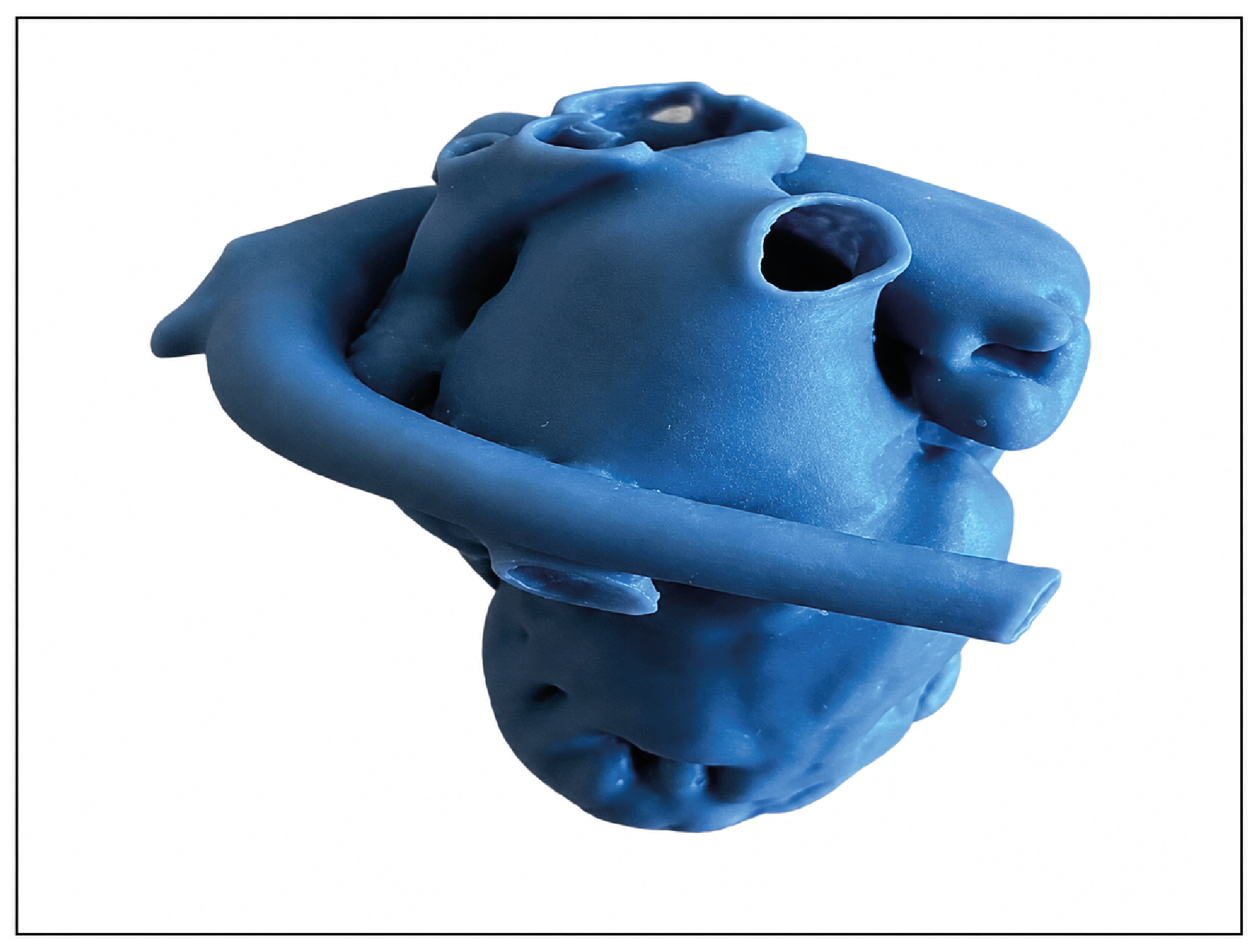
Figure 1. 3D heart model created from CT images
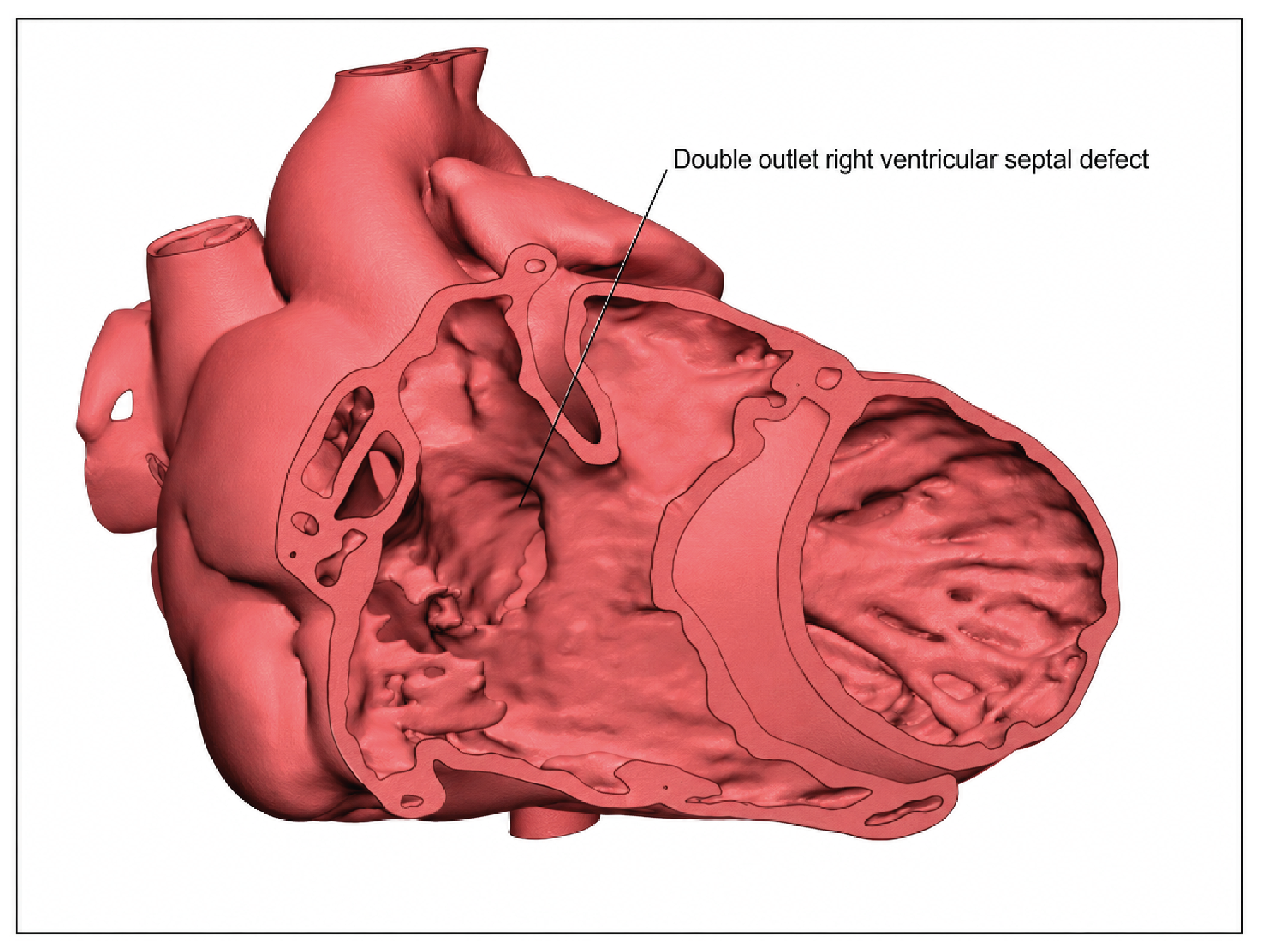
Figure 2. Double outlet right ventricle defect according to from CT images
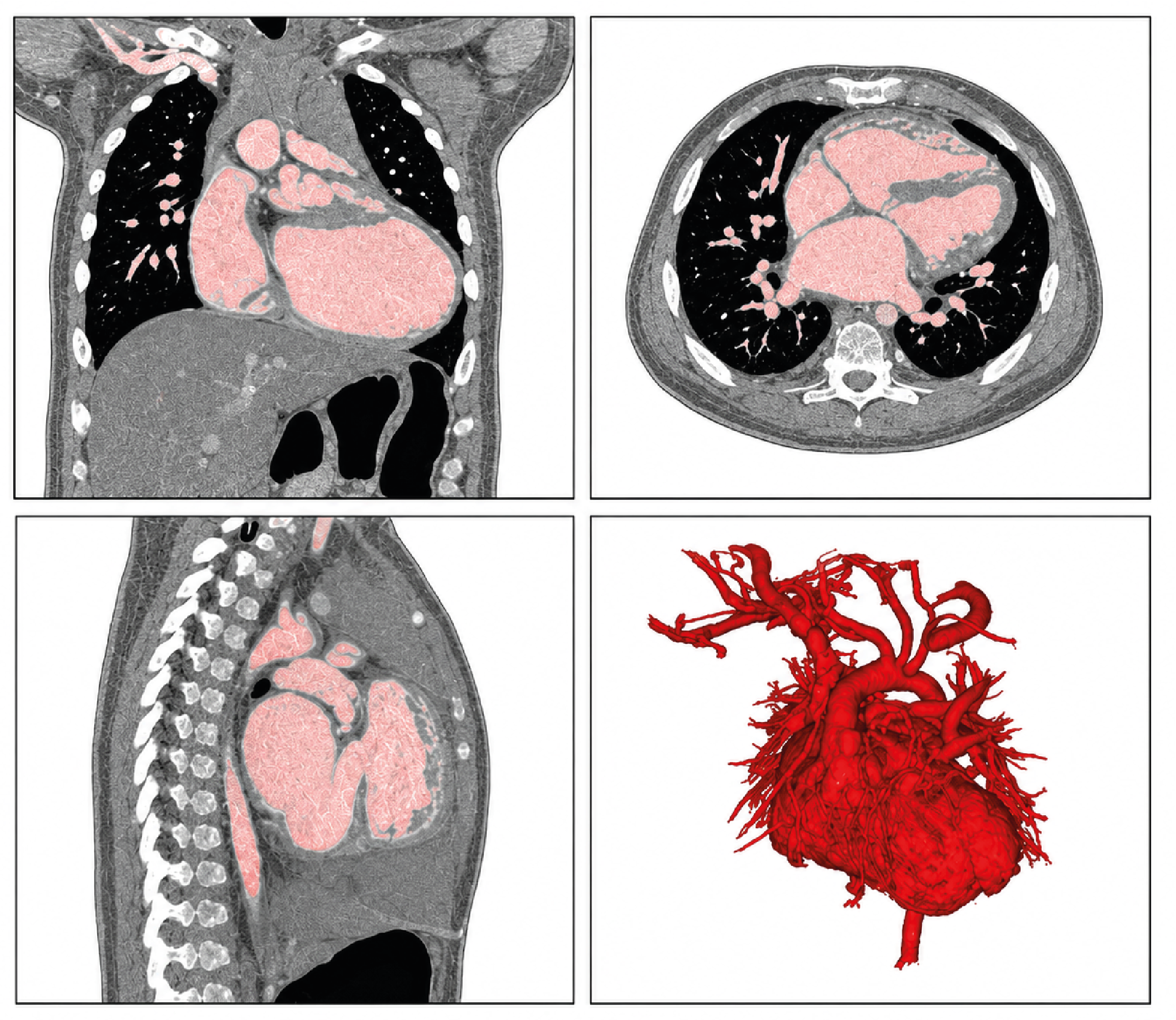
Figure 3. View of the intracardiac structure
Additional Information
Publisher’s Note
Bayrakol MP remains neutral with regard to jurisdictional and institutional claims.
Rights and Permissions

This work is licensed under a Creative Commons Attribution-NonCommercial 4.0 International License (CC BY-NC 4.0). To view a copy of the license, visit https://creativecommons.org/licenses/by-nc/4.0/
About This Article
How to Cite This Article
Ömer Göç, Mehmet Salih Aydın, İsmail Demircioğlu, Reşat Dikme, Osman Tunç. Three-dimensional anatomical modeling of cases with atrial septal defectand ventricular septal defect, which are congenital heart anomalies. Ann Clin Anal Med 2024;15(1):52-56. doi:10.4328/ACAM.22009
Publication History
- Received:
- 06.10.2023
- Accepted:
- 06.11.2023
- Published Online:
- 14.11.2023
- Printed:
- 01.01.2024