
Abstract
AimThe present study was designed to determine the therapeutic effects and immunomodulatory potential of silver nanoparticles (AgNPs) at two doses on cardiotoxicity induced by doxorubicin (DOX).MethodsSixty rats were randomized into six equal groups as follows: G1, Control; G2, NP1, rats received AgNPs (4 mg/kg b.w); G3, NP2, rats received AgNPs (8 mg/kg b.w); G4, cardiotoxicity group (DOX), rats received intraperitoneal single injection of DOX at dose of 30 mg/kg (i.p.); G5, DOX+NP1; G6, DOX+NP2. AgNPs was administered intraperitoneally for 4 weeks.ResultsAgNPs showed immunomodulatory potential as shown by an increase in blood leukocytes, lymphocytes, and neutrophils counts and the decrease in serum proinflammatory cytokines and oxidative stress markers. Cardio-therapeutic effects of AgNPs are shown by the decrease in biochemical markers of cardiac toxicity, namely, CPK, LDH, TropT, and ACL. DOX-treated rats showed significantly higher total cholesterol, triglycerides, total phospholipids, and LDL-c. Administration of AgNPs as therapeutic (4 and 8 mg/kg b.w) improved these parameters.ConclusionDepending on the immunomodulatory activity of AgNPs, therapeutic effect on the heart against cardiotoxicity induced by doxorubicin was shown by an increase in blood leukocytes count, decrease in proinflammatory cytokines, and oxidative stress biomarkers.
Keywords
Introduction
Nanotechnology is a promising technology with potential advantages in treating and preventing disease. Among the metal nanoparticles, silver nanoparticles (AgNPs) are the most widely used nanoparticle in biomedical-related products because of their broad-spectrum antimicrobial activity. Recently, AgNPs have been reported to have antioxidant activity.1 and anti-inflammatory property.2 Several studies have been reporting promising results of AgNPs for the treatment of inflammatory disease, such as for neuropathy in diabetic rats.3 and colitis.4
Nanoparticles have an impact on the immune system and can either stimulate or suppress it. Nanoparticles may be recognized as foreign by immune cells, triggering an unintended immune response against them, potentially leading to toxicity within the body. The physical properties of nanoparticles, including their size, surface charge, and coating, play a crucial role in determining their compatibility with the immune system. Immune cells are likely involved in the disease’s pathophysiology as they receive signals from injured myocytes, endothelial cells, and cardiac progenitor cells.5
Cardiotoxic exposures cause electrophysiological dysfunction or muscle damage to the heart. Typically, most acute cardiotoxic effects are reversible and thought to be the result of myocardial edema.6 While chronic cardiotoxicity has been extensively studied, there is limited research on the manifestations of acute cardiotoxicity, and the development of secondary dilated cardiomyopathy is not fully understood. Doxorubicin (DOX) is a crucial and effective anticancer drug used in the treatment of many childhood cancer patients. However, its use carries potential risks for various complications in the short and long term. One of the most significant complications is the development of cardiotoxicity, which can lead to congestive heart failure.5
Materials and Methods
ChemicalsDoxorubicin was purchased from Sigma Chemical Co., St. Louis MO, USA.
Silver Nanoparticles (AgNPs): Dark gray powder (Particles Size Analysis {APS} 30–50 nm) 1 g dissolved in saline, purchased from Sigma Aldrich (USA).Experimental AnimalsSixty adult male albino rats weighing 198 ±11 g were used in this study. The animals were kept at standard housing facilities (24±1 °C, 45±5% humidity, and 12 h light/dark cycle). They were supplied with standard laboratory chow and water ad libitum and left to acclimatize for 1 week before the experiments.Experimental DesignExperimental rats were classified into six groups (n = 10) as follows:
Group 1 – Control (C): Rats received 1 ml saline solution.
Group 2 – AgNPs: Rats received AgNPs (4 mg/kg b.w) (NP1).
Group 3 – AgNPs: Rats received AgNPs (8 mg/kg b.w) (NP2).
Group 4 – Cardiotoxicity group (DOX): Rats received intraperitoneal single injection of DOX at dose of 30 mg/kg (i.p.) dissolved in saline solution, this dose is well proved to induce cardiotoxic effects.
Group 5 – Cardio-therapeutic1 (DOX+NP1): Four days after DOX administration rats received AgNPs (4 mg/kg b.w).
Group 6 – Cardio-therapeutic2 (DOX+NP2): Four days after DOX administration rats received AgNPs (8 mg/kg b.w).
Treatments with AgNPs were given intraperitoneally for 4 weeks.Sample CollectionBlood was collected through retro-orbital puncture. The collected blood samples were left at a temperature of 24 °C for 30 minutes, followed by centrifugation at 5000 r.p.m. for 20 minutes. The resulting serum was divided into multiple aliquots and stored at −20 °C until analysis. For hematological analysis, heparinized blood was collected in EDTA-coated vials. The leukocytes count (WBC), lymphocytes count, and neutrophils count were determined from the whole blood using an automated hematological analyzer (Beckam Coulter, USA).Serum and Heart AnalysisSeveral serum levels were measured in different experimental groups. These included anti-cardiolipin (ACL), troponin-T (TropT), CRP, albumin (Alb), LDH, CPK, AST, acid phosphatase activity (AP), TC, TG, total phospholipids (PL), and LDL-c. Additionally, the serum levels of tumor necrosis factor alpha (TNF-α), IL-6, NF-κB, IL-1β, WBC count, lymphocytes count, and neutrophils count were measured. A portion of each heart from all groups was taken and a 30% w/v homogenate was prepared in 0.9% buffered KCl (pH 7.4) to estimate GSH, MDA, SOD, and CAT.Ethical ApprovalThis study was approved by the Ethics Committee of Sciences Academy of Experimental Research, AL-Mansoura, Egypt (Date: 15.05.2023, Decision No: 44121).Statistical AnalysisData were shown as means ± standard error of mean (SEM), and a one-way analysis of variance (One-Way ANOVA) test. Differences were considered significant when p≤0.05.Reporting GuidelinesThis study was reported in accordance with the STROBE guideline.
Results
The administration of DOX significantly increased serum levels of cardiac markers, including troponin T (TropT), anti-cardiolipin (ACL), and C-reactive protein (CRP), while decreasing albumin levels (p≤0.05), indicating cardiac damage. However, when AgNPs were administered to cardiotoxic rats, the cardiac damage caused by DOX was reduced. The administration of AgNPs as a therapeutic agent restored TropT, ACL, albumin, and CRP levels (Table 1).
(Table 2) shows the effect of DOX and AgNPs on serum cardiac enzymes markers LDH, CPK, AP, and AST. DOX administration increased serum cardiac enzymes significantly (p≤0.05). It was observed that administering AgNPs as therapeutic restored levels of serum enzymes near to normal levels. The rats treated with doxorubicin showed significantly higher levels of total cholesterol (TC), triglycerides (TG), total phospholipids (PL), and LDL-c compared to the control group (p≤0.05). However, the administration of silver nanoparticles (AgNPs) as a therapeutic intervention (G5, G6) improved these parameters and significantly reduced levels of TC and TG compared to the group experiencing cardiotoxicity (G4) (Figure 1).
The findings indicated a connection between DOX-induced cardiotoxicity and a decrease in white blood cells count (WBCs), neutrophils, and lymphocytes. When compared to the cardiotoxic group (G4), the administration of AgNPs (G5, G6) resulted in an increase in leukocytes, lymphocytes, and neutrophils count. Treating cardiotoxic rats with AgNPs at either 4 mg/kg or 8 mg/kg effectively restored these counts in a dose-dependent manner (Figure 2).
The administration of DOX caused a substantial increase in MDA levels (127.27%) and a significant decrease in GSH (-49.55%), SOD (-57.00%), and CAT (-44.96%) levels compared to the normal control group. However, when DOX-intoxicated rats were treated with AgNPs (4 mg/kg, 8 mg/kg), there was a significant improvement in MDA levels and a notable enhancement in GSH, SOD, and CAT activities compared to the DOX group (Table 3).
The findings indicated that there were notable increases in levels of serum TNF-α, NF-κβ, IL-6, and IL-1β when DOX was administered, in comparison to the control group. Conversely, when cardiotoxic rats were treated with AgNPs at either 4 mg/kg or 8 mg/kg, there was a significant reduction in these inflammatory mediators in a dose-dependent manner. Moreover, the levels of proinflammatory cytokines in the serum were significantly lower in rats given the higher dose of AgNPs (8 mg/kg) compared to those given the lower dose of AgNPs (4 mg/kg) (Figure 3).
Discussion
Nanoparticles have become increasingly important in therapeutic applications in medicine due to their small size and large surface-to-volume ratio. Recent data has raised concerns about the use of silver nanoparticles (AgNPs) in therapeutics, as they have been found to be less toxic compared to other noble metal nanoparticles. Once inside the body, these nanoparticles encounter immune cells in the bloodstream, such as lymphocytes and granulocytes.7 When tissue damage occurs and inflammatory stimuli and chemokines are released, neutrophils are attracted to these signals, leaving the circulation and migrating to the site of damage. The number of neutrophils is related to the size of the infarction and to the development of heart failure.8 Silver could interact with these immune cells and either stimulate or suppress them, leading to different pathological conditions.9
Immune cells recognize silver nanomaterials as foreign particles, which can lead to an inflammatory response involving the activation of neutrophils and helper T cells. This activation results in the production of various cytokines, including tumor necrosis factor-α (TNF-α) and interleukins (IL-1β, IL-6). These cytokines play a crucial role in the body’s natural defense against diseases and are used in immunotherapies and vaccines. In a study conducted by Liu et al.,10 it was demonstrated that carefully designed silver nanomaterials effectively suppressed the recruitment of inflammatory cells to affected tissues, leading to the prevention of inflammation in mice.
Modulation of immune response may be stimulating or suppressive, but it is crucial to avoid excessive stimulation or suppression when using AgNPs in medical products. The effects of AgNPs on immune cells depend on their physicochemical properties and stability in biological environments, and thorough evaluation is necessary regardless of the intended purpose of the nanoparticles.11
In this study, we examined the immunomodulatory effects of AgNPs at non-toxic concentrations. It is important to distinguish between immunotoxicity and immunomodulatory activity of nanomaterials. Previous studies have investigated the anti-inflammatory and antiproliferative effects of AgNPs by assessing cytokine expression using immunoassays with limited sensitivity and specificity.11 The observed decrease in cytokine expression could potentially be attributed to the cytotoxic effects of AgNPs, particularly at higher concentrations.
Notably, coadministration of AgNPs resulted in reduced expression of NF-κB, indicating that the formulation of AgNPs may have a mitigating effect on NF-κB-mediated inflammatory response.12
In the present study, myocardial injury in rats is induced by doxorubicin (DOX) as evidenced by a notable rise in CPK and LDH activities. These elevated activities serve as indicators of leakage of cardiac enzymes. However, in the group treated with silver nanoparticles (AgNPs), CPK activity was significantly reduced compared to the DOX group, suggesting the protective effects of silver against myocardial damage.
In the study conducted by Nordgren and Wallace,13 it was observed that reactive oxygen species (ROS) can function as immune mediators and have an impact on different types of immune cells. When the levels of ROS are increased, immune cells can experience dysfunction, resulting in a state of immunosuppression. The development of cardiotoxicity because of doxorubicin treatment is a complex process that involves the regulation of multiple mechanisms of action.14 The major mechanisms leading to cardiomyocyte cell death include the generation of reactive oxygen species (ROS) and nitrogen species that leads to protein and DNA damage and lipid peroxidation. This leads to regulated or unregulated cell death apoptosis as well as necroptosis and the eventual release of inflammatory mediators.15
In the present study, we demonstrated that doxorubicin (DOX) not only caused a significant disruption in the balance of redox status, but also induced lipid peroxidation, as evidenced by the elevated levels of systemic MDA in the heart. Additionally, this damage was accompanied by an inflammatory response, as indicated by the increased levels of IL-6 after DOX exposure. Previous research has also observed an increase in IL-6 in response to DOX.16 Results of the current study showed a significant increase of MDA level after DOX administration. The protective role of AgNPs against oxidative stress-induced myocardial damage was observed through changes in MDA and SOD values. Co-administration of AgNPs resulted in a significant decrease in MDA levels and an increase in SOD levels. Previous studies have reported the pro-oxidant effects of AgNPs.17
The role of inflammatory cytokines and chemokines in the development of myocardial dysfunction and cardiac remodeling is crucial. These versatile cytokines are increased in patients who have suffered a myocardial infarction as a response to myocardial injury. The initial stage of remodeling involves the secretion of TNF-α, IL-1β, and IL-18, which promote inflammation. The subsequent stage is characterized by the release of anti-inflammatory cytokines. The activation of NF-κB, a major transcription factor in response to oxidative stress, has been established to contribute to cell proliferation and differentiation, thus playing a role in cardioprotective effects.
In addition to its cardioprotective effects, AgNPs have also been implicated in early inflammatory responses through the activation of NF-κB, leading to the production of inflammatory cytokines such as TNF-α, IL-1β, and IL-6. Previous studies have shown that the activation of NF-κB is crucial in the progression of cardiotoxicity induced by doxorubicin.18 In the current study, the administration of doxorubicin resulted in an elevation of NF-κB. However, it was found that co-treatment with AgNPs reduced the expression of NF-κB, indicating that AgNPs may have a mitigating effect on the inflammatory response mediated by NF-κB.
Cardiac troponins are the serum biomarkers of choice for monitoring potential drug-induced myocardial injury in both clinical and preclinical studies. In a study conducted by Lipshultz et al.,19 it was found that children treated with DOX experienced increased levels of cardiac troponin, even after the treatment had stopped. This suggests that there was damage to the heart and irreversible necrosis of cardiomyocytes.
The elevation of serum lipid levels caused by DOX toxicity is a clear indication of its well-known hyperlipidemic effect, as stated by Xiong et al.20 Our study observed notable increases in serum cholesterol and LDL-c levels, indicating that doxorubicin (DOX) hindered the breakdown of lipids. Consequently, our findings suggest that the detrimental effects of DOX, such as hyperlipidaemia, may contribute to the development of doxorubicin-induced heart failure, which negatively impacts heart function.21 High levels of circulating cholesterol and its accumulation in heart tissue are well associated with cardiovascular damage.22 Changes in lipid metabolism can affect cardiac function by altering the characteristics of the cardiac cell membrane.23
The study revealed severe biochemical changes as well as oxidative damage in the cardiac tissue after the administration of DOX. Transaminases such as AST are liberated into the serum after extensive tissue injury. Because the heart muscle is rich in AST, it suggests that the increased level is an indicator of myocardial damage.24 The result of the present study revealed that DOX intoxication caused a significant increase in acid phosphatase (AP) activity in heart tissues. Acid phosphatase activity on endothelial cells is responsible, in part, for the conversion of adenosine nucleotides to adenosine, a potent vasodilator and anti-inflammatory mediator that can protect tissues from the ischemic damage that results from injury.
The cardiac biomarker enzyme LDH is extensively used in clinical practice as markers for the diagnosis of cardiac toxicity. Doxorubicin induces marked cardiotoxicity which was demonstrated by an increase in CPK and LDH activities. The magnitude of LDH activity in blood after myocardial injury reflects the extent of damage in its musculature. Increasing the serum levels of LDH have been indicated to cardiac tissue dysfunctions because these are normally located in the cytoplasm of cardiomyocytes and leakage occurs into the serum after cardiomyocytes damage.25
Limitations
This study was conducted in an experimental rat model with a relatively small sample size.
Conclusion
The findings of this study suggest that silver nanoparticles (AgNPs) may have a therapeutic effect on the heart against cardiotoxicity induced by doxorubicin (DOX). AgNPs showed immunomodulatory potential as shown by an increase in blood leukocytes count, decrease in proinflammatory cytokines, and oxidative stress biomarkers.
Declarations
Animal and Human Rights Statement
All procedures performed in this study were in accordance with the ethical standards of the institutional and/or national research committee and with the 1964 Helsinki Declaration and its later amendments or comparable ethical standards.
Informed Consent
Written informed consent was obtained from all participants.
Data Availability
The datasets used and/or analyzed during the current study are not publicly available due to patient privacy reasons but are available from the corresponding author on reasonable request.
Conflict of Interest
The authors declare that there is no conflict of interest.
Funding
None.
Abbreviations
ACL: Anticardiolipin
ANOVA: Analysis of variance
AP: Acid phosphatase
AST: Aspartate aminotransferase
CAT: Catalase
CPK: Creatine phosphokinase
CRP: C-reactive protein
DOX: Doxorubicin
GSH: Reduced glutathione
IL: Interleukin
LDH: Lactate dehydrogenase
MDA: Malondialdehyde
ROS: Reactive oxygen species
SEM: Standard error of the mean
SOD: Superoxide dismutase
TC: Total cholesterol
TG: Triglycerides
WBC: White blood cell
References
- Docea AO, Calina D, Buga AM, et al. The effect of silver nanoparticles on antioxidant/pro-oxidant balance in a murine model. Int J Mol Sci. 2020;21(4):1233. doi:10.3390/ijms21041233
- Prabakaran AS, Mani N. Anti-inflammatory activity of silver nanoparticles synthesized from Eichhornia crassipes: an in vitro study. J Pharmacogn Phytochem. 2019;8(4):2556-2558.
- Alkhalaf MI, Hussein RH, Hamza A. Green synthesis of silver nanoparticles by Nigella sativa extract alleviates diabetic neuropathy through anti-inflammatory and antioxidant effects. Saudi J Biol Sci. 2020;27(9):2410-2419. doi:10.1016/j.sjbs.2020.05.005
- Ige SF, Adeloye AA, Anifowose PE, Akinola OB, Badmus JA. Therapeutic potential of silver nanoparticles biosynthesized using aqueous extract of Citrus sinensis peel on acetic acid-induced ulcerative colitis in male Wistar rats. Int J BioLife Sci. 2022;1(4):202-219.
- Mancilla TR, Iskra B, Aune GJ. Doxorubicin-induced cardiomyopathy in children. Compr Physiol. 2019;9(3):905-931. doi:10.1002/cphy.c180017
- Dulf PL, Mocan M, Coadă CA, et al. Doxorubicin-induced acute cardiotoxicity is associated with increased oxidative stress, autophagy, and inflammation in a murine model. Naunyn Schmiedebergs Arch Pharmacol. 2023;396(6):1105-1115. doi:10.1007/s00210-023-02382-z
- Mills JA, Liu F, Jarrett TR, Fletcher NL, Thurecht KJ. Nanoparticle-based medicines: approaches for evading and manipulating the mononuclear phagocyte system and potential for clinical translation. Biomater Sci. 2022;10(12):3029-3053. doi:10.1039/d2bm00181k
- Sreejit G, Johnson J, Jaggers RM, et al. Neutrophils in cardiovascular disease: warmongers, peacemakers, or both? Cardiovasc Res. 2022;118(12):2596-2609. doi:10.1093/cvr/cvab302
- Wei L, Lu J, Xu H, Patel A, Chen ZS, Chen G. Silver nanoparticles: synthesis, properties, and therapeutic applications. Drug Discov Today. 2015;20(5):595-601. doi:10.1016/j.drudis.2014.11.014
- Liu X, Gao P, Du J, Zhao X, Wong KK. Long-term anti-inflammatory efficacy in intestinal anastomosis in mice using silver nanoparticle-coated suture. J Pediatr Surg. 2017;52(12):2083-2097. doi:10.1016/j.jpedsurg.2017.08.026
- Ninan N, Goswami N, Vasilev K. The impact of engineered silver nanomaterials on the immune system. Nanomaterials (Basel). 2020;10(5):967. doi:10.3390/nano10050967
- Azeem MN, Ahmed OM, Shaban M, Elsayed KN. In vitro antioxidant, anticancer, anti-inflammatory, antidiabetic, and anti-Alzheimer potentials of innovative macroalgae bio-capped silver nanoparticles. Environ Sci Pollut Res Int. 2022;29(39):59930-59947. doi:10.1007/s11356-022-20039-x
- Nordgren KK, Wallace KB. Disruption of the Keap1/Nrf2-antioxidant response system after chronic doxorubicin exposure in vivo. Cardiovasc Toxicol. 2020;20(6):557-570. doi:10.1007/s12012-020-09581-7
- Wenningmann N, Knapp M, Ande A, Vaidya TR, Ait-Oudhia S. Insights into doxorubicin-induced cardiotoxicity: molecular mechanisms, preventive strategies, and early monitoring. Mol Pharmacol. 2019;96(2):219-232. doi:10.1124/mol.119.115725
- Christidi E, Brunham LR. Regulated cell death pathways in doxorubicin-induced cardiotoxicity. Cell Death Dis. 2021;12(4):339. doi:10.1038/s41419-021-03614-x
- Jiang Y, Zhang Q. Catalpol ameliorates doxorubicin-induced inflammation and oxidative stress in H9C2 cells through PPARγ activation. Exp Ther Med. 2020;20(2):1003-1011. doi:10.3892/etm.2020.8743
- Dziendzikowska K, Wilczak J, Grodzicki W, et al. Coating-dependent neurotoxicity of silver nanoparticles: an in vivo study on hippocampal oxidative stress and neurosteroids. Int J Mol Sci. 2022;23(3):1365. doi:10.3390/ijms23031365
- Yarmohammadi F, Karbasforooshan H, Hayes AW, Karimi G. Inflammation suppression in doxorubicin-induced cardiotoxicity: natural compounds as therapeutic options. Naunyn Schmiedebergs Arch Pharmacol. 2021;394(10):2003-2011. doi:10.1007/s00210-021-02132-z
- Lipshultz SE, Lipsitz SR, Sallan SE, et al. Chronic progressive cardiac dysfunction years after doxorubicin therapy for childhood acute lymphoblastic leukemia. J Clin Oncol. 2005;23(12):2629-2636. doi:10.1200/jco.2005.12.121
- Xiong C, Wu YZ, Zhang Y, et al. The protective effect of berberine on acute cardiomyopathy is associated with doxorubicin treatment. Oncol Lett. 2018;15(4):5721-5729. doi:10.3892/ol.2018.8020
- Sheibani M, Azizi Y, Shayan M, et al. Doxorubicin-induced cardiotoxicity: an overview on preclinical therapeutic approaches. Cardiovasc Toxicol. 2022;22(4):292-310. doi:10.1007/s12012-022-09721-1
- Klinnikova MG, Lushnikova EL, Koldysheva EV, et al. Cardiotoxic and dyslipidemic effects of doxorubicin and betulinic acid amide. Bull Exp Biol Med. 2016;162(2):277-282. doi:10.1007/s10517-016-3594-9
- Zhang ZH, Yang CT, Su XR, et al. CCK1R2R-/- ameliorates myocardial damage caused by unpredictable stress via altering fatty acid metabolism. Stress. 2023;26(1):2254566. doi:10.1080/10253890.2023.2254566
- Aldous SJ. Cardiac biomarkers in acute myocardial infarction. Int J Cardiol. 2013;164(3):282-294. doi:10.1016/j.ijcard.2012.01.081
- Ross JM, Öberg J, Brené S, et al. High brain lactate is a hallmark of aging and caused by a shift in the lactate dehydrogenase A/B ratio. Proc Natl Acad Sci U S A. 2010;107(46):20087-20092. doi:10.1073/pnas.1008189107
Tables
Table 1. Effect of Agnps Administration on Serum Levels of Cardiac Destructive Markers in Dox-Induced Cardiotoxicity
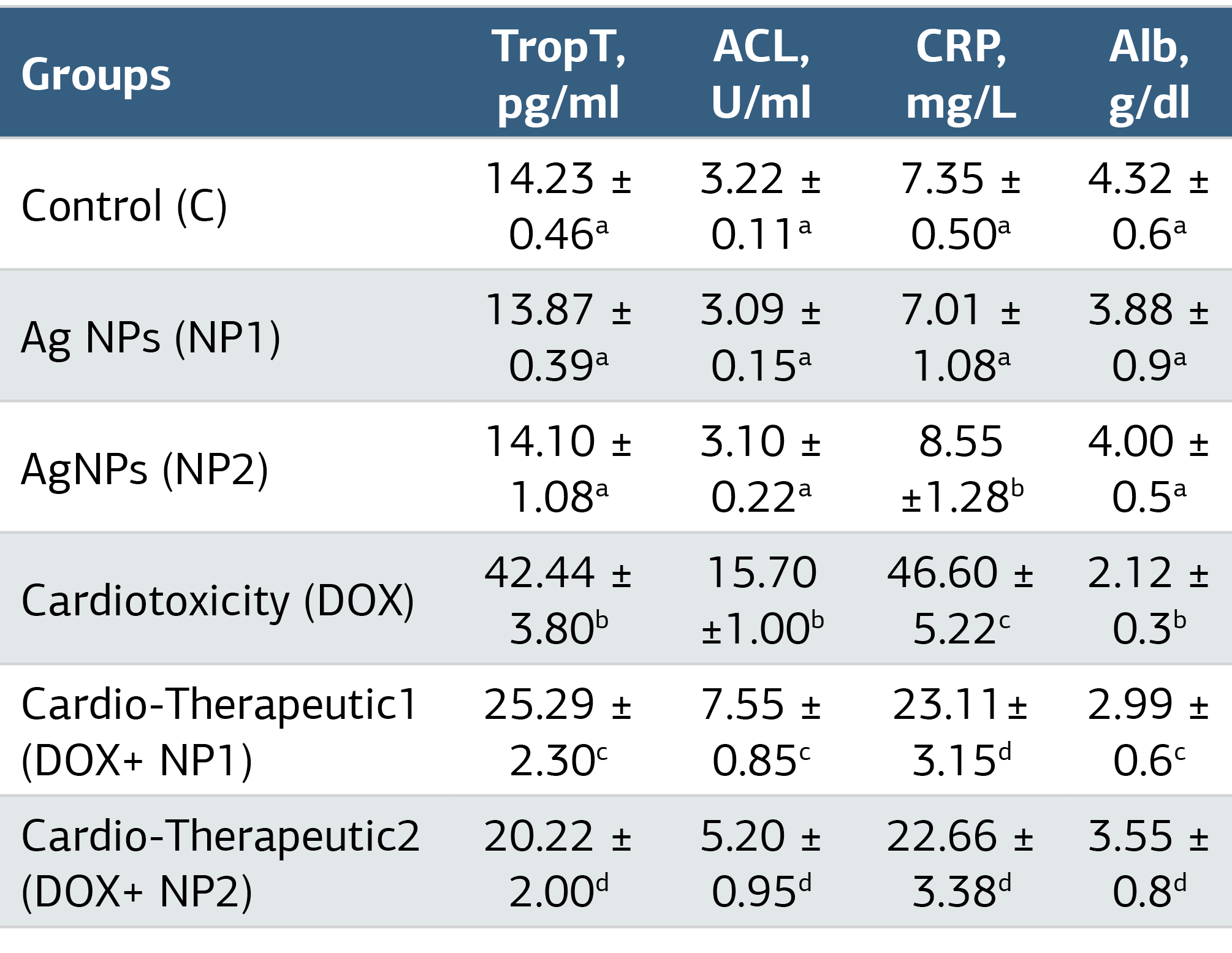
*Data are expressed as mean +/- SE , (n = 10). Significance was made using One-Way ANOVA test (LSD). Means that have different letters are significantly at (P ≤ .05)
Table 2. Effect of AgNPs administration on serum levels of cardiac enzymes in DOX-induced cardiotoxicity
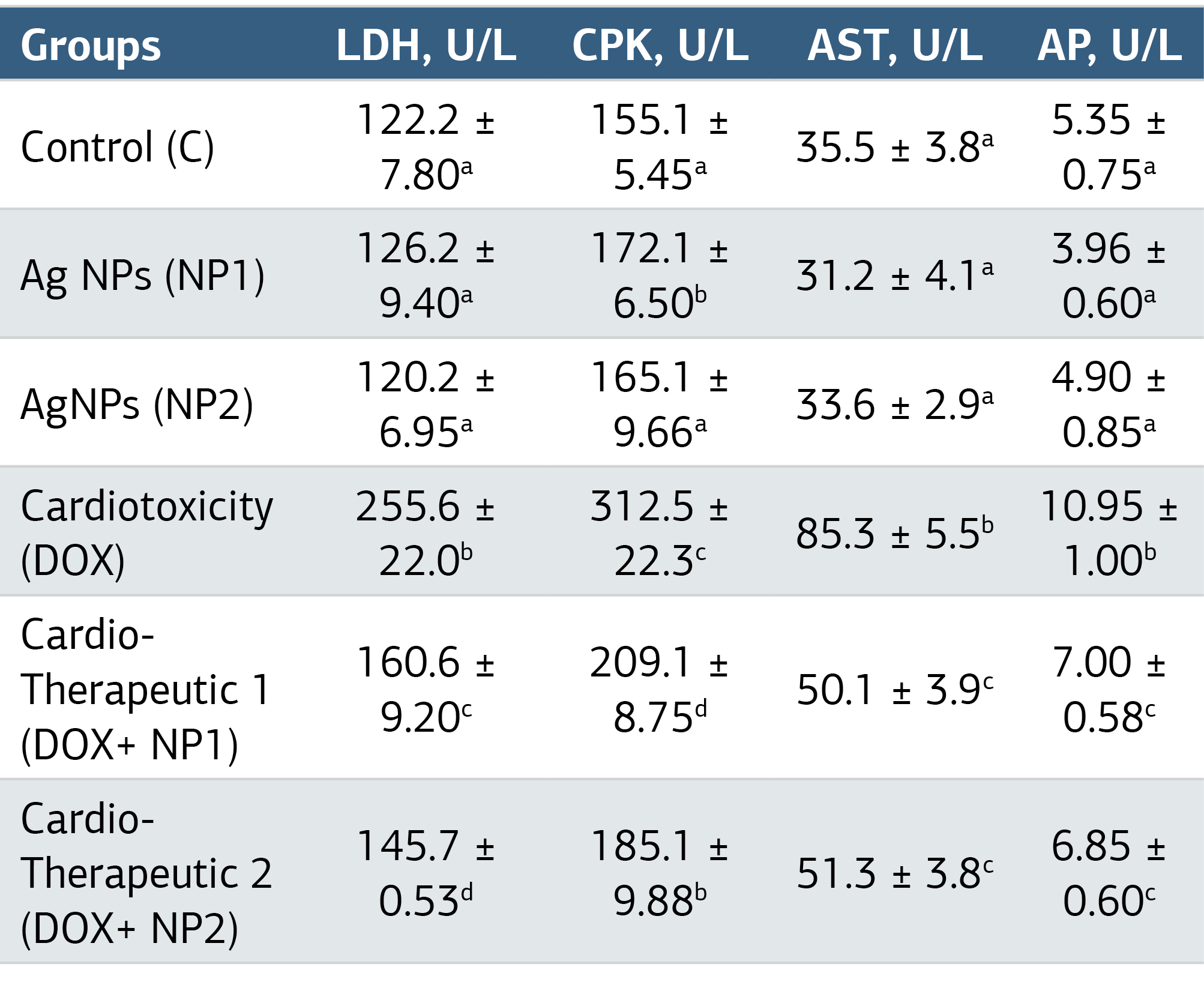
Data are expressed as mean+/- SE, (n = 10). Significance was made using one Way ANOVA test (LSD). Means that have different letters are significantly at (P ≤ .05)
Figures
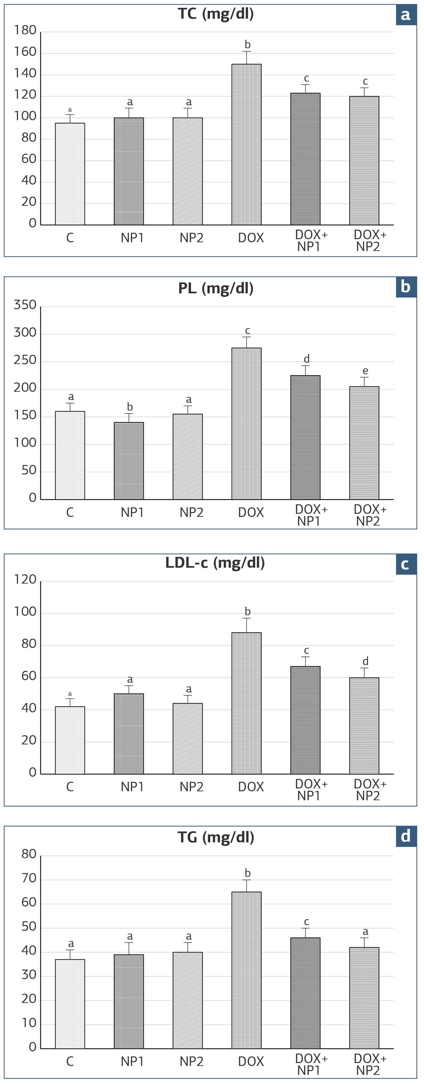
Figure 1. Effect of AgNPs Administration on Serum Lipid Profiles in DOX-Induced Cardiotoxicity
DOX, doxorubicin; NP1, silver nanoparticles (4 mg/kg b.w); NP2, silver nanoparticles (8 mg/kg b.w). Data are expressed as means (P ≤ .05). The P values were calculated using a one-way ANOVA test
Additional Information
Publisher’s Note
Bayrakol MP remains neutral with regard to jurisdictional and institutional claims.
Rights and Permissions

This work is licensed under a Creative Commons Attribution-NonCommercial 4.0 International License (CC BY-NC 4.0). To view a copy of the license, visit https://creativecommons.org/licenses/by-nc/4.0/
About This Article
How to Cite This Article
Nesrin I. Tarbiah, Fares K. Khalifa, Nuha A. Alkhattabi, Reem F. Ghazali, Reem Y. Alzahri, Sahar A. Alkhodair, Reham A. Shindi, Ahd A. Mansour. Immunomodulatory potential of silver nanoparticles and therapeutic effect against doxorubicin-induced-cardiotoxicity in rats. Ann Clin Anal Med 2024;15(3):169-175. doi:10.4328/ACAM.22032
Publication History
- Received:
- 03.11.2023
- Accepted:
- 11.12.2023
- Published Online:
- 12.01.2024
- Printed:
- 01.03.2024