
Abstract
IntroductionPulmonary hypertension (PH) is a potentially fatal condition associated with increased pulmonary vascular resistance and elevated right ventricular pressure after hematopoietic stem cell transplantation (HSCT). Case PresentationHerein we report a 3-year-old girl with stage-IV neuroblastoma who developed PAH at the day 52nd after autologous HSCT. ConclusionShe had good response to oxygen, diuretics, endothelin receptor antagonist and sildenafil.
Keywords
Introduction
Pulmonary arterial hypertension (PAH) is an unusual and life-threatening condition associated with increased pulmonary vascular resistance and elevated right ventricular pressure.1-2 PAH is defined by a pulmonary artery pressure (PAP) that exceeds 25 mmHg at rest and over 30 mmHg during activity.3 Elevated pulmonary artery pressures could cause permanent changes in the pulmonary vasculature, right ventricular failure and death. At the initial time, PAH may be asymptomatic or present with non-specific symptoms such as respiratory distress, fatigue. Therefore, the diagnosis is very challenging, and it could be mis-diagnosed.1,3-5 The diagnosis requires a high degree of suspicion.5 The most commonly reported pathological types of PH in HSCT recipients are pulmonary arterial hypertension (PAH) and pulmonary veno-occlusive disease (PVOD), depending on the site of endothelial injury.1,4 PH therapy can be disease specific or supportive.2 At this moment, the drugs used to treat PH are all accepted for PAH.2,4 Early diagnosis and timely treatment could allow improved clinical outcome with normalisation of PAP.5
Case Presentation
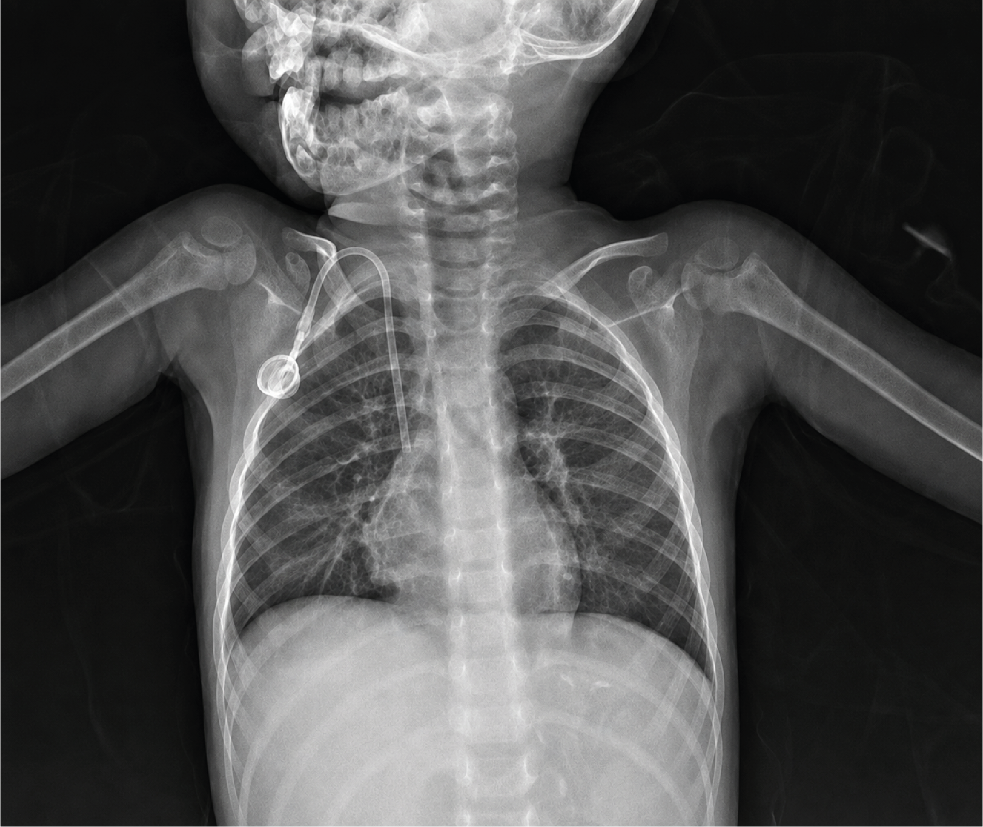
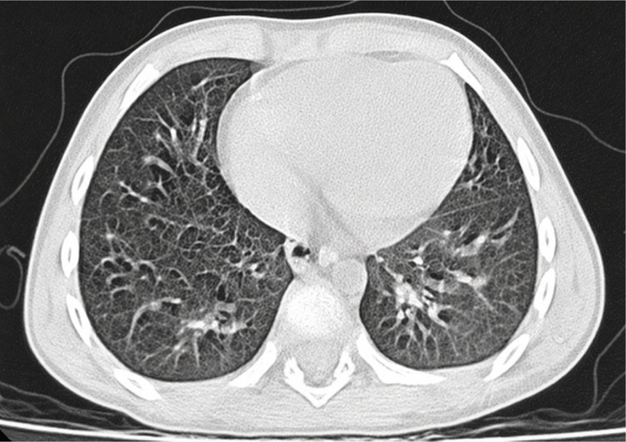
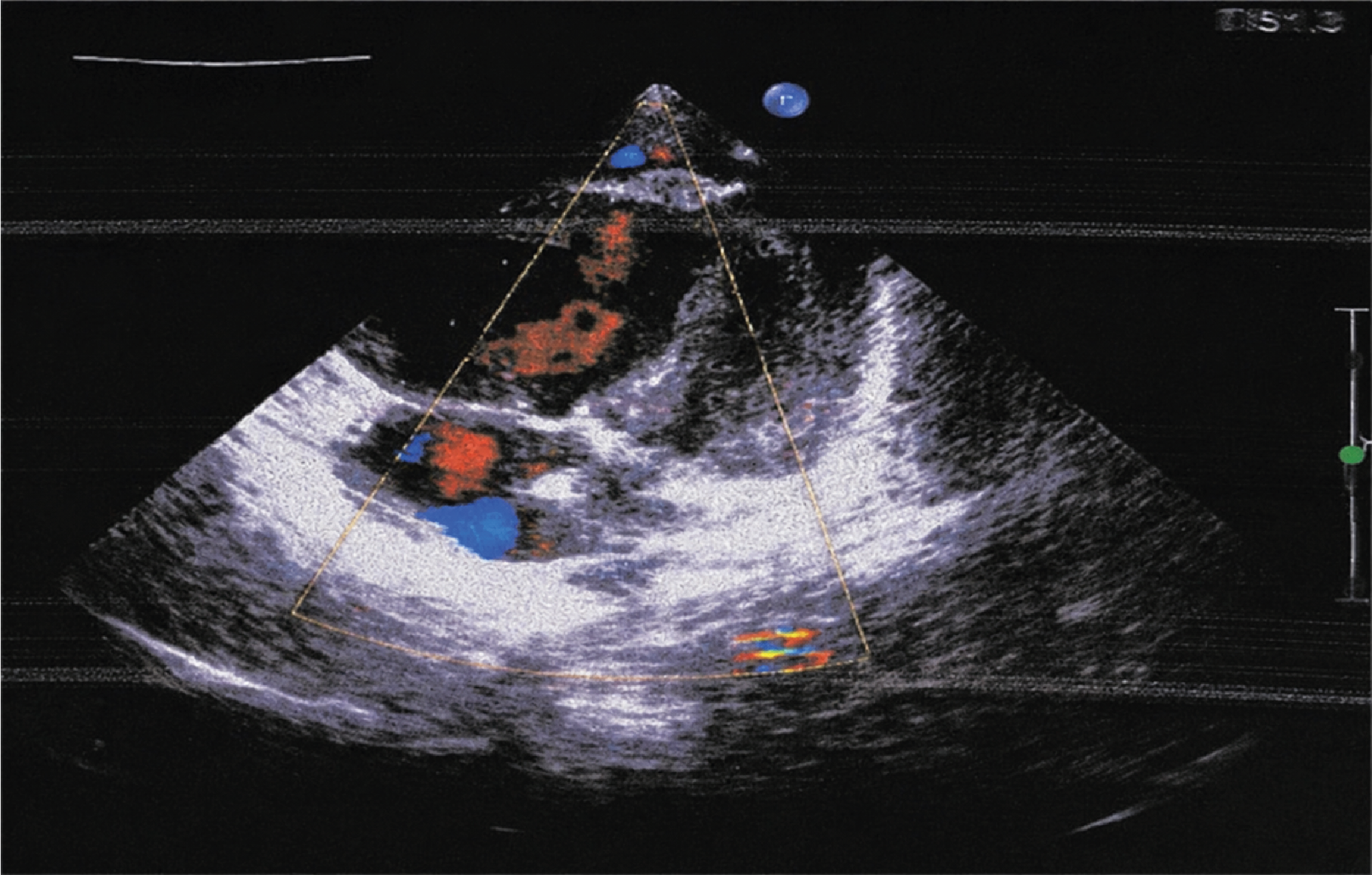
A 3 year-old female with stage IV neuroblastoma achieved partial remission after 8 course rapid-cojec. She underwent autologous transplant with conditioning regimen of busulfan and melphalan. Neutrophile and platelet engraftments were administered on day +25 and +35 respectively. At day +52, she presented with cough, anorexia, vomiting, dyspnea, and fatigue. Physical examination showed the following: temperature 37.5oC, pulse rate 160 bpm, blood pressure 95/60 mmHg and respiratory rate 45 bpm, with nasal, intercostal and subcostal retraction. Lung sounds increased at both sides, and hepatomegaly was 3 cm under subcostal margin. Oxygen saturation was 85% by pulse oximeter on room air. Key lab evaluations showed increased pro-BNP: 9646 pg/ml (cutoff value < 125 pg/ml), full blood count, blood biochemistry (LDH, CRP, liver, kidney function) and coagulation tests were normal. The chest radiography and CT scan didn’t demonstrate any lesions at her lungs (Fig.1 and Fig.2). ECG revealed right axis deviation and heart ultrasound showed an increased pulmonary artery pressure (systolic 80 mmHg and mean 45 mmHg), and dilated right ventricle and right atrium (Fig.3). Before transplant, she was evaluated and the heart ultrasound examination was normal. In addition, pro-BNP was normal. At this time, her peripheral blood smear and biochemical finding revealed no evidence for microangiopathic hemolytic anemia. Therefore, PAH related to primary respiratory disease or chronic thromoboembolic pulmonary hypertension was further excluded. The patient was diagnosed secondary PAH to HSCT, and was treated with diuretics (furosemide 1 mg/kg/day + spironolactone 1 mg/kg/day), endothelin receptor antagonist (bosetan: 2 mg/kg every 12 hours), enalapril (0.1 mg/kg/day), digoxin (30 mcg/kg/day, divided three times) and sildenafil (3 mg/kg/day, divided three times). After 18 days, her pulmonary artery pressure returned normal value. Her heart rate and pro-BNP returned normal. She did not have hepatomegaly and respiratory distress anymore. We stopped oxygen and digoxin, continued to use diuretics + sildenafil + bosetan + enalapril for one month. Then, she received radiotherapy at her tumor bed with 21.6 Gy. After finishing radiation, she was discharged and used 13 cis-retino acid for 6 months according to treatment regimen in high risk neuroblastoma. One year later, she is still healthy.
This case report was prepared in accordance with the CARE guidelines.
Discussion
The first case with PAH as a complication after transplant was reported in 1984 by Troussard et al.6 This was a 12-year-old boy who underwent HSCT for relapsed acute lymphoblastic leukemia and was reported autopsy findings consistent with PH. Since then, there were some other cases were reported.6
The lung is a major target organ for infection, toxicity and inflammation prior and after allogeneic and autologous HSCT. After transplantation, there are some common pulmonary complications, such as infectious pneumonia, acute respiratory distress syndrome, pulmonary edema, diffuse alveolar hemorrhage, heart failure, interstitial pneumonitis, idiopathic pneumonia syndrome, bronchiolitis obliterans, and organizing pneumonia.1 PH is rarely included in the differential diagnosis of respiratory distress post-transplantation and can be easily omitted.
Early identification and diagnosis of PAH is very important so that we could provide the right treatment immediately before right-sided heart failure and irreversible cardiac compromise.1 In HSCT patients, family history could be unrevealing because most risk factors are associated with HSCT therapy or complication in these patients.1 These factors could be infection, malignancy, immune dysregulation, epithelial damage (caused by radiation, conditioning regimen), endothelial damage, genetic/biochemical, pro-inflammatory cytokine release and cell-mediated damage.5
The initial respiratory symptoms of PAH are shortness of breath, fatigue, weakness, hypoxemia, and these signs could be vague and difficult to attribute specifically to PAH in the post-transplantation patient. Edema and ascites could develop in the later stages from increased venous congestion.7
In our case, at D+52 after autologous transplant, the patient was presented with cough, anorexia, vomiting, dyspnea, and fatigue. Examination showed increased respiratory rate and increased lung sound at both sides. Her oxygen saturation was 85%. With these symptoms, at the beginning, we thought patient had viral pneumonia. However, after receiving the normal result of chest radiography, we had to think of another diagnosis. Particularly when combining with other symptoms, such as: elevated heart rate, hepatomegaly, we ordered some tests including heart ultrasound, ECG, pro-BNP, LDH, CRP, liver, kidney function and bilirubine. With the result of ECG showed right axis deviation, heart ultrasound demonstrated an increased pulmonary artery pressure (systolic 80 mmHg and mean 45 mmHg), dilated right ventricle and right atrium and pro-BNP increased (9646 pg/ml), we concluded that the patient had PAH secondary to HSCT. For this case, in the beginning, we did not expect to diagnose PAH with her symptoms (since this was the first case with PAH we met in our hospital), and we had to wait the result of some lab tests. However, the time to wait PAH diagnosis was short and could be acceptable. Analysing the reasons to cause PAH in our case, we thought about conditioning regimen with busulfan and melphalan which have been described to cause endothelial injury.5
Regarding treatment, the initial purpose of therapy aims at optimizing cardiac function, especially if PAH has resulted in right ventricular compromise. The treatment could be a combination of some medicines, such as diuretics, afterload-reducing agents, intravenous inotropes, pulmonary vasodilator therapy, oxygen therapy, inhaled nitric oxide, calcium channel blockers, phosphodiesterase-5 inhibitors, endothelin receptor antagonists, prostanoids. For our patient, she was treated with diuretics, endothelin receptor antagonist, enalapril, digoxin and sildenafil. After 18 days, her pulmonary artery pressure returned normal value. We stopped oxygen and digoxin and continued to use diuretics + sildenafil + bosetan + enalapril for one month. She had good response.
According to Ozyoruk, he reported one case with high risk neuroblastoma appeared PAH after autologous transplant with bul+mel conditioning. The patient was treated with a similar therapy, including enalapril, furosemide, sildenafil and iloprost inhaler, and he had good response. It has been reported that sildenafil effectively reduces pulmonary artery pressure, improves cardiac function and does not cause adverse reactions in children.4 However, if diagnosis delays, the prognosis is very poor. Levy reported 22 patients who were diagnosed severe pulmonary hypertension after HSCT with a fatal outcome in 32%. All the patients who were diagnosed died later, whereas pulmonary hypertension always resolved in those who had a rapid diagnosis allowing timely administration of targeted therapy.5 Therefore, the most important thing is how to diagnose timely and provide treatment properly to normalise pulmonary pressure and improve survival.3,5
Aggressive and timely up-front combination therapy allowed normalisation of pulmonary pressure and improved survival.
Limitations
The findings of this report are based on the clinical course of a single patient and may not be applicable to other patients undergoing HSCT.
Conclusion
Pulmonary arterial hypertension is rare complication of cardiorespiratory failure after pediatric hematopoietic stem cell transplant. It is crucial to diagnose and provide treatment immediately before right-side heart failure and irreversible cardiac compromise.
Declarations
Animal and Human Rights Statement
All procedures performed in this study were in accordance with the ethical standards of the institutional and/or national research committee and with the 1964 Helsinki Declaration and its later amendments or comparable ethical standards.
Informed Consent
Written informed consent for publication was obtained from the patient’s legal guardian.
Data Availability
The datasets used and/or analyzed during the current study are not publicly available due to patient privacy reasons but are available from the corresponding author on reasonable request.
Conflict of Interest
The authors declare that there is no conflict of interest.
Funding
None.
Abbreviations
HSCT: Hematopoietic stem cell transplantation
PAH: Pulmonary arterial hypertension
PAP: Pulmonary artery pressure
PH: Pulmonary hypertension
References
- Dandoy CE, Hirsch R, Chima R, Davies SM, Jodele S. Pulmonary hypertension after hematopoietic stem cell transplantation. Biol Blood Marrow Transplant. 2013;19(11):1546-1556. doi:10.1016/j.bbmt.2013.07.017
- Beghetti M, Gorenflo M, Ivy DD, Moledina S, Bonnet D. Treatment of pediatric pulmonary arterial hypertension: a focus on the NO-sGC-cGMP pathway. Pediatr Pulmonol. 2019;54(10):1516-1526. doi:10.1002/ppul.24442
- Houtchens J, Martin D, Klinger JR. Diagnosis and management of pulmonary arterial hypertension. Pulm Med. 2011;2011:845864. doi:10.1155/2011/845864
- Ozyoruk D, Kibar AE, Surucu M, et al. Pulmonary arterial hypertension in a child with stage IV neuroblastoma after autologous hematopoietic stem cell transplantation and review of the literature. Pediatr Transplant. 2015;19(7).
- Levy M, Moshous D, Szezepanski I, et al. Pulmonary hypertension after bone marrow transplantation in children. Eur Respir J. 2019;54(5). doi:10.1183/13993003.00612-2019
- Troussard X, Bernaudin JF, Cordonnier C, et al. Pulmonary veno-occlusive disease after bone marrow transplantation. Thorax. 1984;39(12):956-957. doi:10.1136/thx.39.12.956
- Galie N, Hoeper MM, Humbert M, et al. Guidelines for the diagnosis and treatment of pulmonary hypertension: the Task Force for the Diagnosis and Treatment of Pulmonary Hypertension of the European Society of Cardiology and the European Respiratory Society, endorsed by the International Society of Heart and Lung Transplantation. Eur Heart J. 2009;30(20):2493-2537.
Figures
Additional Information
Publisher’s Note
Bayrakol MP remains neutral with regard to jurisdictional and institutional claims.
Rights and Permissions

This work is licensed under a Creative Commons Attribution-NonCommercial 4.0 International License (CC BY-NC 4.0). To view a copy of the license, visit https://creativecommons.org/licenses/by-nc/4.0/
About This Article
How to Cite This Article
Tran Kiem Hao, Nguyen Thi Kim Hoa, Bui Binh Bao Son. Pulmonary arterial hypertension in a child with stage IV neuroblastoma after autologous stem cell transplant: A Case Report. Ann Clin Anal Med 2024;15(Suppl 1):32-35. doi:10.4328/ACAM.22056
Publication History
- Received:
- 21.11.2023
- Accepted:
- 22.12.2023
- Published Online:
- 26.12.2023
- Printed:
- 20.03.2024