
Abstract
Introduction Sarcoidosis is a multisystem inflammatory illness with no recognised cause. Non-caseating granulomas are a characteristic of this illness. Case PresentationBilateral hilar lymphadenopathy and lung infiltration are the most typical presentations. However, the disease is quite diverse and has an unexpected clinical outcome. Less often, there is bone involvement but considered serious and significant. Involvement of the bone is present in 1–15% of sarcoidosis patients. Hand involvement in sarcoidosis is uncommon and Sarcoid lesions of the hand occur in only around 0.2 percent of cases. ConclusionWe describe a case of sarcoidosis in Palestine with a history of finger swelling, radiographic and magnetic resonance imaging (MRI) findings showed bony involvement for increasing the awareness of the medical team and population.
Keywords
Introduction
Sarcoidosis is an inflammatory disorder with non-caseating granulomas that affects multiple organ systems and has no known cause, tuberculosis, leprosy, syphilis, wegener granulomatosis, multiple myeloma, lymphoma, and other diseases are among the differential diagnoses of cases.1 Bilateral hilar lymphadenopathy and lung infiltration are the most typical presentations; however, the disease is quite diverse and has an unexpected clinical outcome.2 There are frequent musculoskeletal symptoms. Less often, there is bone involvement.3 Involvement of the osseous system is present in 1–15% of sarcoidosis patients.2,4
Hand involvement in sarcoidosis is uncommon and when it occurs, it usually presents as tenosynovitis, dactylitis, nodules, and osteoarticular bone deterioration.5 Occasionally, however, sarcoidosis is diagnosed based on irregular symptoms. Soft-tissue swelling in the hands, for example, without serious systemic disease is an uncommon symptom, but it may suggest subcutaneous sarcoid involvement. These swellings can appear in a variety of ways, posing challenges for orthopedic surgeons who encounter them.1
Sarcoid lesions of the hand occur in just around 0.2 percent of cases. Bone lytic lesions are seen in the majority of these cases,6 with tendon, muscle, and subcutaneous soft-tissue involvement occurring infrequently. In addition, sarcoid dactylitis, which is characterized by non-caseating granulomas invading the phalanges and adjacent soft tissue, is well recognized.3 Skin involvement is seen in 9 percent to 37 percent of systemic sarcoidosis patients, subcutaneous involvement is a less common manifestation.1 Diagnostic imaging methods are crucial. Radiological examinations indicated cortical anomalies, sclerotic or destructive lesions (including also joints), and cystic and punched-out lesions.2
In Palestine, sarcoidosis is commonly recognized as a rare disorder, with bone involvement even rarer. Herein, we report a case of hand bony involvement of sarcoidosis in a 58-year-old woman. This case was diagnosed using clinical findings, and correlated radiographic imaging.
Case Presentation
A 58-year-old lady presented with progressive, painful swellings in the left index finger of her hand for four months. She had a pre-existing diagnosis of sarcoidosis and had been consistently taking medication. Although she discontinued corticosteroids 3 month ago, her finger was swollen completely (Figure 1). It was tough, tender, firm, and had well-demarcated borders. Routine laboratory studies indicated a uric acid level of 4.3mg/dl, urea of 33.6 mg/dl, normal triglyceride, and normal kidney and liver function test and total cholesterol. Hand X-ray and MRI were done. The findings were matching with dactylitis as a diagnosis for this case (Figure 2). Chest X-ray was also done and confirmed the diagnosis (Figure 3).Ethical ApprovalEthical committee approval of the polytechnic university was taken, and a written consent form the patient was also taken, the patient also given her consent for her image and clinical information to be reported in the journal, the patient understand that her name will not be published.
This case report was prepared and reported in accordance with the CARE (Case Report) reporting guideline.
Discussion
Most patients are aged 20–45 years, although recently an increase in new onset of sarcoidosis in people older than 60, especially women, has been reported.2 Sarcoidosis can affect multiple organs, most often the lungs, lymph nodes, eyes, and skin. Bone involvement is rare7 which is present in 1–15% of sarcoidosis patients.2,4 Skin involvement is seen in 9 percent to 37 percent of systemic sarcoidosis patients. Subcutaneous involvement is a less common manifestation.1 Sarcoid lesions of the hand occur in just around 0.2 percent of cases. Bone lytic lesions are seen in the majority of these cases,6 with tendon, muscle, and subcutaneous soft-tissue involvement occurring infrequently. In addition, sarcoid dactylitis, which is characterized by non-caseating granulomas invading the phalanges and adjacent soft tissue, is well recognized.3
The most frequent manifestation of the bone in sarcoidosis patients is dactylitis. It appears in about 90% of the cases. The condition is generally bilateral and asymmetric, occurring most often in the middle and distal phalanges of the second and third digits.8 Various circumstances can lead to dactylitis discovery. It is the most common clinical presentation, with swelling of one or more fingers that become purplish and may or may not be painful. Fluid accumulation in the skin and subcutaneous tissue, along with nail splitting, has also been reported.3 In rare cases, it is discovered suddenly when radiography is performed for any other indications.
In our case, there is a female patient who had bone changes that were characteristic of osseous sarcoidosis and was diagnosed using an X-ray of the hand, which showed dactylitis. MRI showed extensive soft tissue mass lesion involving the whole index finger and extensive abnormal right bone marrow.
In terms of laboratory testing, higher erythrocyte sedimentation rate, and increased acetylcholinesterase levels are observed in a variable and nonspecific manner, independent of where the disease is located.4
In our case, the patient doesn’t have any abnormal laboratory test results. However, in radiograph images, the lesion is found mainly in the index phalange. This kind of lesion most often occurs bilateral and asymmetric.3 MRI mainly provides detailed information about fluid accumulation in the bone and mainly bone marrow and extension to soft tissues.7 The involvement of small bones in the hands may be evident with MRI, as MRI may show a much more extensive process than one would see on the radiographs.7
Limitations
This report is limited to a single case.
Conclusion
This presented case demonstrate a bone lesions typical of osseous sarcoidosis. These lesions were severe and may cause noticeable bone damage with fluid accumulation in the soft tissues. This case is rare in this manifestation of sarcoidosis. The possibility of osseous sarcoidosis should be considered when a patient with radiographic signs also has a suggestive clinical context. With the correct diagnosis, suitable treatment can be provided early on, thereby avoiding unnecessary surgery that compromises the functional prognosis.From an Orthopedic Point of ViewThe significance of this case is that, although sarcoidosis commonly presents with typical signs and symptoms, unusual manifestations should not preclude the diagnosis. An atypical soft-tissue swelling in the hands may arouse the suspicion of sarcoid involvement of subcutaneous tissue so orthopedics must be aware of that all the time.
Declarations
Animal and Human Rights Statement
All procedures performed in this study were in accordance with the ethical standards of the institutional and/or national research committee and with the 1964 Helsinki Declaration and its later amendments or comparable ethical standards.
Informed Consent
Written informed consent was obtained from the patient for publication of this case and accompanying images.
Data Availability
The datasets used and/or analyzed during the current study are not publicly available due to patient privacy reasons but are available from the corresponding author on reasonable request.
Conflict of Interest
The authors declare that there is no conflict of interest.
Funding
None.
Acknowledgements
The authors thank Dr.Abdulrahman Marridi for their efforts and cooperation.
Abbreviations
MRI: Magnetic resonance imaging
References
- Sheila M, Stefano S, Elena BB, Roberto G. Unusual subcutaneous swellings on the hand as a primary presenting feature of sarcoidosis. J Rheumatol. 2011;38(3):519-521.
- Kucharz EJ. Osseous manifestations of sarcoidosis. Reumatologia. 2020;58(2):93-100. doi:10.5114/reum.2020.95363
- Aptel S, Lecocq-Teixeira S, Olivier P, Regent D, Teixeira PG, Blum A. Multimodality imaging evaluation of musculoskeletal sarcoidosis: imaging findings and literature review. Diagn Interv Imaging. 2016;97(1):5-18. doi:10.1016/j.diii.2014.11.038
- Kobak S. Sarcoidosis: a rheumatologist’s perspective. Ther Adv Musculoskelet Dis. 2015;7(5):196-205. doi:10.1177/1759720x15591310
- Liew SK, Park JK, Gong HS. Intrinsic contracture of the hands in sarcoid myopathy. J Rheum Dis. 2023;30(3):204-207. doi:10.4078/jrd.2023.0007
- Duckworth AD, Hill AT, Beggs I, Patton JT, Salter DM, Porter DE. Sarcoidosis presenting as a proximal phalangeal bony swelling: a case and review of the literature. Hand (N Y). 2010;5(1):90-94. doi:10.1007/s11552-009-9207-6
- Moore SL, Teirstein AE. Musculoskeletal sarcoidosis: spectrum of appearances at MR imaging. Radiographics. 2003;23(6):1389-1399. doi:10.1148/rg.236025172
- Jeffrey AS, Jakob IM, Nehal S, et al. Osseous sarcoidosis: clinical characteristics, treatment, and outcomes—experience from a large academic hospital. Semin Arthritis Rheum. 2014;44(3):371-379.
Figures
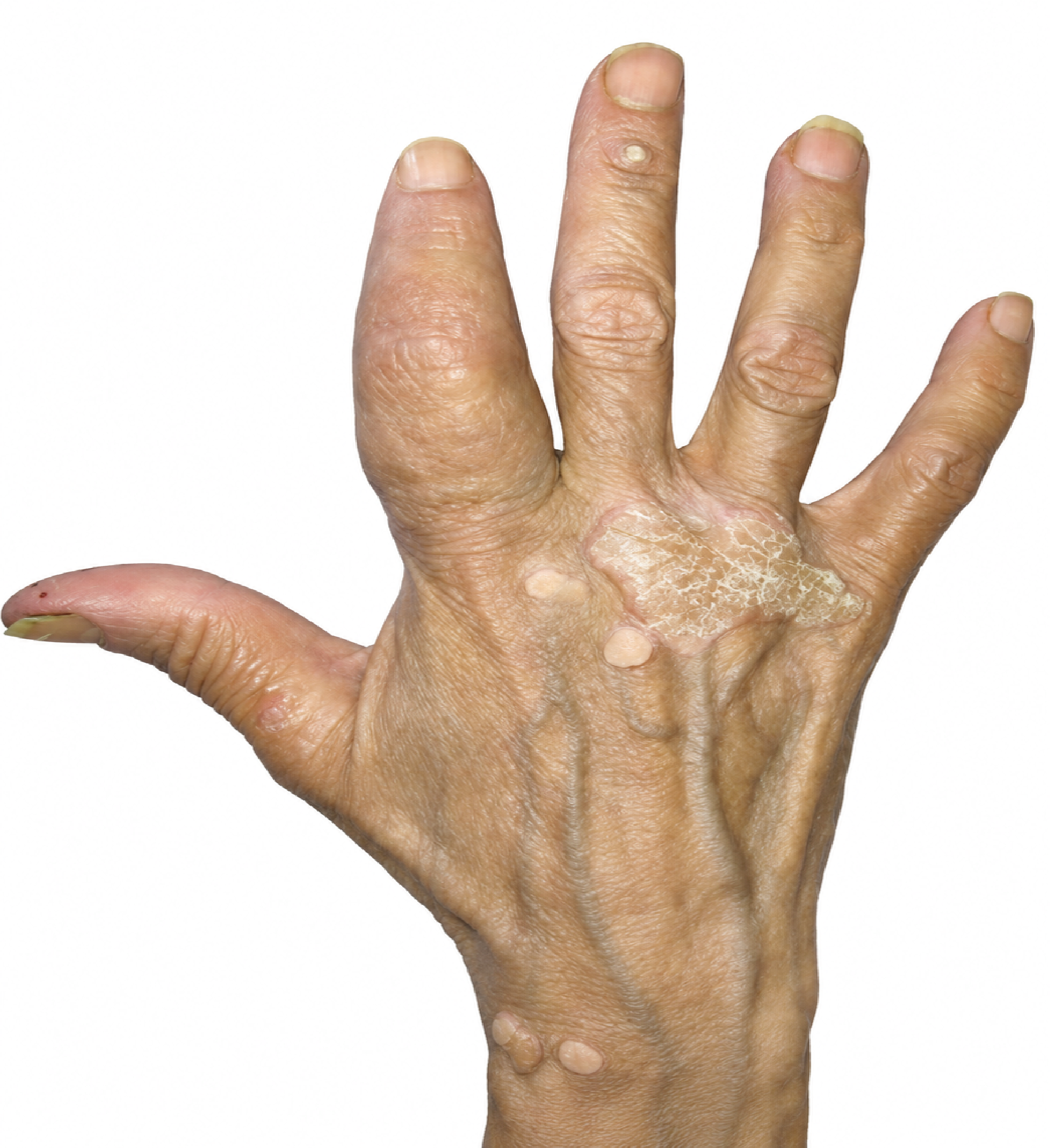
Figure 1. The Swollen Finger of the Patient
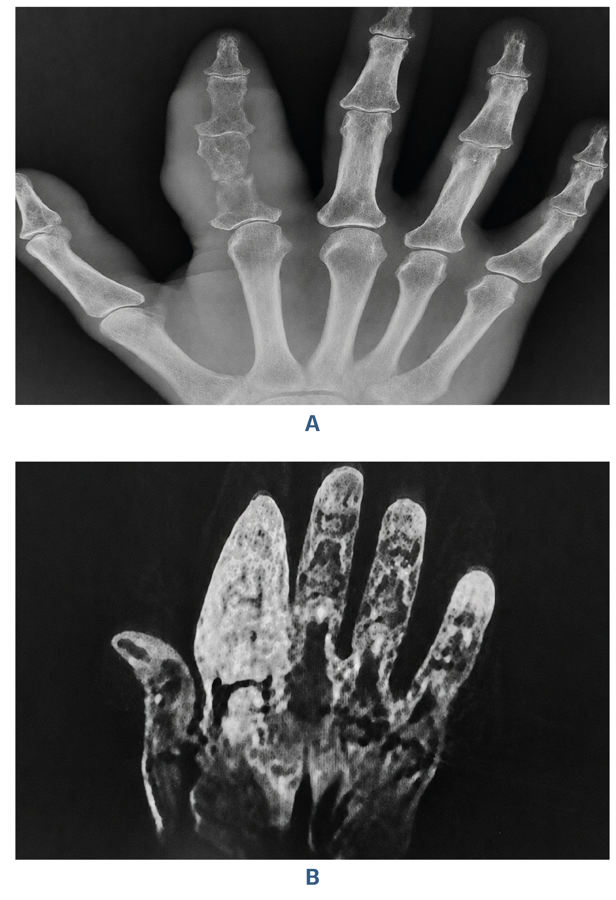
Figure 2. X- RAY of the Hand
A; dactylitis, inflammation of the index digit. B; MRI, extensive soft tissue mass lesion involving the whole index finger and extensive abnormal right bone marrow.
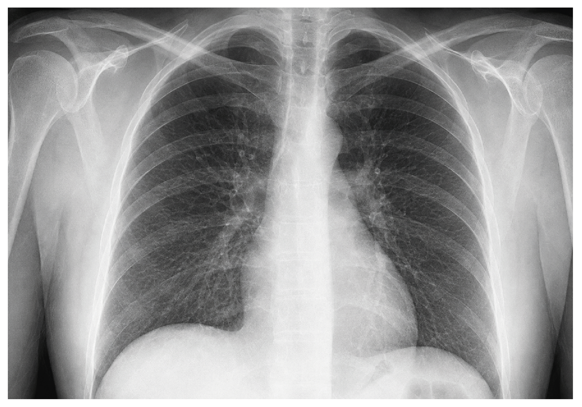
Figure 3. Chest X-ray, Bilateral Hilar Lymphadenopathy
Additional Information
Publisher’s Note
Bayrakol MP remains neutral with regard to jurisdictional and institutional claims.
Rights and Permissions

This work is licensed under a Creative Commons Attribution-NonCommercial 4.0 International License (CC BY-NC 4.0). To view a copy of the license, visit https://creativecommons.org/licenses/by-nc/4.0/
About This Article
How to Cite This Article
Neveen Shalalfa, Saleh Shalalfa, Zuhair Nazar Mohammed, Salam Noman Hattawi, Javid Mohammadzadeh Azarabadi. Rare case of hand bony involvement of sarcoidosis in a palestinian woman: A Case Report. Ann Clin Anal Med 2024;15(Suppl 1):39-42. doi:10.4328/ACAM.22064
Publication History
- Received:
- 05.12.2023
- Accepted:
- 15.01.2024
- Published Online:
- 02.02.2024
- Printed:
- 20.03.2024