
Abstract
AimCOVID-19 prevalence in clinical practice is still significantly high even if it isn’t as a pandemic. This study aims to investigate the relationship between chest computed tomography (CT) severity scores and hemogram parameters in inpatients due to SARS-CoV2.MethodsThis study is a retrospective analysis of 70 inpatients who were hospitalised due to COVID-19 in several services of 750-bed referral hospital in 2021. The positive SARS-CoV-2 RT-PCR test, available thorax CT scans, and data of platelet (PLT), White blood cells(WBC), Procalcitonin(PCT), (Erythrocyte Sedimentation Rate (ESR), International Normalized Ratio (INR), Ferritin, and Fibrinogen) obtained initially and 1 week after hospitalisation were analyzed. The associations between semiquantitative radiological score and age, gender, comorbidity, and clinical and laboratory parameters were examined. CT imaging was independently reevaluated by a radiologist who was blinded to the patients’ age, laboratory and clinical findings.ResultsThe mean age of the patients was 60.20 ± 12.62 years and CT severity score of them was 16.24 ± 6.47. A positive significant correlation was observed between the CT severity score and PLT (r = 0.263, P = .028) as well as PCT (r = 0.292, P = .014) in the first visit and the second one after a week (PLT (r = 0.377, P = .001) and PCT (r = 0.375, P = .001).ConclusionWe believe that using readily available measures like PLT and PCT can be valuable in estimating the extent of lung damage in COVID-19 patients with viral pneumonia, especially when CT scans are not feasible.
Keywords
Introduction
COVID-19 is a contagious virus that causes respiratory tract infections, with the development of viral pneumonia being the main determinant of severity of this disease. As it spread rapidly in large populations at the maximum point of infection, WHO declared it a “pandemic”. The clinical feature and course of the infection vary based on individuals’ age, comorbidity, immune condition and other risk factors.1
The primary goal should be quick diagnosis using available tests and effective treatment for critical cases. In this respect, Polymerase Chain Reaction (PCR) test, laboratory parameters and imaging methods are very important in detecting COVID-19.2 However, the importance of simple tests that result quickly when the PCR test takes about 3-8 hours to complete and can be found everywhere is vital in cases where it is necessary to act quickly, such as a pandemic.
The biomarkers associated with the host response may play an effective role in stratifying the risk for disease progression, given the burden of the epidemic on health systems and economies. Cost-effective, fast, reliable and widely available blood parameters are needed to detect the disease, and it is very valuable to establish their relationship with the severity of lung involvement.3 Although Computarized Tomography (CT) scan is essential for diagnosing cases with viral pneumonia and assessing the severity of involvement, which impacts mortality, it is unfortunately not readily accessible under all circumstances.1
Therefore, in this study we aimed to determine whether a simple test can predict the severity of lung involvement measured by CT scale in COVID-19 patients.
Materials and Methods
Study PopulationThis is a retrospective study that reviewed the medical records of patients older than 18 years with SARS-CoV-2 PCR positivity who had been hospitalized in a referral hospital in the capital city of the country between 01.01.2021 and 30.11.2021. We excluded 72 patients with a negative PCR test, 5 patients younger than 18 years old, 67 patients whose blood parameters were not measured simultaneously with thorax CT, and 23 patients for whom sufficient clinical data could not be obtained. Additionally, we excluded 41 patients who did not have a second control blood test within 1 week and 7 patients who died during hospitalization. Ultimately, data from 70 COVID-19 patients who met the inclusion criteria were obtained for the study.Data CollectionThe cases had initial RT-PCR positivity in nasopharyngeal swab samples for the diagnosis of COVID-19. Thorax CT scans and blood tests (complete blood count, procalcitonin, C-reactive protein (CRP), D-dimer, International Normalized Ratio (INR), ferritin, and fibrinogen), which were evaluated simultaneously with the CT scan, as well as the same blood test results checked for the second time in the first week of hospitalization, were extracted from medical records.Chest CT and CT Image ReviewThoracic CT scans at the first admission to the hospital were interpreted using the scoring system used by Ran Yang et al., and the CT severity score was calculated. The CT scans of the cases were independently re-evaluated by a radiologist who was blinded to both demographic information and clinical issues. The lungs were divided into 20 segments, with 10 segments for the right lung and 10 segments for the left lung, according to Boyden bronchial segmentation. The total CT score was calculated as the sum of segmental scores, with 0 points assigned for normal segments, 1 point for segments with ≤50% involvement, and 2 points for segments with >50% involvement. The total score ranged from 0 to 40.4Ethical ApprovalThis study was approved by the Ethics Committee of Dışkapı Yıldırım Beyazıt Training and Research Hospital (Date: 27.12.2021, Decision No: 127/15).Statistical AnalysisIn the analysis of the data, IBM SPSS Statistics for Windows, Version 23.0 (Armonk, NY: IBM Corp.) was used. There are three methods for determining whether the distribution is normal: skewness and kurtosis coefficients, histogram and Normal Q-Q graph, and Shapiro-Wilk and Kolmogorov-Smirnov tests. The frequency, percentage, mean and standard deviation values were included for information about the CT score and blood values of the patients, and Pearson and Spearman correlation analysis was performed between CT scores and blood test results. The level of significance was set as P = .05.Reporting GuidelinesThis study was reported in accordance with the STROBE guideline.
Results
A total of 70 patients (51.4% male, mean age: 60.20 ± 12.62 years) were included in the study. The mean thorax CT scores were found to be 16.24 ± 6.47. The findings regarding the blood measurements of the patients are given in Table 1. The results of the correlation between the CT scores of the patients and the hemogram measurements in the first and second examinations are shown in Table 2.
There was a significant correlation between the CT score and PLT (r = 0.263, P = .028) as well as PCT (r = 0.292, P = .014), unlike the other hemogram subparameters. A significant correlation was also found at the second hemogram subparameters of PLT (r = 0.377, P = .001) and PCT (r = 0.375, P = .001).
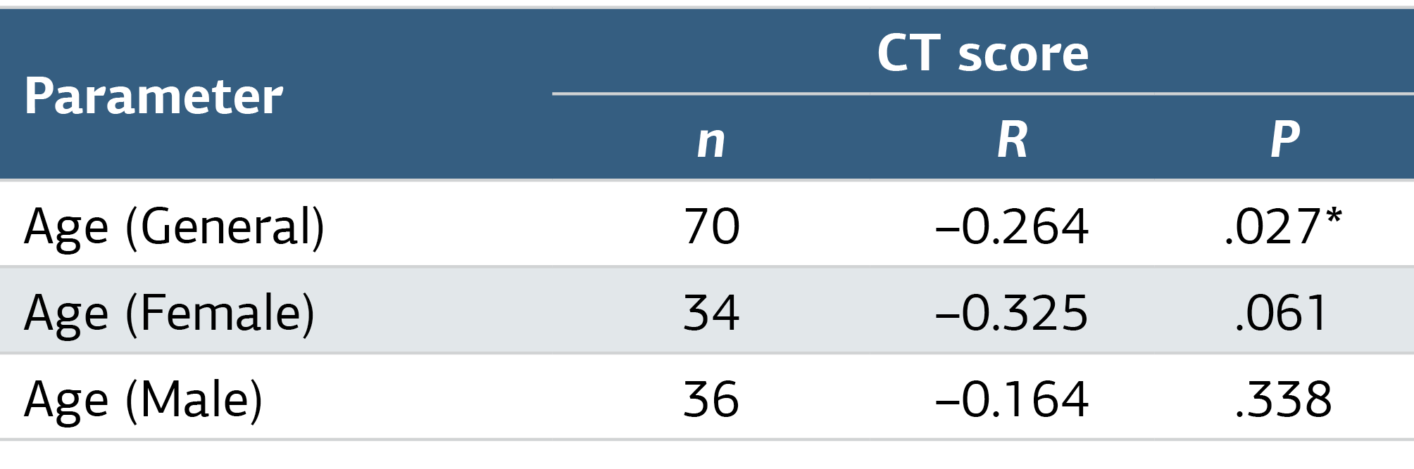
There was a low, negative and significant relationship between CT score and age (r = -0.264, P < .05). Accordingly, as age increases, CT score decreases. When examined separately by gender, there was no significant relationship between age and CT score in either women or men (t = 1.875, P = .065) (Table 3).
Discussion
It was aimed to evaluate the compatibility of CT severity score with platelet and platelet indices in viral pneumonia in the current study. We assessed the correlation between thorax CT scans and hemogram subparameters taken concurrently. We tried to examine these patients by including a week following measurements.
There was no significant relationship between the CT score of the inpatients and the laboratory measurements other than PLT and PCT obtained in the first week. A significant relationship between CT score and PLT as well as PCT was shown in the present study. However, there was a low, positive and significant relationship between the CT score and PLT and PCT. As PLT and PCT measurements increase, the CT score also increases. When the CT score was compared with the second time PLT and PCT measurements, a more significant relationship was found between the CT score and PLT and PCT.
Several studies have highlighted the importance of platelet count in COVID-19 patients. For instance, Qu et al. reported that patients presenting with a peak in PLT during the course of the disease had worse clinical outcomes. They concluded that PLT and its dynamic changes during treatment can make a recommendation about the criticality and prognosis of the disease. They suggested that this elevation in PLT may be related to the cytokine storm occurring in patients. IL-6, which is released as a result of cytokine storm, stimulates the increase of TPO levels and promotes the formation of megakaryocytes.5
Another study found that patients with severe pneumonia caused by SARS-CoV-2 had higher platelet counts compared to patients with non-COVID-19 pneumonia. Furthermore, when comparing these two groups, the 28-day mortality in the COVID-19 group was approximately twice as high as the mortality in the non-COVID group.6
In an analysis examining the correlations of routine blood parameters, viral load, and lung imaging in patients with SARS-CoV-2, hospitalized patients’ routine laboratory tests were compared to those taken 1 week later. They observed a significant increase in WBC and platelets compared to the base measurements.7
Considering the critical cases of SARS-CoV-2 pneumonia in Wuhan, it was noticed that those who died due to the disease during hospitalization had a higher PLT than those who survived.8
A review focusing on a small number of critically ill pediatric COVID-19 patients aged 2 months to 15 years treated in an Intensive Care Unit in China revealed that these children’s PLT were either within the normal range or elevated.9 Anemia and increased coagulation are significant factors that influence the prognosis of SARS-CoV-2 infection. Helin et al. reported elevated platelet and CRP levels in 68% of the patients in the intensive care unit. Additionally, the platelet count was found to be significantly higher in patients admitted to the intensive care unit compared to other patients.10
In a single-center, retrospective study conducted with 191 patients with COVID-19 in Pakistan, high PLT measurements were observed during critical phases of the infection, including the moment of admission to the hospital, hospitalization in the intensive care unit, and death.11
On the other hand, contrary to the findings of our study, there are studies in the literature that associate low PLT (thrombocytopenia) with COVID-19. Thrombocytopenia is characterized by a reduction or destruction of PLT. Some studies suggest that SARS-CoV-2 may inhibit hematopoiesis by using CD13 receptors, leading to a decrease in various blood cells, especially platelets, ultimately resulting in thrombocytopenia.12 In a study where thrombocytopenia was associated with disease severity, researchers observed that PLT increased just before reaching the severe disease stage, peaked within 11-15 days, and then declined rapidly.13 In a study of patients transferred to the intensive care unit, no significant difference in platelet count at hospitalization was found compared to those who did not require intensive care. However, they observed that platelet levels increased on the 7th day after hospitalization in patients with stable vital status, while patients who were referred to the intensive care unit consistently exhibited low PLT levels.14
It can be concluded that increased PLT and PCT in COVID-19 patients are indicators of poor prognosis, especially in healthcare centers without access to CT scans. However, further comprehensive studies are required to confirm and better understand the association between PLT parameters and the severity of COVID-19 lung damage.
Limitations
Despite overlaps with the literature, there are also contradictory resuts of the present study. It is a single-centered and retrospective, cross-sectional designed study with limited number of cases. So that, it cannot be generalized to reflect the whole population. In additon, the results couldn’t meet the expections that the inflammatory markers measured together with hemogram as guide. Still, we think that the data we obtained may be useful for future studies.
Conclusion
This study aimed to investigate the correlation between Thorax CT involvement severity scores in patients with positive PCR tests and simultaneous changes in blood biomarkers. Based on the data we obtained in our study for this purpose, as the PLT and PCT measurements increased, the CT severity score Therefore, we believe that simple hemogram subparameters can be used to identify critical, initiate early treatment, and estimate the CT severity score a positive correlation. It has been observed that multicenter, large-scale prospective studies should be warranted.
Declarations
Animal and Human Rights Statement
All procedures performed in this study were in accordance with the ethical standards of the institutional and/or national research committee and with the 1964 Helsinki Declaration and its later amendments or comparable ethical standards.
Informed Consent
Not applicable.
Data Availability
The datasets used and/or analyzed during the current study are not publicly available due to patient privacy reasons but are available from the corresponding author on reasonable request.
Conflict of Interest
The authors declare that there is no conflict of interest.
Funding
None.
Acknowledgements
We extend our gratitude to Dr. Muhammed Selim Demir, Prof. Dr. Süleyman Görpelioğlu for their valuable contributions to our study. There is no conflict of interest between authors.
Abbreviations
CRP: C-reactive protein
CT: Computed tomography
EOS: Eosinophil
ESR: Erythrocyte sedimentation rate
HCT: Hematocrit
HGB: Hemoglobin
INR: International normalized ratio
LYMPH: Lymphocyte
MCV: Mean corpuscular volume
MONO: Monocyte
MPV: Mean platelet volume
NEU: Neutrophil
PCT: Plateletcrit
PDW: Platelet distribution width
PLT: Platelet
PROC: Procalcitonin
RBC: Red blood cell
RDW: Red blood cell distribution width
RT-PCR: Reverse transcription-polymerase chain reaction
WBC: White blood cell
References
- Ochani R, Asad A, Yasmin F, et al. COVID-19 pandemic: from origins to outcomes: a comprehensive review of viral pathogenesis, clinical manifestations, diagnostic evaluation, and management. Infez Med. 2021;29(1):20-36.
- Tang B, Shojaei M, Wang Y, et al. Prospective validation study of prognostic biomarkers to predict adverse outcomes in patients with COVID-19: a study protocol. BMJ Open. 2021;11(1):1-7. doi:10.1136/bmjopen-2020-044497
- Komurcuoglu B, Susam S, Batum Ö, et al. Correlation between chest CT severity scores and clinical and biochemical parameters of COVID-19 pneumonia. Clin Respir J. 2022;16(7):497-503. doi:10.1111/crj.13515
- Yang R, Li X, Liu H, et al. Chest CT severity score: an imaging tool for assessing severe COVID-19. Radiol Cardiothorac Imaging. 2020;2(2). doi:10.1148/ryct.2020200047
- Qu R, Ling Y, Zhang Y, et al. Platelet-to-lymphocyte ratio is associated with prognosis in patients with coronavirus disease 2019. J Med Virol. 2020;92(9):1533-1541. doi:10.1002/jmv.25767
- Yin S, Huang M, Li D, Tang N. Difference of coagulation features between severe pneumonia induced by SARS-CoV-2 and non-SARS-CoV-2. J Thromb Thrombolysis. 2021;51(4):1107-1110. doi:10.1007/s11239-020-02105-8
- Javadi A, Dabiri S, Meymandi MS, et al. Changes of routine hematological parameters in COVID-19 patients: correlation with imaging findings, RT-PCR, and outcome. Iran J Pathol. 2022;17(1):37-47. doi:10.30699/ijp.2021.533645.2675
- Yang X, Yu Y, Xu J, et al. Clinical course and outcomes of critically ill patients with SARS-CoV-2 pneumonia in Wuhan, China: a single-centered, retrospective, observational study. Lancet Respir Med. 2020;8(5):475-481. doi:10.1016/s2213-2600(20)30079-5
- Sun D, Li H, Lu XX, et al. Clinical features of severe pediatric patients with coronavirus disease 2019 in Wuhan: a single-center observational study. World J Pediatr. 2020;16(3):251-259. doi:10.1007/s12519-020-00354-4
- Helin TA, Lemponen M, Lahtiharju T, Koskinen M, Lassila R, Joutsi-Korhonen L. Anaemia and enhancement of coagulation are associated with severe COVID-19 infection. Scand J Clin Lab Invest. 2021;81(8):653-660. doi:10.1080/00365513.2021.2001845
- Asghar MS, Khan NA, Haider Kazmi SJ, et al. Hematological parameters predicting severity and mortality in COVID-19 patients of Pakistan: a retrospective comparative analysis. J Community Hosp Intern Med Perspect. 2020;10(6):514-520. doi:10.1080/20009666.2020.1816276
- Xu P, Zhou Q, Xu J. Mechanism of thrombocytopenia in COVID-19 patients. Ann Hematol. 2020;99(6):1205-1208. doi:10.1007/s00277-020-04019-0
- Bi X, Su Z, Yan H, et al. Prediction of severe illness due to COVID-19 based on an analysis of initial fibrinogen-to-albumin ratio and platelet count. Platelets. 2020;31(5):674-679. doi:10.1080/09537104.2020.1760230
- Baranovskii DS, Klabukov ID, Krasilnikova OA, et al. Prolonged prothrombin time as an early prognostic indicator of severe acute respiratory distress syndrome in patients with COVID-19-related pneumonia. Curr Med Res Opin. 2021;37(1):21-25. doi:10.1080/03007995.2020.1853510
Tables
Table 1. Laboratory Results of Inpatients (n = 70)
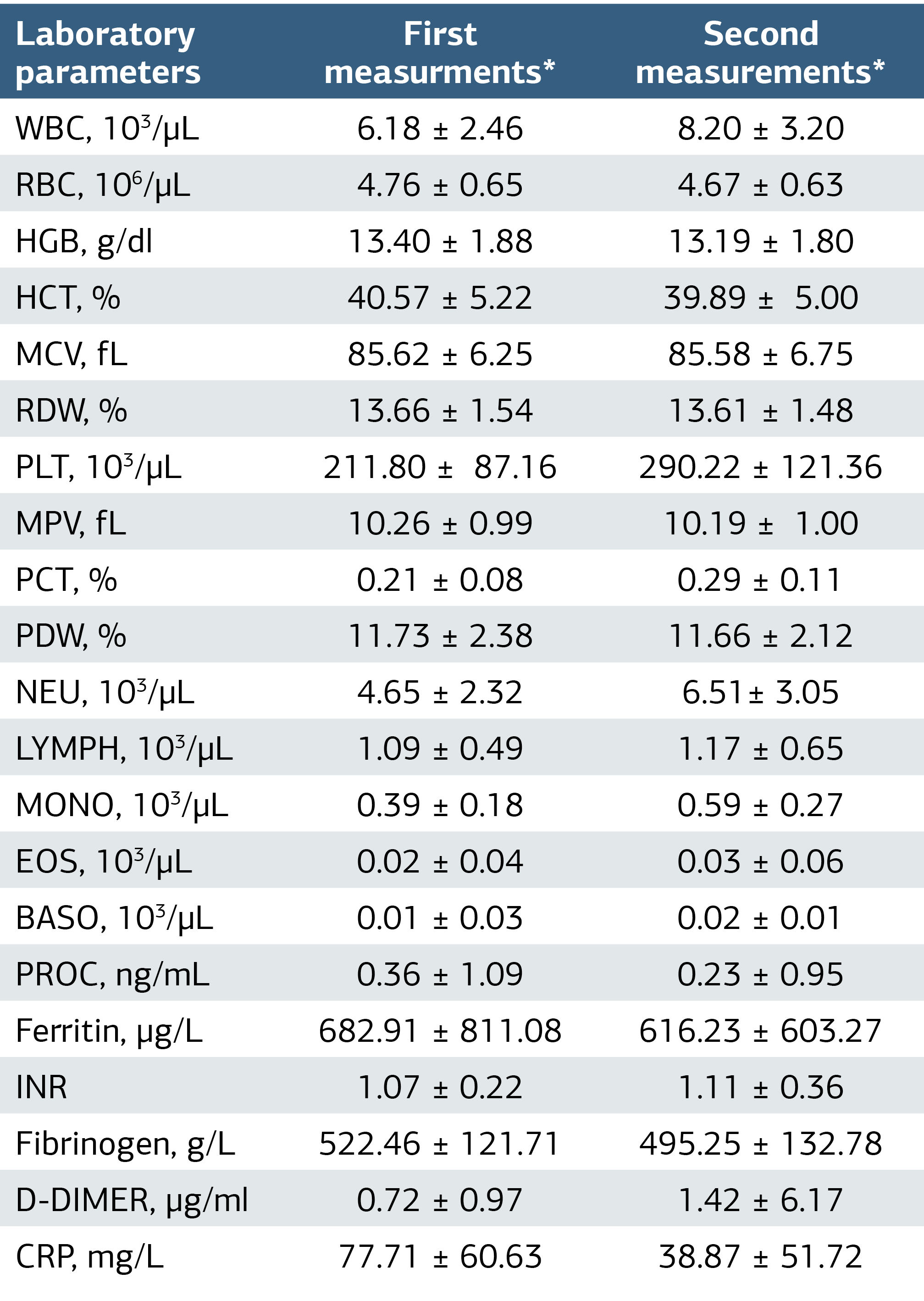
*The first measurement, the initial results; the second measurement, the results obtain a week later form the blood tests simultaneously with thorax CT. WBC, white blood cell; RBC, Red Blood Cell; HGB, hemoglobin; HCT, hematocrit; MCV, Mean Corpuscular Volume; RDW, red blood cell distribution width; PLT, platelet; MPV, Mean Platelet Volume; PCT, platelet crit; PDW, Platelet Distribution Width; NEU, neutrophil; LYMPH, lymphocyte; MONO, monocyte; EOS, eosinophil; BASO, basophil; PROC, procalcitonin; INR, International normalized ratio; CRP, C-reactive protein.
Table 2. The Association between CT Score and Hemogram Parameters of Inpatients
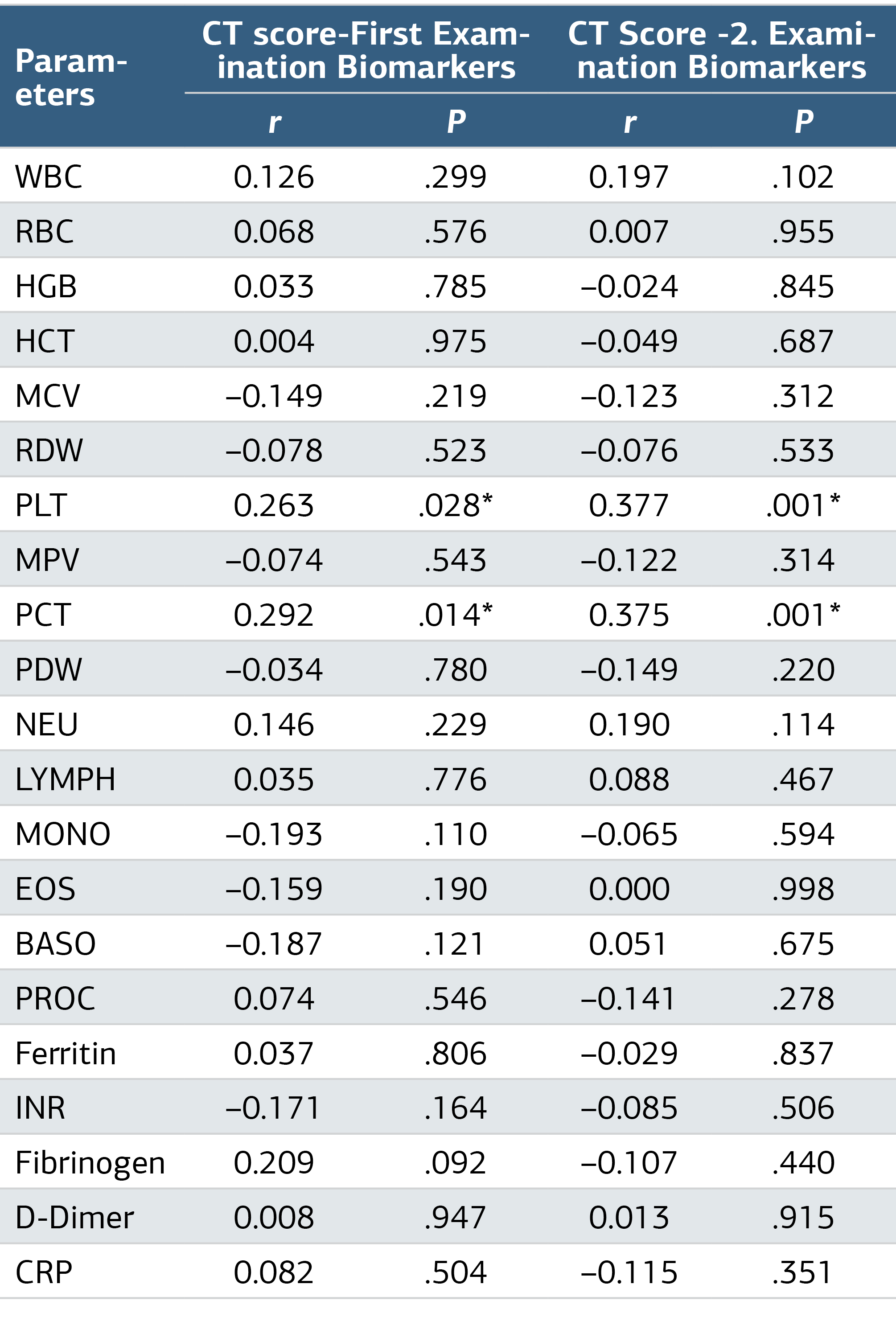
*Pearson correlation analysis.
Additional Information
Publisher’s Note
Bayrakol MP remains neutral with regard to jurisdictional and institutional claims.
Rights and Permissions

This work is licensed under a Creative Commons Attribution-NonCommercial 4.0 International License (CC BY-NC 4.0). To view a copy of the license, visit https://creativecommons.org/licenses/by-nc/4.0/
About This Article
How to Cite This Article
Gamze Demir, Özlem Güç Suvak, Yeliz Aktürk, Cenk Aypak. Association between hemogram and chest CT severity scores in COVID-19 inpatients. Ann Clin Anal Med 2024;15(9):596-599. doi:10.4328/ACAM.22102
Publication History
- Received:
- 23.01.2024
- Accepted:
- 19.03.2024
- Published Online:
- 23.07.2024
- Printed:
- 01.09.2024