
Abstract
AimTo evaluate the effect of concomitant heart failure on diabetic retinopathy (DR) in patients with Diabetes Mellitus (DM).MethodsIn this cross-sectional study, 40 eyes of 20 patients with DM alone (control group) and 70 eyes of 35 patients with heart failure and DM were included. Anterior segment and dilated fundus examination were performed in all patients. DR was classified as mild, moderate, severe non-proliferative DR and proliferative DR. According to the ejection fraction (EF), the stage of heart failure was classified as decreased (HFrEF) if LVEF <40%, moderate (HFmrEF) if LVEF was 40-49%, and preserved (HFpEF) if LVEF ≥50%. After this staging of HF patients; 13 were classified as HFpEF, 12 as HFmrEF and 10 as HFrEF ResultsIn our cross-sectional study, the mean age of the patients in the HF+DM+ group consisting of 35 patients was 54.4 ± 12.6 and the mean age of the control group consisting of 20 patients was 51.7 ± 6.8. PDR and DR in the HFrEF group were found to be significantly higher than the control (P = .02, P = .04 respectively. In addition, as a result of examining the relationship between the factors affecting the severity of DR, the duration of DM (P = .01, OR=1.62), HbA1c (P = .03, OR=2.95) and HF severity (P = .02, OR=1.86).ConclusionOur study shows that the risk of PDR is increased in the HFrEF stage in patients with HF accompanying DM, in addition to blood glucose regulation, HF treatment may contribute positively to DR.
Keywords
Introduction
Diabetes mellitus (DM) is a disease that threatens visual acuity and can cause microvascular complications, which is one of the leading causes of blindness.1 The prevalence of diabetic retinopathy (DR) in individuals with DM is approximately 35%, and this rate reaches up to 60% in individuals with diabetes for 20 years.2,3 Disease duration and severity of hyperglycemia can be counted among the main risk factors associated with diabetic retinopathy.4,5 Diabetic Retinopathy is a disease that occurs as a result of chronically high blood sugar and can be seen in retinal hemorrhages, exudates, macular edema, neovascularizations and occlusions in retinal vessels.6 DR can be classified as Non-proliferative diabetic retinopathy (NPDR) and proliferative diabetic retinopathy (PDR), and it is diagnosed as PDR in the presence of neovascularization (NV), preretinal hemorrhage, intravitreal hemorrhage findings, and as NPDR in the absence of this findings.7 The major factor in the emergence of the PDR phase is hypoxia in the body.8-10
Heart failure (HF) is a prevalent public health issue; with reportedly affects 26 million people worldwide.11 HF is categorized based on the left ventricular ejection fraction (LVEF). Patients are most often classified as having HF with reduced (HFrEF; LVEF <40%), mid-range (HFmrEF; LVEF 40–49%), or preserved ejection fraction (HFpEF; LVEF ≥50%).12 HF and sleep disturbances are two common conditions that frequently coexist and overlap.13 Sleep apnea, a reported prevalence as high as 40% in HF patients, is a worse prognosis predictor due to hypoxia.14
This study aims to evaluate in detail whether the stages of diabetic retinopathy are affected by the stage of HF.
Materials and Methods
In this cross-sectional study, 35 patients with DM accompanying HF disease (DM+HF+) who applied to the cardiology outpatient clinic between 06.08.2021 and 08.02.2022 were included in the study. Forty eyes (DM+HF-) of 20 patients with DM who were sent to the Cardiology outpatient clinic and confirmed as not having HF were taken as the control group. All study procedures were performed in accordance with the Declaration of Helsinki. In addition, informed consent was obtained from all patients before they participated in the study.
Thirty-two patients with HF and twenty patients without HF (control group) were enrolled in the study. Patients were divided into three groups according to LVEF: HFrEF (n = 7), HFmrEF (n = 12), and HFpEF (n = 13).
Hypertension and coronary artery disease which can affect Diabetic Retinopathy, presence of anterior and posterior segment pathologies that reduce the visibility of the retina, and pregnancy were determined as exclusion criteria. In addition, diseases such as thyroid diseases and anemia were excluded from the differential diagnosis of heart failure.
Data such as age, gender, duration of diabetes mellitus, glycosylated hemoglobin (HbA1c) level, and stage of heart failure of the patients included in the study were recorded. After a detailed anterior segment examination with biomicroscopy (Topconsl-D7, SN:1613331, Japan), both pupils were dilated with mydriatic eye drops (0.1% tropicamide). After 30 minutes, detailed retinal examinations including the peripheral retina were performed. Patients were classified according to the International Clinical Diabetic Retinopathy Disease Severity Scale as mild NPDR (microaneurysm only), moderate NPDR (more than a single microaneurysm but less than severe NPDR), severe NPDR (severe intraretinal hemorrhages and microaneurysms, more severe NPDR in each of the four quadrants, two or more venous pilling in and moderate IRMA in one or more quadrants) and PDR (Neovascularization, one or both vitreous/preretinal hemorrhages). The degree of diabetic retinopathy was evaluated for the right and left eyes separately in the patients, and the result was classified according to the worst eye. Fundus fluorescein angiography was performed in patients whose neovascularization could not be differentiated on fundus examination.Ethical ApprovalThis study was approved by the Ethics Committee of Aksaray University (Date: 27.12.2019, Decision No: 2019/12-41).Statistical AnalysisStatistical Package for the Social Sciences (SPSS) 23.0-Windows (SPSS Inc., Chicago, IL) was used for statistical analysis. Shapiro-Wilk test was used to test whether the sample had a normal distribution. ANOVA test was used to compare the means of the groups. The Chi-square test was performed to compare the means of categorical variables in the groups. Binomial logistic regression analysis was performed to calculate the correlation ratios between the variables. Statistical significance was accepted as P < .05.
Reporting GuidelinesThis study was reported in accordance with the STROBE guideline.
Results
The mean age of the patients in the HF group was 54.4±12.6, and the group consisted of 17 females and 18 males in terms of gender. The mean age of the patients in the control group was 51.7±6.8 years and the group consisted of 11 females and 9 males. When the groups were compared in terms of mean age and gender, the statistically significant difference was not determined between them (P > .05) (Table 1).
When mild and moderate groups were compared separately with the control group, a significant difference was not found between them in terms of DR, NPDR, PDR, HbA1c and DM duration (P > .05). There was no difference between the severe group and the control group in terms of NPDR, HbA1c and DM duration (P > .05). A statistically significant difference was found between the severe group and the control group in terms of the incidence of PDR and DR (P = .02, P = .04 respectively). Detailed information is shown in Binomial logistic regression analysis was performed to investigate its association with the factors affecting the stage of DRP. There was a positive association between DM duration (P = .03, OR=1.49), and HbA1c (P = .02, OR=2.62) in the NPDR group, and in addition the association was not found in terms of age and gender (P > .05). A positive association was found between DM duration (P = .02, OR=1.62), HbA1c (P = .01, OR=2.95) and HF severity (P = .02, OR=1.86) in the PDR group, and no association was found in terms of age and gender (P > .05). There was a positive association between DM duration (P = .03, OR=1.27), HbA1c (P = .01, OR=2.37) and HF severity (P = .04, OR=1.21) in the DR group, and no association was found between age and gender (P > .05). As a result, as the degree of HF increased, the risk of PDR increased 1.86 times and the risk of DR increased 1.21 times, while there was no significant increase in the risks of NDRP(P > .05). Detailed information is shown in Table 2.
Discussion
Because of DM is a vascular disease, if glucose regulation is not good years after the onset of the disease, it affects many organs and one of them is the eye. Diabetic retinopathy is a disease that is a complication of diabetes and is associated with chronic hyperglycemia.1 Although the main factor affecting diabetic retinopathy is blood sugar level, it can also contribute to this retinopathy through secondary causes. The main result of our study is that one of the secondary causes affecting diabetic retinopathy in patients with severe HF was determined as advanced heart failure.
In many studies, the duration of DM and the severity of hyperglycemia (HBA1c) have been reported as the main risk factors for DR.4-7 In addition, although glucose is regulated sometimes, diabetic retinopathy due to secondary causes has been observed. Among the secondary causes, HT, hypercholesterolemia, CAD, heart failure, smoking, alcohol, Obstructive Sleep Apnea Syndrome, advanced nasal septum deviation, and other causes are still under investigation.15-21 In this study, since these related diseases were determined as exclusion criteria to prevent diabetic retinopathy from being affected, a comparison with these diseases could not be made. As far as we know, there is no study in the literature investigating the detailed association between HF stages and DR stages.
Diabetic retinopathy can be classified as PDR and NPDR, and NPDR itself is classified according to the degree of retinopathy. Fundus findings in the early stage of NPDR are microaneurysms and microhemorrhages. The development of venous pilling, soft exudate and intraretinal microvascular abnormalities seen in the later progressive stage are findings suggestive of decreased capillary perfusion. When neovascularizations in the optic disc and retinal surface or preretinal hemorrhage or intravitreal hemorrhage due to these develops, our definition is PDR in which retinal ischemia is at the forefront.10 The major factor in the PDR phase is hypoxia, and in this phase, VEGF and other growth factors released secondary to hypoxia increase the permeability of retinal vessels and neovascularizations and related hemorrhages occur.10,22
The angiogenesis of neovascularizations seen in PDR is known as VEGF-A, which is regulated by hypoxia-induced vascular endothelial growth factor-A. This growth factor is secreted by Ganglion cells, Müller cells, and RPE cells and binds to high-affinity VEGF receptors on endothelial cells and pericytes to act.10 VEGF is an important growth factor especially for hypoxia-induced angiogenesis and is closely associated with the formation of new pathological vessels, and this angiogenesis is controlled by angiogenic inducers and inhibitors.10,23 These newly formed pathological vessels are fragile and permeable and may grow along the retinal surface or into the posterior hyaloid. In addition, these vessels can be easily ruptured by vitreous traction and may cause bleeding in the vitreous or preretinal cavity, and may also cause tractional retinal detachment if not treated.23 With a similar mechanism in our study, PDR findings may have occurred as a result of hypoxia caused by severe HF.
In patients with advanced HF, hypoxia occurs as a result of left ventricular enlargement and decreased ejection fraction.24 We think that retinal hypoxia and ischemia may have occurred as a result of systemic hypoxia in this severe HF. When PDR is detected, the first thing that comes to mind is hyperglycemia, which causes retinal ischemia. However, in some patients, DRP findings are seen even though diabetes is regulated. Therefore, other systemic diseases that may cause retinal ischemia and hypoxia should be kept in mind. This study will ensure that advanced HF and other risk factors are taken into account in the etiology of PDR.
The limitations of this study are that it was conducted with a relatively low number of patients. However, the number of our patients remained relatively low since it is very difficult to find patients who have both heart failure (3 different stages) and DM at the same time and do not have other accompanying diseases. Two cardiology specialists took part in the study to reach this number of patients. In addition, the fact that two eyes of the patients were included in the study for the staging of diabetic retinopathy can be said to be a statistical limitation.
Limitations
The main limitations of this study are the relatively small sample size and the inclusion of both eyes of the patients in the evaluation of diabetic retinopathy, which may represent a statistical limitation.
Conclusion
Our study is valuable in that only HF accompanying DM is not accompanied by other diseases that may affect diabetic retinopathy. This study showed that diabetic and HF patients with decreased EF are at risk for proliferative stage DR. Therefore, in these patients, besides blood sugar regulation, HF treatment may also contribute positively to proliferative retinopathy.
Declarations
Animal and Human Rights Statement
All procedures performed in this study were in accordance with the ethical standards of the institutional and/or national research committee and with the 1964 Helsinki Declaration and its later amendments or comparable ethical standards.
Informed Consent
Written informed consent was obtained from all participants before their participation in the study.
Data Availability
The datasets used and/or analyzed during the current study are not publicly available due to patient privacy reasons but are available from the corresponding author on reasonable request.
Conflict of Interest
The authors declare that there is no conflict of interest.
Funding
None.
Abbreviations
DM: Diabetes mellitus
DR: Diabetic retinopathy
HF: Heart failure
LVEF: Left ventricular ejection fraction
NPDR: Non-proliferative diabetic retinopathy
PDR: Proliferative diabetic retinopathy
References
- Tan TE, Wong TY. Diabetic retinopathy: looking forward to 2030. Front Endocrinol (Lausanne). 2023;13:1077669. doi:10.3389/fendo.2022.1077669
- Yau JW, Rogers SL, Kawasaki R, et al. Global prevalence and major risk factors of diabetic retinopathy. Diabetes Care. 2012;35(3):556-564. doi:10.2337/dc11-1909
- Teo ZL, Tham YC, Yu M, et al. Global prevalence of diabetic retinopathy and projection of burden through 2045: systematic review and meta-analysis. Ophthalmology. 2021;128(11):1580-1591. doi:10.1016/j.ophtha.2021.04.027
- Yin L, Zhang D, Ren Q, Su X, Sun Z. Prevalence and risk factors of diabetic retinopathy in diabetic patients: a community-based cross-sectional study. Medicine (Baltimore). 2020;99(9).
- Simó-Servat O, Hernández C, Simó R. Diabetic retinopathy in the context of patients with diabetes. Ophthalmic Res. 2019;62(4):211-217. doi:10.1159/000499541
- Azad R, Sinha S, Nishant P. Asymmetric diabetic retinopathy. Indian J Ophthalmol. 2021;69(11):3026-3034. doi:10.4103/ijo.ijo_1525_21
- Solomon SD, Goldberg MF. ETDRS grading of diabetic retinopathy: still the gold standard? Ophthalmic Res. 2019;62(4):190-195. doi:10.1159/000501372
- Stitt AW, Curtis TM, Chen M, et al. The progress in understanding and treatment of diabetic retinopathy. Prog Retin Eye Res. 2016;51:156-186. doi:10.1016/j.preteyeres.2015.08.001
- Bandello F, Lattanzio R, Zucchiatti I, Del Turco C. Pathophysiology and treatment of diabetic retinopathy. Acta Diabetol. 2013;50(1):1-20. doi:10.1007/s00592-012-0449-3
- Aouiss A, Anka Idrissi D, Kabine M, Zaid Y. Update of inflammatory proliferative retinopathy: ischemia, hypoxia and angiogenesis. Curr Res Transl Med. 2019;67(2):62-71. doi:10.1016/j.retram.2019.01.005
- Shiga T, Suzuki A, Haruta S, et al. Clinical characteristics of hospitalized heart failure patients with preserved, mid-range, and reduced ejection fractions in Japan. ESC Heart Fail. 2019;6(3):475-486. doi:10.1002/ehf2.12418
- Ponikowski P, Voors AA, Anker SD, et al. 2016 ESC guidelines for the diagnosis and treatment of acute and chronic heart failure: the Task Force for the diagnosis and treatment of acute and chronic heart failure of the European Society of Cardiology (ESC): developed with the special contribution of the Heart Failure Association (HFA) of the ESC. Eur J Heart Fail. 2016;18(8):891-975. doi:10.1002/ejhf.592
- Lévy P, Naughton MT, Tamisier R, Cowie MR, Bradley TD. Sleep apnoea and heart failure. Eur Respir J. 2022;59(5):2101640. doi:10.1183/13993003.01640-2021
- Sanderson JE, Fang F, Lu M, Ma CY, Wei YX. Obstructive sleep apnoea, intermittent hypoxia and heart failure with a preserved ejection fraction. Heart. 2021;107(3):190-194. doi:10.1136/heartjnl-2020-317326
- Sabanayagam C, Banu R, Chee ML, et al. Incidence and progression of diabetic retinopathy: a systematic review. Lancet Diabetes Endocrinol. 2019;7(2):140-149. doi:10.1016/s2213-8587(18)30128-1
- Tarasewicz D, Conell C, Gilliam LK, Melles RB. Quantification of risk factors for diabetic retinopathy progression. Acta Diabetol. 2023;60(3):363-369.
- Pradeepa R, Surendar J, Indulekha K, Chella S, Anjana RM, Mohan V. Relationship of diabetic retinopathy with coronary artery disease in Asian Indians with type 2 diabetes: the Chennai Urban Rural Epidemiology Study (CURES) Eye Study—3. Diabetes Technol Ther. 2015;17(2):112-118. doi:10.1089/dia.2014.0141
- Chang AC, Fox TP, Wang S, Wu AY. Relationship between obstructive sleep apnea and the presence and severity of diabetic retinopathy. Retina. 2018;38(11):2197-2206. doi:10.1097/iae.0000000000001848
- Wu YB, Wang CG, Xu LX, Chen C, Zhou XB, Su GF. Analysis of risk factors for progressive fibrovascular proliferation in proliferative diabetic retinopathy. Int Ophthalmol. 2020;40(10):2495-2502. doi:10.1007/s10792-020-01428-y
- Lin KY, Hsih WH, Lin YB, Wen CY, Chang TJ. Update in the epidemiology, risk factors, screening, and treatment of diabetic retinopathy. J Diabetes Investig. 2021;12(8):1322-1325. doi:10.1111/jdi.13480
- Yaşar E, Kayabasi S. Can nasal septum deviation be one of the factors affecting diabetic retinopathy? J Surg Med. 2020;4(12):1215-1218.
- Al-Kharashi AS. Role of oxidative stress, inflammation, hypoxia and angiogenesis in the development of diabetic retinopathy. Saudi J Ophthalmol. 2018;32(4):318-323. doi:10.1016/j.sjopt.2018.05.002
- Arrigo A, Aragona E, Bandello F. VEGF-targeting drugs for the treatment of retinal neovascularization in diabetic retinopathy. Ann Med. 2022;54(1):1089-1111. doi:10.1080/07853890.2022.2064541
- Jung M, Riley P, Drozdzewski A, Pressler SJ. Mild to moderate hypoxemia among stable heart failure patients with reduced ejection fraction: 24-hour oxygen monitoring. J Card Fail. 2017;23(8). doi:10.1016/j.cardfail.2017.07.233
Tables
Table 1. The Comparison of the Control Group and Heart Failure Stage Groups in Terms of Diabetic Retinopathy
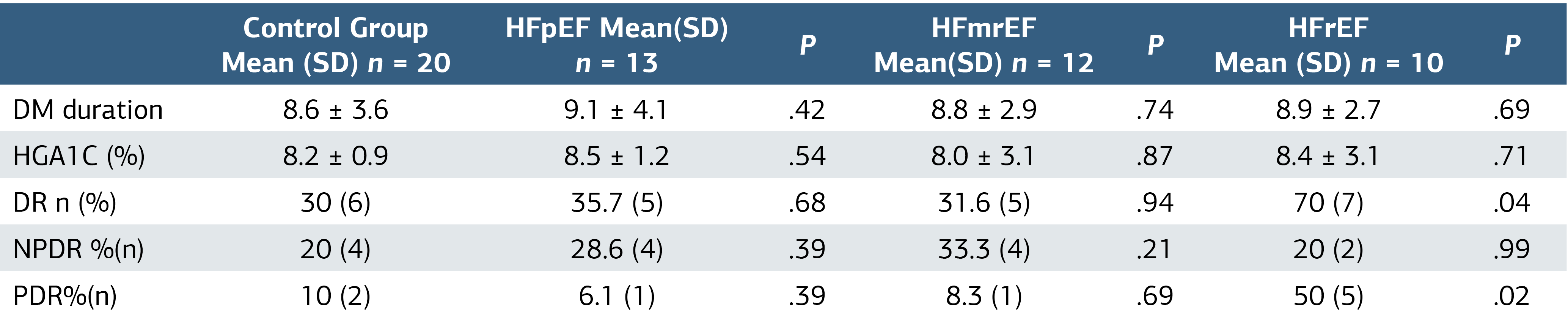
DM, Diabetes Mellitus; DR, Diabetic Retinopathy; NPDR, Non-Proliferative Diabetic Retinopathy; PDR, Proliferative Diabetic Retinopathy; mean, mean; SD, Standard Deviation; HFpEF, HF with Preserved Ejection Fraction; HFmrEF, HF with Mid-Range Ejection Fraction; HFrEF, HF with Reduced Ejection Fraction
Table 2. The Factors Affecting the Stage of Diabetic Retinopathy
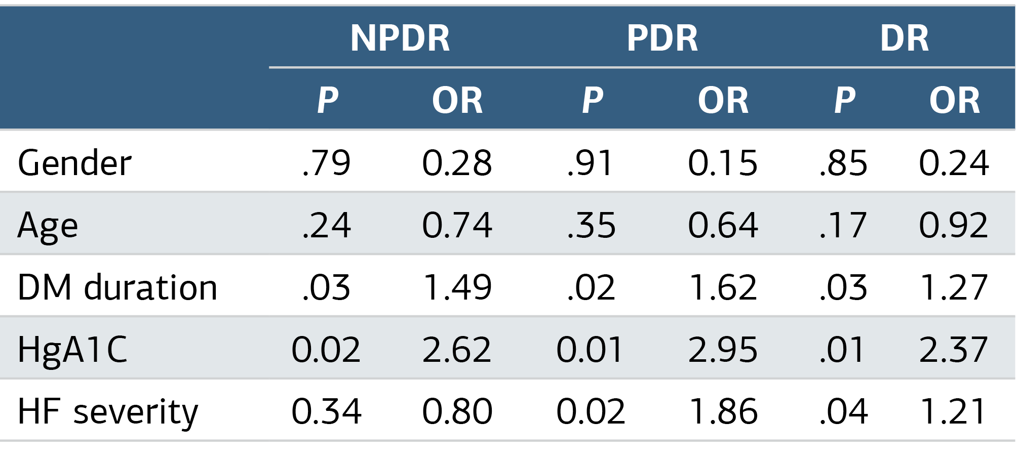
OR, Odds Ratio; DM, Diabetes Mellitus; DR, Diabetic Retinopathy; NPDR, Non-Proliferative Diabetic Retinopathy; PDR, Proliferative Diabetic Retinopathy; HF, Heart Failure
Additional Information
Publisher’s Note
Bayrakol MP remains neutral with regard to jurisdictional and institutional claims.
Rights and Permissions

This work is licensed under a Creative Commons Attribution-NonCommercial 4.0 International License (CC BY-NC 4.0). To view a copy of the license, visit https://creativecommons.org/licenses/by-nc/4.0/
About This Article
How to Cite This Article
Erdoğan Yaşar, Halil Aktaş, Murat Gül, Uğur Gürlevik. Does heart failure have an effect on the progress of diabetic retinopathy?. Ann Clin Anal Med 2024;15(4):293-296. doi:10.4328/ACAM.22116
Publication History
- Received:
- 17.01.2024
- Accepted:
- 05.03.2024
- Published Online:
- 19.03.2024
- Printed:
- 01.04.2024