
Abstract
AimThe objective of this study is to identify the risk variables associated with lymph node metastasis (LNM) in individuals diagnosed with cervical squamous cell carcinoma (SCC).MethodsCervical cancer patients who underwent type II/III radical hysterectomy and pelvic lymphadenectomy +/- para-aortic lymphadenectomy were analyzed.ResultsIn total, 422 SCC patients were included in the cohort. Three hundred-twenty-three (76.5%) patients were stage IB1, 59 (14%) were stage IB2, 33 (7.8%) were stage IIA1, and 7 (1.7%) were stage IIA2. Eighty-seven (20.6%) patients had LNM only in the pelvic region, 2 (0.5%) patients in the paraaortic region, and 8 (1.9%) patients in both regions. In the multivariate logistic analysis, parametrial invasion (hazard ratio [HR]: 2.182, 95% confidence interval [CI]: 1.090–4.366, P = .027), lymphovascular space invasion (LVSI) (HR: 6.300, 95% CI: 2.968–13.370, P < .001) and deep stromal invasion (HR: 2.122, 95% CI: 1.088–4.139, P = .027) were identified as significant risk factors for LNM.ConclusionIn summary, age, FIGO stage, tumor size, vaginal invasion, surgical border involvement, and uterine involvement were not identified as independent risk factors for LNM. However, parametrial invasion, LVSI, and deep stromal invasion are independent risk factors for LNM in stage IB1-IIA2 cervical SCC patients.
Keywords
Introduction
Cervical cancer is a prominent malignancy affecting women globally, occupying the fourth position in terms of prevalence.1 Squamous cell carcinoma (SCC) is the most prevalent histologic subtype of cervical cancer. Radical hysterectomy with pelvic lymphadenectomy is the standard approach for early-stage disease, while chemoradiotherapy is the standard treatment for advanced stages of the disease.2 The addition of pelvic lymphadenectomy in the surgical management of early-stage cervical cancer is widely accepted. However, the appropriateness of para-aortic lymphadenectomy remains a subject of debate due to the associated risk of severe complications and the requirement for a high degree of surgical proficiency.3,4
Cervical cancer can spread directly to the vagina, parametrium, uterus, and nearby organs, such as the rectum and bladder. Furthermore, it disseminates through the lymphatic vessels to the nearby lymph nodes, such as the obturator, external iliac, and internal iliac nodes, and subsequently to the common iliac and paraaortic nodes. A late manifestation of the disease is hematogenous metastasis to the lungs, liver, and skeleton.5
The presence of lymph node metastasis (LNM) is an important factor of prognosis in patients with cervical cancer.6 The occurrence of pelvic LNM in individuals diagnosed with early-stage cervical cancer (stages IA1–IB1) has been documented to be approximately 15–20%.7,8 These patients have a poorer prognosis, and their 5-year survival rates are lower than those of patients without LNM.9,10 LNM is associated with the histological subtype, tumor size, parametrial invasion, deep stromal invasion, and lymphovascular space invasion (LVSI).10,11 LNM was not included in the International Federation of Gynecology and Obstetrics (FIGO) cervical cancer staging system until 2018. In the latest classification, evaluation of lymph nodes by imaging and/or pathology is incorporated as stage IIIC disease. Stage IIIC1 is defined if LNM is in the pelvic lymph nodes, and stage IIIC2 if LNM is in the para-aortic lymph nodes.12
In the present study, patients with stages IB1, IB2, IIA1, and IIA2 SCC had radical hysterectomy and pelvic ± paraaortic lymphadenectomy. Our objective was to identify the prognostic markers associated with lymph node metastasis in patients with cervical SCC.
Materials and Methods
The study was designed retrospectively. Cervical cancer patients who underwent type II/III radical hysterectomy and pelvic lymphadenectomy ± para-aortic lymphadenectomy were analyzed at 6 gynecologic oncology centers between 1993 and 2022. The electronic database system, medical files, and pathology reports were utilized to obtain patient information. Patients who were not treated with a radical hysterectomy, those with microinvasive cancer, a non-squamous cell component in their tumors, synchronous primary tumors, and those receiving neo-adjuvant chemotherapy were excluded.
The following clinical data were collected: age, FIGO stage, the pathologic data (parametrial invasion, vaginal invasion, surgical border involvement, depth of stromal invasion, LVSI, uterine invasion, size of the tumor, lymph node status, adnexal metastasis). The tumor size was determined by the greatest tumor diameters. The clinical stage was evaluated in accordance with the FIGO 2014 criteria. Deep stromal invasion was defined as ≥½ stromal invasion. In hematoxylin and eosin stained pathologic sections, LVSI was described as tumor cells or cell clusters adhering to vascular walls containing both tumor and surrounding healthy tissue. The spread of the disease to the endometrium and/or myometrium above the level of the internal ostium was defined as uterine invasion. The criterion for surgical border involvement was established as a measurement of less than 0.5 cm between the tumor and the termination point of the specimen. Vaginal metastasis was defined as the detection of the tumor in any location within the vagina. The decision to include a bilateral salpingo-oophorectomy in the surgical procedure was based on factors such as the patient’s age, the state of the ovaries, and the professional judgment of the surgeon. Depending on the presence of suspicious lymph nodes in the paraaortic region and the preference of the senior surgeon, paraaortic lymphadenectomy was added to the surgical procedure and was performed up to the level of the inferior mesenteric artery or left renal vein. All surgical procedures were performed by gynecological oncologists.Ethical ApprovalThis study was approved by the Ethics Committee of Ankara City Hospital (Date: 09.11.2022, Decision No: E2-22-2777).Statistical AnalysisThe effects of categorical variables on LNM were evaluated using Fisher’s exact test or Pearson’s chi-square test, as applicable, to determine statistical significance. With factors that were statistically significant in univariate analysis, a model for multivariate analysis was developed. Backward stepwise multivariate Cox proportional hazard regression analysis was utilized to identify the effects of LNM-influencing variables. A P value <0.05 was determined to be statistically significant. Statistical Package for the Social Sciences (SPSS) for Windows version 22.0 was used to conduct statistical analyses.Reporting GuidelinesThis study was reported in accordance with the STROBE guideline.
Results
In total, 422 SCC patients were included in the cohort. The size of the median tumor was 30 mm (range, 6–80) and the mean age of the patients was 52 ± 10.2 years (range, 26–80). Three hundred twenty-three (76.5%) patients were stage IB1, 59 (14%) were stage IB2, 33 (7.8%) were stage IIA1, and 7 (1.7%) were stage IIA2. All patients underwent pelvic lymphadenectomy, while 389 patients (92.2%) underwent paraaortic lymphadenectomy. The median number of lymph nodes excised was 44. Ninety-seven (23%) patients had metastatic lymph nodes. The mean number of metastatic lymph nodes was 3 ± 4.1 (range, 1–31). Eighty-seven (20.6%) patients had LNM only in the pelvic region, 2 (0.5%) patients in the paraaortic region, and 8 (1.9%) patients in both regions. Bilateral salpingo-oophorectomy was performed in 339 (80.3%) patients. There were 64 (15.2%) cases with positive parametrial invasion, 24 (5.7%) with surgical border involvement, 70 (16.6%) with vaginal invasion, 57 (13.5%) uterine invasion, 241 (57.1%) with LVSI, and 265 (62.8%) with deep stromal invasion. The clinical characteristics and surgical pathological outcomes of the patients were detailed in Table 1.
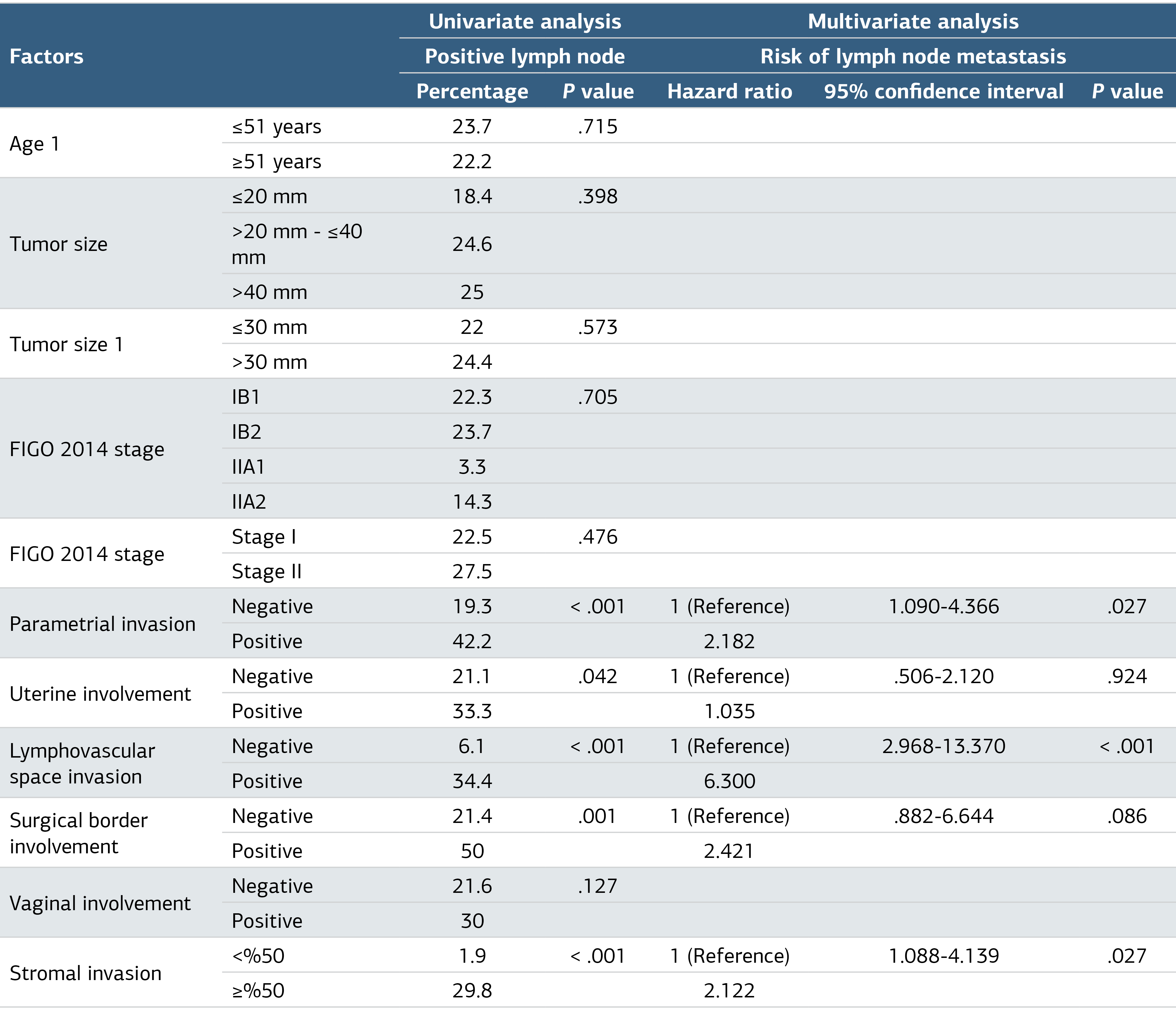
In the univariate analysis, parametrial invasion, surgical border involvement, uterine invasion, deep stromal invasion, and positive LVSI were found to be associated with LNM (Table 2). However, age, tumor size, FIGO stage, and vaginal involvement were not associated with LNM. In the multivariate logistic analysis, parametrial invasion (hazard ratio [HR]: 2.182, 95% confidence interval [CI]: 1.090–4.366, P = .027), LVSI (HR: 6.300, 95% CI: 2.968–13.370, P < .001), and deep stromal invasion (HR: 2.122, 95% CI: 1.088–4.139, P = .027) were identified as significant risk factors for LNM (Table 2).
Discussion
This investigation analyzes the clinical and pathological characteristics of 422 patients diagnosed with stage IB1–IIA2 cervical SCC who underwent radical hysterectomy and lymphadenectomy. Additionally, the study investigates the relationship between LNM and these parameters. Deep stromal invasion, LVSI, and parametrial invasion were independent factors for LNM. Deep stromal invasion, LVSI, and parametrial invasion increased the incidence of LNM by more than twofold, sixfold, and twofold, respectively.
As predictors of LNM in cervical cancer, the following have been identified: advanced stage.8,13,14 histology.8 larger tumor size.8,13-17 LVSI.8,13-18 deep stromal invasion.13-16,18 parametrial invasion.13,15,16 older age.13,17 and high-level serum squamous cell carcinoma antigens (SCC-Ag).13,18 In contrast, our study found no association between age, tumor size, or FIGO stage and LNM.
The present study examined the prevalence of lymph node metastasis (LNM) in patients diagnosed with stage IB1, IB2, IIA1, and IIA2 cervical squamous cell carcinoma (SCC). The findings revealed LNM rates of 22.3%, 23.7%, 30.3%, and 14.3% for each stage. In the cervical SCC study presented by Yang et al., LNM metastasis rates in stage IB1–2 and IIA1–2 were similar to our study.13 In contrast to previous studies.8,13,14 stage was not found to be associated with LNM in our study. This may be explained by the fact that the stage IA2 group, where LNM is rarely seen, was not included in our study.
Tumors larger than 2 cm were associated with an increased risk of LNM in a study by Togami et al. of patients with stages IA2–IIB cervical cancer.19 Yang et al. found that a tumor diameter greater than 2 cm was statistically significant in the univariate analysis for LNM but not in the multivariate analysis.13 In our study, however, tumor diameter was not found to be significant.
In accordance with our study, Wang et al. reported that deep stromal invasion and LVSI were significantly associated with LNM in a multivariate analysis of cervical SCC.18 Similarly, Zhou et al. demonstrated in a multivariate study of stages IA2, IB1, and IIA1 epithelial cervical carcinoma that deep stromal invasion and LVSI had a significant association with LNM.14
In a multivariate analysis of cervical cancer, Nanthamongkolkul et al. and Togami et al. also reported that parametrial invasion was significantly associated with LNM.15,19 According to the study by Benedetti-Panici et al.20 The parametrium is the initial location of extracervical spread, which could explain these results.
Limitations
The main limitation of this study is its retrospective design. In addition, the locations of lymph node involvement (external iliac, internal iliac, obturator, common iliac, and paraaortic nodes) are not specified. However, the study’s strengths are its multicentric design and large patient population. Secondly, patients with only squamous cell types were included, whereas microinvasive patients were excluded. Thirdly, all surgical procedures were conducted by gynecological oncology specialists, and pathologic assessments were performed by expert gyneco-pathologists.
Conclusion
In summary, age, FIGO stage, tumor size, vaginal invasion, surgical border involvement, and uterine involvement were not identified as independent risk factors for LNM. However, parametrial invasion, LVSI, and deep stromal invasion are independent risk factors for LNM in stage IB1-IIA2 cervical SCC patients. Further clinical research can help to strengthen these results.
Declarations
Animal and Human Rights Statement
All procedures performed in this study were in accordance with the ethical standards of the institutional and/or national research committee and with the 1964 Helsinki Declaration and its later amendments or comparable ethical standards.
Informed Consent
Informed consent was waived due to the retrospective study.
Data Availability
The datasets used and/or analyzed during the current study are not publicly available due to patient privacy reasons but are available from the corresponding author on reasonable request.
Conflict of Interest
The authors declare that there is no conflict of interest.
Funding
None.
Abbreviations
CI: Confidence interval
FIGO: International federation of gynecology and obstetrics
HR: Hazard ratio
LNM: Lymph node metastasis
LVSI: Lymphovascular space invasion
SCC: Squamous cell carcinoma
SPSS: Statistical package for the social sciences
References
- Arbyn M, Weiderpass E, Bruni L, et al. Estimates of incidence and mortality of cervical cancer in 2018: a worldwide analysis. Lancet Glob Health. 2020;8(2). doi:10.1016/s2214-109x(19)30482-6
- Cibula D, Pötter R, Planchamp F, et al. The European Society of Gynaecological Oncology/European Society for Radiotherapy and Oncology/European Society of Pathology guidelines for the management of patients with cervical cancer. Virchows Arch. 2018;472(6):919-936. doi:10.1007/s00428-018-2362-9
- Rob L, Pluta M, Strnad P, et al. A less radical treatment option to the fertility-sparing radical trachelectomy in patients with stage I cervical cancer. Gynecol Oncol. 2008;111(2). doi:10.1016/j.ygyno.2008.07.021
- Koh W, Abu-Rustum N, Bean S. NCCN Clinical Practice Guidelines in Oncology: Cervical Cancer. Version 1. National Comprehensive Cancer Network; 2018.
- Bhatla N, Aoki D, Sharma DN, Sankaranarayanan R. Cancer of the cervix uteri: 2021 update. Int J Gynaecol Obstet. 2021;155:28-44. doi:10.1002/ijgo.13865
- Huang BX, Fang F. Progress in the study of lymph node metastasis in early-stage cervical cancer. Curr Med Sci. 2018;38(4):567-574. doi:10.1007/s11596-018-1915-0
- Salvo G, Ramirez PT, Levenback CF, et al. Sensitivity and negative predictive value for sentinel lymph node biopsy in women with early-stage cervical cancer. Gynecol Oncol. 2017;145(1):96-101. doi:10.1016/j.ygyno.2017.02.005
- Ferrandina G, Pedone Anchora L, Gallotta V, et al. Can we define the risk of lymph node metastasis in early-stage cervical cancer patients? A large-scale, retrospective study. Ann Surg Oncol. 2017;24(8):2311-2318. doi:10.1245/s10434-017-5917-0
- Li A, Wang L, Jiang Q, Wu W, Huang B, Zhu H. Risk stratification based on metastatic pelvic lymph node status in stage IIIC1p cervical cancer. Cancer Manag Res. 2020;12:6431. doi:10.2147/cmar.s253522
- Sakuragi N. Up-to-date management of lymph node metastasis and the role of tailored lymphadenectomy in cervical cancer. Int J Clin Oncol. 2007;12(3):165-175. doi:10.1007/s10147-007-0661-2
- Sakuragi N, Satoh C, Takeda N, et al. Incidence and distribution pattern of pelvic and para-aortic lymph node metastasis in patients with stages IB, IIA, and IIB cervical carcinoma treated with radical hysterectomy. Cancer. 1999;85(7):1547-1554.
- Bhatla N, Berek JS, Cuello Fredes M, et al. Revised FIGO staging for carcinoma of the cervix uteri. Int J Gynaecol Obstet. 2019;145(1):129-135. doi:10.1002/ijgo.12749
- Yang S, Zhao J, Zhao H, Hu Y, Zhu H. Development of a nomogram for predicting pelvic lymph node metastasis in cervical squamous cell carcinoma. Int J Gynaecol Obstet. 2022.
- Zhou J, Ran J, He ZY, et al. Tailoring pelvic lymphadenectomy for patients with stage IA2, IB1, and IIA1 uterine cervical cancer. J Cancer. 2015;6(4):377. doi:10.7150/jca.10968
- Nanthamongkolkul K, Hanprasertpong J. Predictive factors of pelvic lymph node metastasis in early-stage cervical cancer. Oncol Res Treat. 2018;41(4):194-198. doi:10.1159/000485840
- Turan T, Yüksel N, Kıyka Ş, et al. Erken evre serviks kanserinde lenf nodu tutulumunu belirleyen faktörler [Factors determining lymph node involvement in early-stage cervical cancer]. Turk Jinekolojik Onkoloji Derg. 2007;10(1):9-15.
- Aziz MF, Andrijono A, Nuranna L, et al. Clinical-pathologic factors as predictors of lymph node metastasis in cervical cancer stage IB and IIA. Med J Indones. 2004;13(2):113-118. doi:10.13181/mji.v13i2.574
- Wang Y, Yao T, Yu J, Li J, Chen Q, Lin Z. Can pelvic lymphadenectomy be omitted in patients with stage IA2, IB1, and IIA1 squamous cell cervical cancer? Springerplus. 2016;5(1):1-7.
- Togami S, Kamio M, Yanazume S, Yoshinaga M, Douchi T. Can pelvic lymphadenectomy be omitted in stage IA2 to IIB uterine cervical cancer? Int J Gynecol Cancer. 2014;24(6). doi:10.1097/igc.0000000000000163
- Benedetti-Panici P, Maneschi F, D’Andrea G, et al. Early cervical carcinoma: the natural history of lymph node involvement redefined on the basis of thorough parametrectomy and giant section study. Cancer. 2000;88(10):2267-2274.
Tables
Table 1. Clinical Features
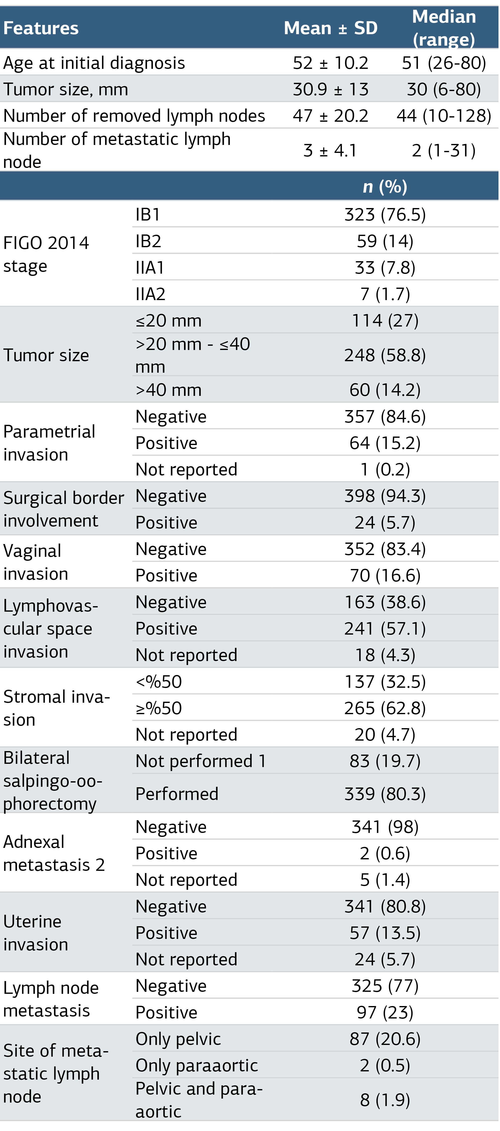
SD, Standard Deviation; 1: Seventy-four patients received bilateral ovarian transposition and 9 patients had unilateral salpingo-oophorectomy and unilateral ovarian transposition 2: Seventy-four patients who underwent bilateral ovarian transposition were excluded and 348 patients were evaluated.
Table 2. Factors Predicting the Lymph Node Metastasis
Additional Information
Publisher’s Note
Bayrakol MP remains neutral with regard to jurisdictional and institutional claims.
Rights and Permissions

This work is licensed under a Creative Commons Attribution-NonCommercial 4.0 International License (CC BY-NC 4.0). To view a copy of the license, visit https://creativecommons.org/licenses/by-nc/4.0/
About This Article
How to Cite This Article
Okan Aytekin, Necim Yalçın, Hande Esra Koca Yıldırım, Mehmet Ünsal, Okan Oktar, Fatih Çelik, Abdurrahman Alp Tokalıoğlu, Mustafa Gökkaya, Dilek Yüksel, Caner Çakır, Çiğdem Kılıç, İlker Selçuk, Günsu Kimyon Cömert, Tayfun Toptaş, Vakkas Korkmaz, Işın Üreyen, Alper Karalok, Derman Başaran, Sevgi Koç, Tolga Taşçı, Taner Turan. Predictive factors of lymph node metastasis in patients with 2014 FIGO stage Ib1-IIa2 cervical squamous cell cancer: a multicentric study. Ann Clin Anal Med 2024;15(6):409-413. doi:10.4328/ACAM.22128
Publication History
- Received:
- 29.01.2024
- Accepted:
- 19.03.2024
- Published Online:
- 26.04.2024
- Printed:
- 01.06.2024