
Abstract
AimInvestigate the relationship between gestational thrombocytopenia (GT) and adverse maternal and fetal outcomes in pregnant women.Methods97 patients who were diagnosed with gestational thrombocytopenia during pregnancy, had no other disease that causes thrombocytopenia and delivered in our clinic were included in the study. The patients were divided into two groups according to their platelet values. Group-1 platelet count was <70.000/mm3 and Group-2 platelet count was 70.000- 150.000/mm3. In the analysis of negative perinatal outcomes, logistic regression analysis was performed. P < .05 was considered significant in the analysis results. ResultsFrom the laboratory data of the patients diagnosed with gestational thrombocytopenia, the mean platelet count was 85.28 ± 27.08 (min-max: 22-142) and mean hematocrit was 35.91 ± 3.63 (min-max: 21-45). When the groups were evaluated considering their hematocrit values; the mean hematocrit value of the patients in Group-2 (36.42 ± 3.22) was higher than the mean hematocrit value (34.26 ± 4.39) of the patients in Group-1, and a significant difference was found between them (p:0.03). This significant difference was also present when the platelet value was taken as the limit of 110,000 (p:0.039).ConclusionApgar scores are lower in patients diagnosed with gestational thrombocytopenia and with platelet values below 70,000, and the need for blood transfusion (erythrocyte suspension, FFP (Fresh Frozen Plasma) and platelet product) is higher. The need for erythrocyte transfusion continues even when the platelet limit is 80,000, and the need for platelet transfusion is 100,000.
Keywords
Introduction
Thrombocytopenia is defined as a platelet count of less than 150,000/mm³ and is observed in 7-10% of pregnancies.1 Thrombocytopenia can be classified as severe (<50,000/mm³), moderate (50-100,000/mm³), and mild (>100,000/mm³) according to platelet count. Although platelet count decreases slightly in normal pregnancies, the level remains within normal limits.2 Most studies have reported a decrease in platelet count during pregnancy, leading to levels that are approximately 10% lower at term than pre-pregnancy levels.3,4
Thrombocytopenia is the most common hematologic disease after anemia in pregnancy. Although there are many causes of thrombocytopenia observed in pregnancy, the gestational week is very important in the differential diagnosis. Autoimmune causes are mostly involved in the etiology of thrombocytopenia observed in early gestational weeks.5 Thrombocytopenia may be associated with pregnancy-related conditions such as preeclampsia and HELLP syndrome (hemolysis, elevated liver enzymes, low platelet count), which account for approximately 18% of cases, or various diseases such as idiopathic thrombocytopenic purpura, which accounts for approximately 5% of cases.6 Thrombocytopenia may cause some adverse perinatal outcomes. Babies of mothers with moderate and severe thrombocytopenia have a high risk of low 5th minute Apgar score, fetal growth restriction (FGR), and stillbirth.7,8
In our study, we aimed to investigate the maternal and fetal outcomes of gestational thrombocytopenia and the relationship between these outcomes and the degree of thrombocytopenia by forming subgroups according to platelet counts.
Materials and Methods
This study is a hospital-based retrospective case study including pregnant women with gestational thrombocytopenia who were admitted to the Gynecology and Obstetrics Clinic of Dicle University Faculty of Medicine Hospital between January 2010 - May 2021 and whose delivery took place in our clinic. Demographic and laboratory data of the patients were analyzed. Patients were called by phone and information was obtained on whether they had a recurrence in the next pregnancy. Patients with a platelet count of <150,000/mm³ as a result of a complete blood count performed 2 times in our clinic were included in the study. Patients were divided into two groups according to platelet counts. Group-1 was formed as platelet count <70,000/mm³ and Group-2 as platelet count 70,000-150,000/mm³, and the subgroups were compared among themselves. Diagnosis of thrombocytopenia before pregnancy, bleeding profile disorder, ITP, use of acetic salicylic acid and/or low molecular weight heparin or unfractionated heparin, hepatic or renal dysfunction, venous thrombosis, hepatitis, sepsis, viral and/or bacterial infection, autoimmune disease, use of drugs affecting platelet function or production, pregnant women with hereditary factor deficiency such as vWF, bone marrow diseases such as leukemia, lymphoma, patients receiving IVIG and/or steroids, pregnancy-induced preeclampsia, gestational cholestasis, HELLP, disseminated intravascular coagulation (DIC), and diseases affecting liver and kidney function were excluded. Spontaneous singleton pregnancies without fetal ultrasonographic and/or cytogenetic fetal anomalies were included in the study. Pregnant women with fetal anomalies, intracranial hemorrhage on ultrasound and/or fetal MRI, intrauterine fetal intervention, and multiple pregnancies were excluded. Ethical ApprovalThis study was approved by the Ethics Committee of the Dicle University, Faculty of Medicine Non-Invasive Clinical Research (Date: 21.09.2021, Decision No: 38).Statistical AnalysisThe data obtained from the study were analyzed using the SPSS package program (Statistical Package for Social Sciences; IBM SPSS Statistics for Macintosh, Armonk, NY) version 25. Descriptive analyses were presented as number (n) and percentage (%) for categorical data and mean ± standard deviation for continuous data. Pearson chi-square test was used to compare categorical variables between groups. The compatibility of continuous variables with normal distribution was evaluated by Kolmogorov-Smirnov test. Mann-Whitney U-test was used to compare variables that did not show the normal distribution in two groups. In the comparison of more than two groups, One-Way ANOVA test was used for parametric variables and Kruskal-Wallis test was used for non-parametric variables. The statistical significance level was accepted as P < .05 in the analyses. Reporting GuidelinesThis study was reported in accordance with the STROBE guideline.
Results
The mean age of all patients included in the study was 29.36 ± 5.32 (min-max: 18-41) years. The mean gravida was 3.82 ± 2.31 (min-max: 1-11), parity 2.26 ± 1.96 (min-max: 0-10), abortion 0.58 ± 1.02 (min-max: 0-5), and number of live births 2.15 ± 1.79 (min-max: 0-8).
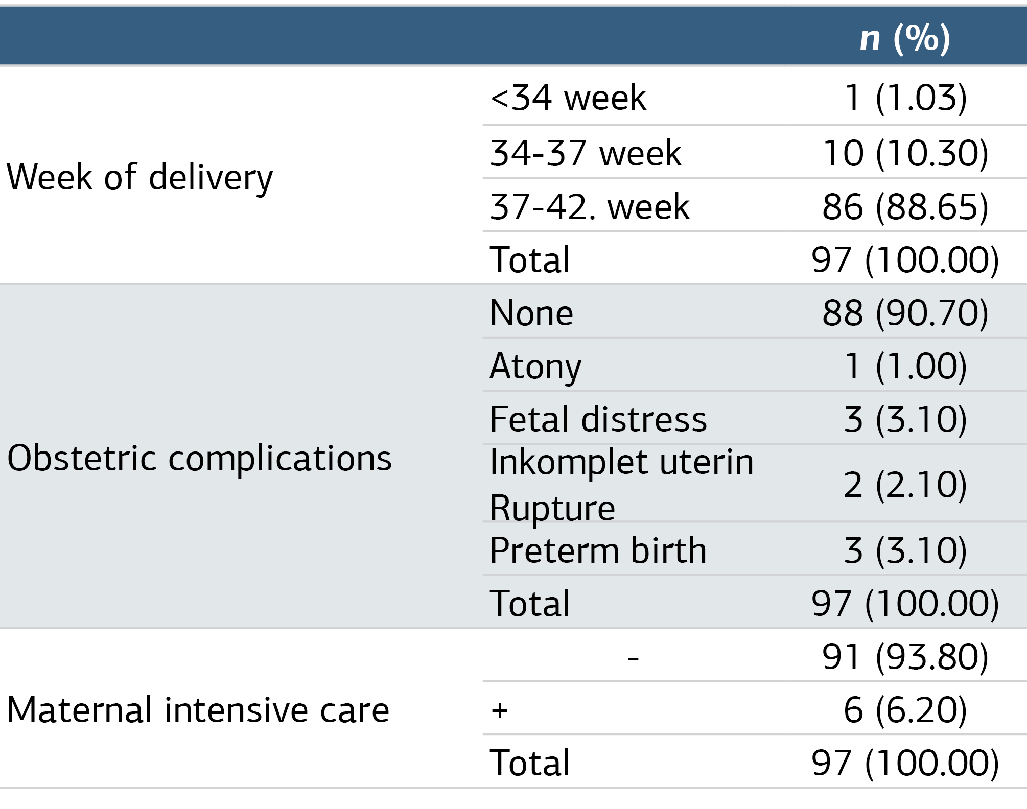
Of a total of 97 patients included in the study, 88.65% (n = 86) delivered between 37-42 weeks, i.e. at term, 10 patients (10.30%) delivered between 34-37 weeks (early term), and 1 patient (1.03%) delivered at <34 weeks, i.e. preterm. There were no obstetric complications in 90.70% (n = 88) of our patients. A total of 9 patients (9.30%) had complications, including preterm-early term labor and fetal distress in three patients each, an incomplete uterine rupture in two patients, and atony in one patient. Of our 97 patients, 91 (93.80%) did not require intensive care, while 6 (6.20%) patients required maternal intensive care. Patients’ gestational week of delivery, obstetric complication status, and maternal intensive care needs are shown in Table 1.
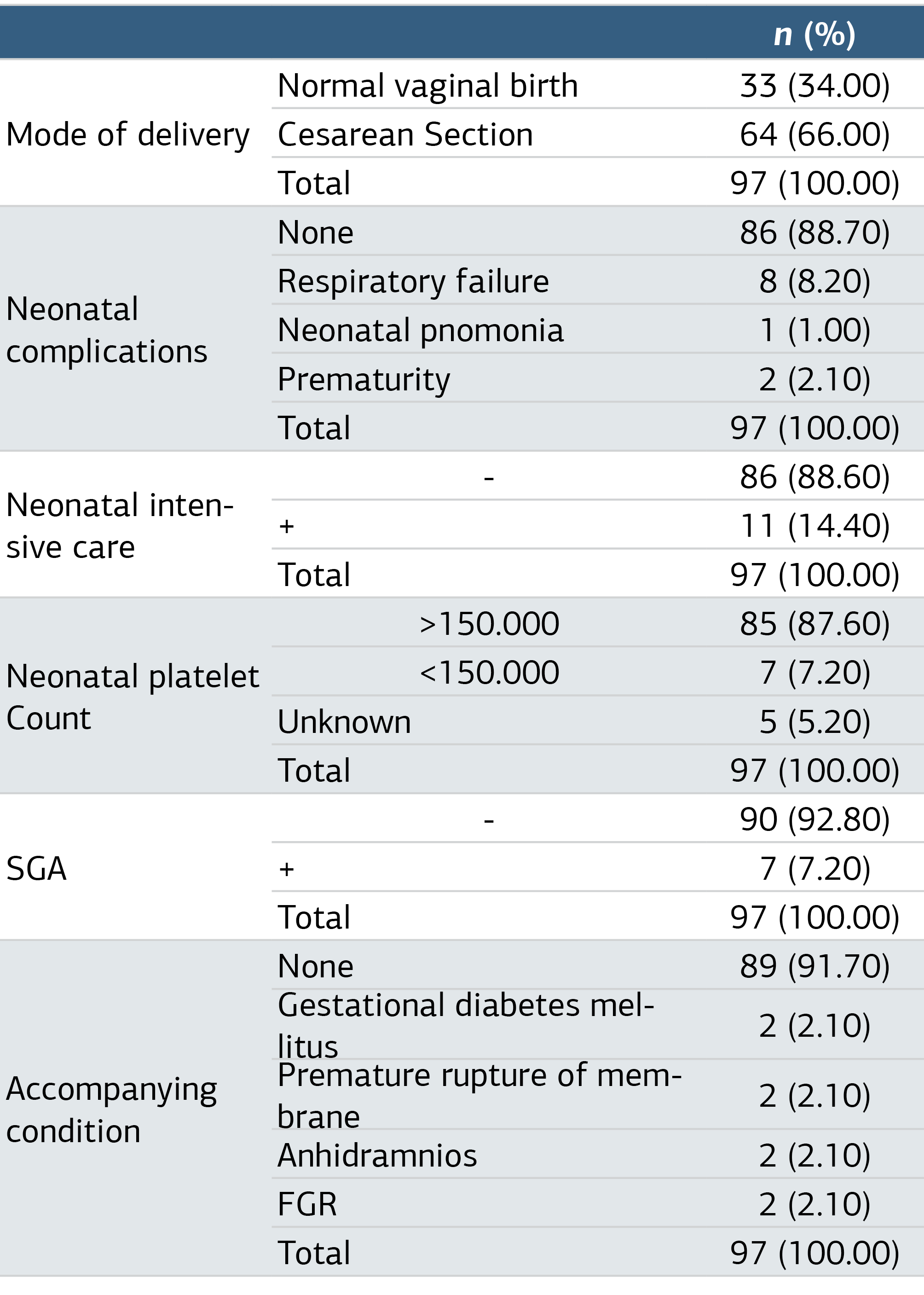
Neonatal intensive care was not needed in 86 (88.60%) neonates, while 11 (11.40%) patients needed neonatal intensive care. When the neonatal platelet value was analyzed, it was determined that this information was not available for 5 babies, platelet value was above 150,000 in 85 babies, whereas this value was lower than 150,000 in 7 babies. Small for gestational age (SGA) was present in 7 of the newborns, and 90 babies (92.80%) did not have SGA (Table 2). When the need for blood transfusion was analyzed in our study, it was determined that 75 (77.30%) patients did not receive blood transfusion and 22 (22.70%) patients received blood transfusion. Erythrocyte, FFP, and platelet suspension were given as blood transfusions. When the distribution of these transfusions was analyzed, it was determined that 88 patients received no RBCs, 5 patients (5.2%) received one unit of RBCs, 2 patients (2.1%) received two units of RBCs, and one patient each received 3 and 6 units of RBCs. 92 patients received no FFP, 3 patients received 2 units, and one patient each received 3 and 4 units of FFP. 77 patients did not receive platelets, 12 patients received one unit, 5 patients received 2 units, 2 patients received 3 units, and 1 patient received 5 units of platelets. The 1st and 5th minute Apgar scores of the babies born from the patients included in the study were analyzed. The mean 1st minute Apgar score was 6.1 ± 1.5 (min-max: 3-10), while the mean 5th minute Apgar score was 8.4 ± 1.0 (min-max: 6-10).
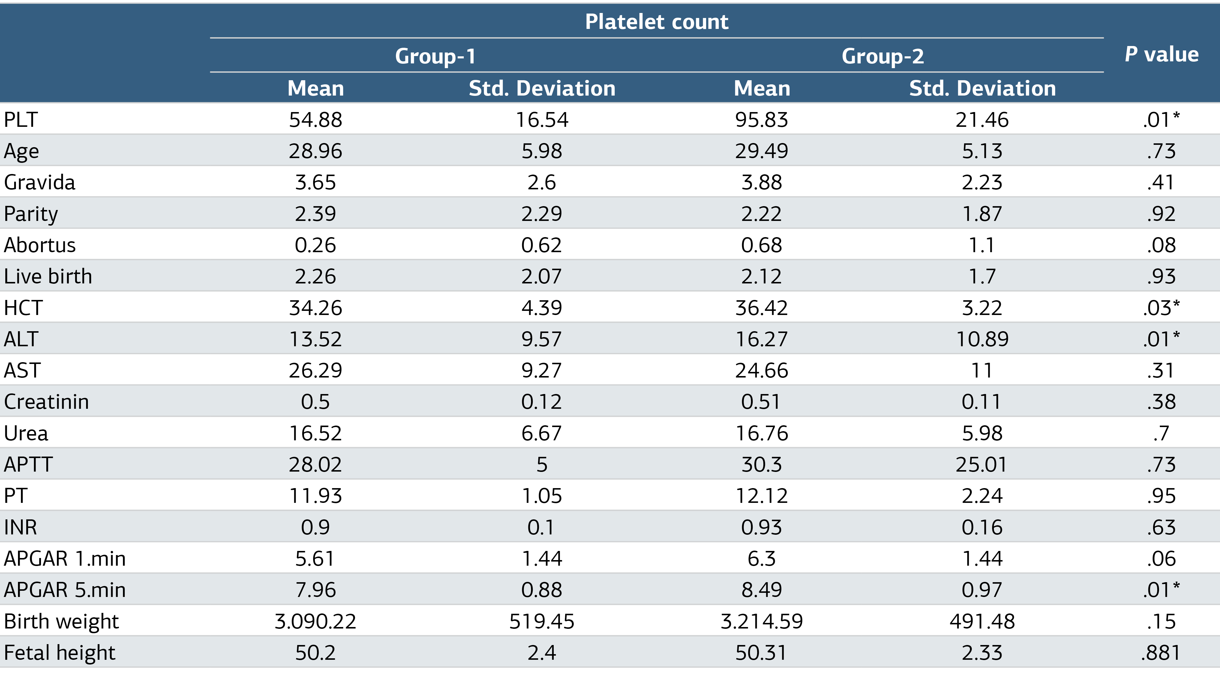
There was a significant difference in hematocrit values, and the mean hematocrit value of the patients in Group-2 (36.42 ± 3.22) was higher than the mean hematocrit value of the patients in Group-1 (34.26 ± 4.39) (P = .03). Similarly, Group-2 ALT values (16.27 ± 10.89) were significantly higher than Group-1 ALT values (13.52 ± 9.57) (P = .01). The mean Apgar at 5 minutes was significantly higher in Group-2 (8.49 ± 0.97) compared to Group-1 (7.96 ± 0.88) (P = .01) (Table 3).
We also compared the need for blood transfusion between Group-1 and Group-2 and which blood products were administered for transfusion in transfused patients. The rate of need for blood transfusion in Group-1 patients (60.90%) was significantly higher than in Group-2 patients (P = .001). The need for erythrocyte suspension in patients in Group-1 was statistically significantly higher (P = .033). Even when an 80,000 platelet count was taken as the limit, the need for erythrocyte transfusion was found to be significantly higher (P = .002).
Discussion
With this study, in parallel with the literature, we concluded that GT does not cause preterm delivery. In the study published by Sadulla et al. in 2020, which included 802 pregnant women who gave birth with a diagnosis of gestational thrombocytopenia, the mean age of the patients was found to be 27 years.9 According to a study published in 2021 by Fogerty et al., 460 (12%) of 3691 women who gave birth were diagnosed with GT and compared with a control group of 3231 patients without thrombocytopenia. The mean age of patients diagnosed with GT was 33 years. When compared with the control group, no significant difference was found between them in terms of maternal age and gravida status.10 In a study published by Şahin et al. in 2019, in which 240 healthy pregnant women and 80 pregnant women diagnosed with gestational thrombocytopenia were compared, the mean age of patients diagnosed with GT was 29, mean gravida was 3, mean parity was 1.9, and no significant difference was found between the control group in terms of maternal age and gravidity-parity.11 Low platelet count in GT was not associated with gravidity and parity. Vaginal delivery is recommended in GT cases, and cesarean section should be decided according to obstetric indications.8 In the study conducted by Fogerty et al., 460 patients with a diagnosis of GT, 68% of the patients had NVB, 28% had C/S, and 3% had vaginal delivery after cesarean section. When compared with the control group with normal platelet count, the mode of delivery was found to be similar.10 In another study by Sadulla et al., it was reported that 572 (71.3%) of 802 pregnant women with a diagnosis of GT delivered vaginally. In contrast, 230 (28.7%) patients were delivered by cesarean section due to obstetric indications (non-progressive trauma, fetal distress, oligohydramnios, breech presentation, and repeat cesarean section).9 In our study, it was found that 33 (34%) of 97 pregnant women underwent vaginal delivery and 64 (66%) underwent cesarean section (C/S). As in the literature, indications for cesarean section in our study were determined according to obstetric reasons. However, contrary to other studies, the reason why the cesarean section rate was higher than the normal delivery rate was that patients with a history of repeated cesarean sections were more likely to apply to our hospital, which is a tertiary center.
According to current guidelines, vaginal delivery is considered safe when the platelet count is >30,000/mm³. For operative vaginal delivery or cesarean section, the safe platelet count should be at least 50,000/mm³.12 Severe thrombocytopenia detected during pregnancy is not an indication of termination of pregnancy. With etiology-directed treatment, pregnancy can be followed up without any risk for the mother-fetus.13 In a study conducted by Elveđi-Gašparović et al. and published in 2016, the platelet count was found to be 50,000-100,000/mm³ in 38 and <50,000/mm³ in 12 of 50 patients diagnosed with gestational thrombocytopenia. When compared with the control group with platelet counts within normal limits, it was found that GT had no significant effect on mode of delivery and preterm delivery, although the platelet counts of the patients with GT included in the study were <100,000/mm³.14
Although there was a statistically significant difference, we think that platelet count was not related to ALT values because the values were within the normal range. We found that the mean Apgar score was within the normal range in both groups. There was a significant difference in Apgar 5 min score at a platelet count of 70,000/mm³, whereas there was no statistically significant difference when the cut-off value was 80,000/mm³. Therefore, we think that a platelet count of 70,000/mm³ can be accepted as the cut-off value for Apgar 5 min.
In our study, even when a platelet count of 50,000/mm³ was taken as the cut-off value, no significant difference was observed in terms of neonatal thrombocytopenia. As a result of the findings of our study, we concluded that GT may cause neonatal thrombocytopenia, but maternal platelet count did not affect neonatal thrombocytopenia.
In addition, although there is no data in the literature on blood transfusion (erythrocyte-platelet-FFP) in pregnant women diagnosed with gestational thrombocytopenia in the intrapartum or early postpartum period, in our study, it was observed that platelet transfusion was needed in patients with plt <100,000, erythrocyte transfusion was needed in the patient group with <80,000, and FFP transfusion was needed in those with <70,000. We think that in patients with gestational thrombocytopenia, a platelet count of 80,000 can be considered as the cut-off value for the need for erythrocyte transfusion, a platelet count of 100,000 for platelet product transfusion, and a platelet count of 70,000 for FFP transfusion.
Limitations
The findings of our study are consistent with the literature and the features that increase the power of our study are that the study was conducted in a single center, focused specifically on gestational thrombocytopenia, and was one of the studies that included a large number of patients (n = 97) grouped according to platelet counts. The main limitations of our study are its retrospective nature and the lack of a control group.
Conclusion
Since there is an increase in the need for platelet transfusion in patients with platelet count <100,000/mm³, erythrocyte transfusion in patients with platelet count <80,000/mm³, and FFP transfusion in patients with platelet count <70,000/mm³, delivery in a more equipped center where blood can be obtained will minimize the risks for mother and baby.
Declarations
Animal and Human Rights Statement
All procedures performed in this study were in accordance with the ethical standards of the institutional and/or national research committee and with the 1964 Helsinki Declaration and its later amendments or comparable ethical standards.
Informed Consent
Informed consent was obtained from all participants.
Data Availability
The datasets used and/or analyzed during the current study are not publicly available due to patient privacy reasons but are available from the corresponding author on reasonable request.
Conflict of Interest
The authors declare that there is no conflict of interest.
Funding
None.
Acknowledgements
The authors would like to thank Dr. Erhan Okuyan contribution.
Abbreviations
ALT: Alanine aminotransferase
C/S: Cesarean section
DIC: Disseminated intravascular coagulation
FFP: Fresh frozen plasma
FGR: Fetal growth restriction
GT: Gestational thrombocytopenia
HELLP: Hemolysis, elevated liver enzymes, and low platelet count
ITP: Idiopathic thrombocytopenic purpura
IVIG: Intravenous immunoglobulin
MRI: Magnetic resonance imaging
RBC: Red blood cell
SGA: Small for gestational age
SPSS: Statistical package for the social sciences
References
- Myers B. Diagnosis and management of maternal thrombocytopenia in pregnancy. Br J Haematol. 2012;158(1):3-15. doi:10.1111/j.1365-2141.2012.09135.x
- Adams TM, Allaf MB, Vintzileos AM. Maternal thrombocytopenia in pregnancy: diagnosis and management. Clin Lab Med. 2013;33(2):327-334. doi:10.1016/j.cll.2013.01.002
- Boehlen F, Hohlfeld P, Extermann P, Perneger TV, De Moerloose P. Platelet count at term pregnancy: a reappraisal of the threshold. Obstet Gynecol. 2000;95(1):29-33. doi:10.1016/s0029-7844(99)00537-2
- Jensen JD, Wiedmeier SE, Henry E, Silver RM, Christensen RD. Linking maternal platelet counts with neonatal platelet counts and outcomes using the data repositories of a multihospital health care system. Am J Perinatol. 2011;28(8):597-604. doi:10.1055/s-0031-1276733
- Özsürmeli M, Sucu M, Çetin C, Khatib G, Demir C. Gebelikte trombositopeni [Thrombocytopenia in pregnancy]. Arsiv Kaynak Tarama Derg. 2016;25(3):420-432. doi:10.17827/aktd.248424
- Gill KK, Kelton JG. Management of idiopathic thrombocytopenic purpura in pregnancy. Semin Hematol. 2000;37(3):275-289. doi:10.1016/s0037-1963(00)90106-9
- Parnas M, Sheiner E, Shoham-Vardi I, et al. Moderate to severe thrombocytopenia during pregnancy. Eur J Obstet Gynecol Reprod Biol. 2006;128(1-2):163-168. doi:10.1016/j.ejogrb.2005.12.031
- Webert KE, Mittal R, Sigouin C, Heddle NM, Kelton JG. A retrospective 11-year analysis of obstetric patients with idiopathic thrombocytopenic purpura. Blood. 2003;102(13):4306-4311. doi:10.1182/blood-2002-10-3317
- Sadulla SMS, Mahmood AY, Shamdeen MY. Gestational thrombocytopenia: maternal and fetal outcomes in Duhok. Duhok Med J. 2020;14(2):86-96.
- Fogerty AE, Dzik W. Gestational thrombocytopenia: a case-control study of over 3500 pregnancies. Br J Haematol. 2021;194(2):433-438. doi:10.1111/bjh.17611
- Şahin ME, Şahin E, Madendağ Y, Çöl İ, Açmaz G. Effect of gestational thrombocytopenia on negative fetal and maternal outcomes in low-risk pregnancies. Cumhur Med J. 2019;41(3):620-625.
- Ekéus C, Högberg U, Norman M. Vacuum-assisted birth and risk for cerebral complications in term newborn infants: a population-based cohort study. BMC Pregnancy Childbirth. 2014;14(1):1-10.
- Wang DP, Liang MY, Wang SM. Clinical analysis of pregnancy complicated with severe thrombocytopenia. Zhonghua Fu Chan Ke Za Zhi. 2010;45(6):401-405.
- Elveđi-Gašparović V, Beljan P, Gverić-Ahmetašević S, Schuster S, Škrablin S. Fetomaternal complications and their association with gestational thrombocytopenia. Ginekol Pol. 2016;87(6):454-459. doi:10.5603/gp.2016.0025
Tables
Table 1. Evaluation of Patients’ Delivery Week, Obstetric Complication Status and Maternal Intensive Care
*Results were analyzed by Pearson chi-square test (percentage; %).
Table 2. Evaluation of Mode of Delivery, Neonatal Complications, Need for Neonatal Intensive Care, SGA, Neonatal Platelet Value and Accompanying Conditions During Pregnancy
*Results were analyzed by Pearson chi-square test (percentage; %).
Table 3. Comparison of Demographic Characteristics, Laboratory Values, APGAR Score, Height and Birth Weight of Newborns in Group-1 and Group-2 Patients
*Mann-Whitney U test was used in the comparisons and significant values are marked with*.
Additional Information
Publisher’s Note
Bayrakol MP remains neutral with regard to jurisdictional and institutional claims.
Rights and Permissions

This work is licensed under a Creative Commons Attribution-NonCommercial 4.0 International License (CC BY-NC 4.0). To view a copy of the license, visit https://creativecommons.org/licenses/by-nc/4.0/
About This Article
How to Cite This Article
Nebahat Sunar, Mulaim Sizer, Hamdin Günsel, Gülten Çirkin Tekeş, Dicle Akkılıç Dönmez, Mehmet Sait İçen. Evaluation of maternal and fetal outcomes in pregnant women with gestational thrombocytopenia. Ann Clin Anal Med 2024;15(6):439-443. doi:10.4328/ACAM.22186
Publication History
- Received:
- 20.03.2024
- Accepted:
- 13.05.2024
- Published Online:
- 30.05.2024
- Printed:
- 01.06.2024