
Abstract
IntroductionInitially termed geometric phagedenism, pyoderma gangrenosum represents a rare, destructive, and highly painful ulcerative lesion affecting the integumentary system. Clinical and pathological manifestations lack distinctiveness. Case PresentationDiagnosis primarily relies on a differential approach, excluding alternative etiologies contributing to the formation of cutaneous ulcers. Given the exclusive reliance on immunosuppressive therapy for Pyoderma gangrenosum, meticulous consideration must be given to excluding other potential causes of ulcerative skin lesions, particularly those stemming from infectious agents where immunosuppression is contraindicated. Frequently, underlying pathologies are implicated, exemplified in our scenario by inflammatory bowel disease. In our study, we present a case involving multiple ulcerated skin lesions. During the evaluation process, we not only diagnosed pyoderma gangrenosum but also identified the underlying etiology as previously misdiagnosed Crohn’s disease.ConclusionTreatment with anti-tumor necrosis factor (anti-TNF) therapy resulted in complete resolution of the skin lesions and remission of the inflammatory bowel disease.
Keywords
Introduction
Pyoderma gangrenosum (PG) is an uncommon inflammatory skin disorder marked by pustules that evolve into non-infectious, ulcerative skin lesions. While most occurrences happen spontaneously and are linked to hematologic, rheumatologic, or gastrointestinal conditions, these lesions can also develop at sites of trauma or surgery.1 It falls under the spectrum of neutrophilic dermatoses, with an incidence of approximately 0.63 per 100,000 individuals. In up to fifty percent of cases, an underlying pathology is identified. There is no pathognomonic feature, necessitating a thorough differential diagnosis.2 We present an intriguing case of inflammatory bowel disease, initially misdiagnosed as ulcerative colitis and subsequently corrected to Crohn’s disease, accompanied by disseminated pyoderma gangrenosum skin lesions. Successful treatment was achieved with anti-tumor necrosis factor (anti-TNF) therapy.
Case Presentation
A 63-year-old female patient was admitted to the general surgery ward with acute abdominal symptoms. In addition to abdominal pain, the patient presented with ulcerated lesions localized on the dorsal aspects of the left foot, right shoulder, and vertex of the scalp (Figure 1).
Three months prior to admission, the patient had been diagnosed with ulcerative colitis and was initiated on 5-aminosalicylate therapy. Despite treatment, there was no alleviation of symptoms, including persistent diarrhea (4-5 episodes per day), ongoing abdominal pain, and significant weight loss (~10 kilograms). A physical examination revealed mild abdominal tenderness without signs of rigidity or rebound. Additionally, a perianal fistula with spontaneous drainage was noted. Abdominal computed tomography and perianal magnetic resonance imaging demonstrated thickening of the ileocecal region and the presence of an intersphincteric perianal fistula (Figure 2). Colonoscopy revealed ulceration in the ileocecal region (Figure 3), an internal opening of the perianal fistula in the rectum, and normal mucosa elsewhere in the colon. Biopsies were obtained from the terminal ileum, as well as from the ulcerated skin lesions. Microbiological analyses of the skin lesions and ileal biopsies did not yield evidence of specific microbial pathogens. Histopathological examination of the skin biopsy revealed findings consistent with Pyoderma Gangrenosum (PG), while biopsy of the ileum demonstrated features indicative of active chronic colitis. Following a discussion with the Dermatology department, based on the clinical, radiological, endoscopic, and histopathological findings, the patient was diagnosed with Pyoderma Gangrenosum secondary to perianal fistulizing Crohn’s disease involving the ileocolonic region. Following the initiation of anti-tumor necrosis factor (Anti-TNF) therapy, the ulcerated skin lesions showed marked improvement (Figure 1), and the patient achieved remission in her Crohn’s disease. Informed consent was obtained from the patient.Ethical ApprovalNot applicable.Statistical AnalysisNot applicable.
This case report was prepared in accordance with the CARE guidelines.
Discussion
Pyoderma Gangrenosum, initially referred to as Geometric Phagedenism by Louis-Anne-Jean Brocq, is an infrequent, noninfectious neutrophilic dermatosis characterized by the presence of destructive skin ulcerations.2
PG histology is non-specific and varies depending on the stage of the lesion. The early observations reveal the presence of a profound suppurative folliculitis characterized by a dense infiltration of neutrophils. The diagnosis relies exclusively on clinical observations and the elimination of other skin conditions that cause ulcers. PG is commonly observed in conjunction with an underlying medical condition, such as inflammatory bowel disease, inflammatory arthritis, hematological illnesses, and solid malignancies, in around 50% to 70% of instances.3
Approximately 40% of individuals with inflammatory bowel disease (IBD) experience an extraintestinal manifestation of the condition, with the skin being the organ most frequently affected. Common skin manifestations often include erythema nodosum or pyoderma gangrenosum. Pyoderma gangrenosum affects approximately 1-2% of individuals with inflammatory bowel disease (IBD). PG has a yearly occurrence of up to 10 per million and has a tendency to impact women more frequently than men.4
In our case, the patient was a female with underlying Crohn’s disease, consistent with findings in existing literature. This association between pyoderma gangrenosum and Crohn’s disease has been noted in previous studies, highlighting the importance of recognizing such comorbidities in clinical practice.
The cornerstone of therapeutic intervention lies within immunosuppression, encompassing both topical and systemic modalities. It is imperative to underscore that prior to embarking on the treatment regimen for PG, meticulous exclusion of alternative etiologies is essential. This precautionary measure is paramount due to the contraindication of immunosuppressive therapy in the majority of other etiological factors precipitating cutaneous ulceration.5
After anti-TNF therapy was initiated, we observed successful healing of the pyoderma gangrenosum ulcerations along with remission of the patient’s Crohn’s disease. This treatment outcome underscores the effectiveness of anti-TNF agents in managing both conditions concurrently, highlighting the potential benefit of this therapeutic approach for patients presenting with this dual pathology.
Limitations
The main limitation of this report is that it presents a single case with limited follow-up.
Conclusion
Pyoderma gangrenosum continues to be an uncommon condition that poses challenges in diagnosis and is seldom encountered by clinicians. Healthcare providers should keep a high level of suspicion, especially in patients with a history of underlying immune conditions, negative culture results, lack of response to antibiotics, or a worsening response to debridement. Timely identification and collaboration with a multidisciplinary team enable proper treatment and help avoid unnecessary procedures.
Declarations
Animal and Human Rights Statement
All procedures performed in this study were in accordance with the ethical standards of the institutional and/or national research committee and with the 1964 Helsinki Declaration and its later amendments or comparable ethical standards.
Informed Consent
Informed consent was obtained from the patient.
Data Availability
The datasets used and/or analyzed during the current study are not publicly available due to patient privacy reasons but are available from the corresponding author on reasonable request.
Conflict of Interest
The authors declare that there is no conflict of interest.
Funding
None.
Abbreviations
IBD: Inflammatory bowel disease
PG: Pyoderma gangrenosum
References
- Binus AM, Qureshi AA, Li VW, Winterfield LS. Pyoderma gangrenosum: a retrospective review of patient characteristics, comorbidities, and therapy in 103 patients. Br J Dermatol. 2011;165(6):1244-1250. doi:10.1111/j.1365-2133.2011.10565.x
- George C, Deroide F, Rustin M. Pyoderma gangrenosum: a guide to diagnosis and management. Clin Med (Lond). 2019;19(3):224-228. doi:10.7861/clinmedicine.19-3-224
- Hobbs MM, Ortega-Loayza AG. Pyoderma gangrenosum: from historical perspectives to emerging investigations. Int Wound J. 2020;17(5):1255-1265. doi:10.1111/iwj.13389
- Chakiri R, Baybay H, Hatimi AE, Gallouj S, Harmouch T, Mernissi FZ. Clinical and histological patterns and treatment of pyoderma gangrenosum. Pan Afr Med J. 2020;36:59. doi:10.11604/pamj.2020.36.59.12329
- Shahid S, Myszor M, De Silva A. Pyoderma gangrenosum as a first presentation of inflammatory bowel disease. BMJ Case Rep. 2014;2014. doi:10.1136/bcr-2014-204853
Figures
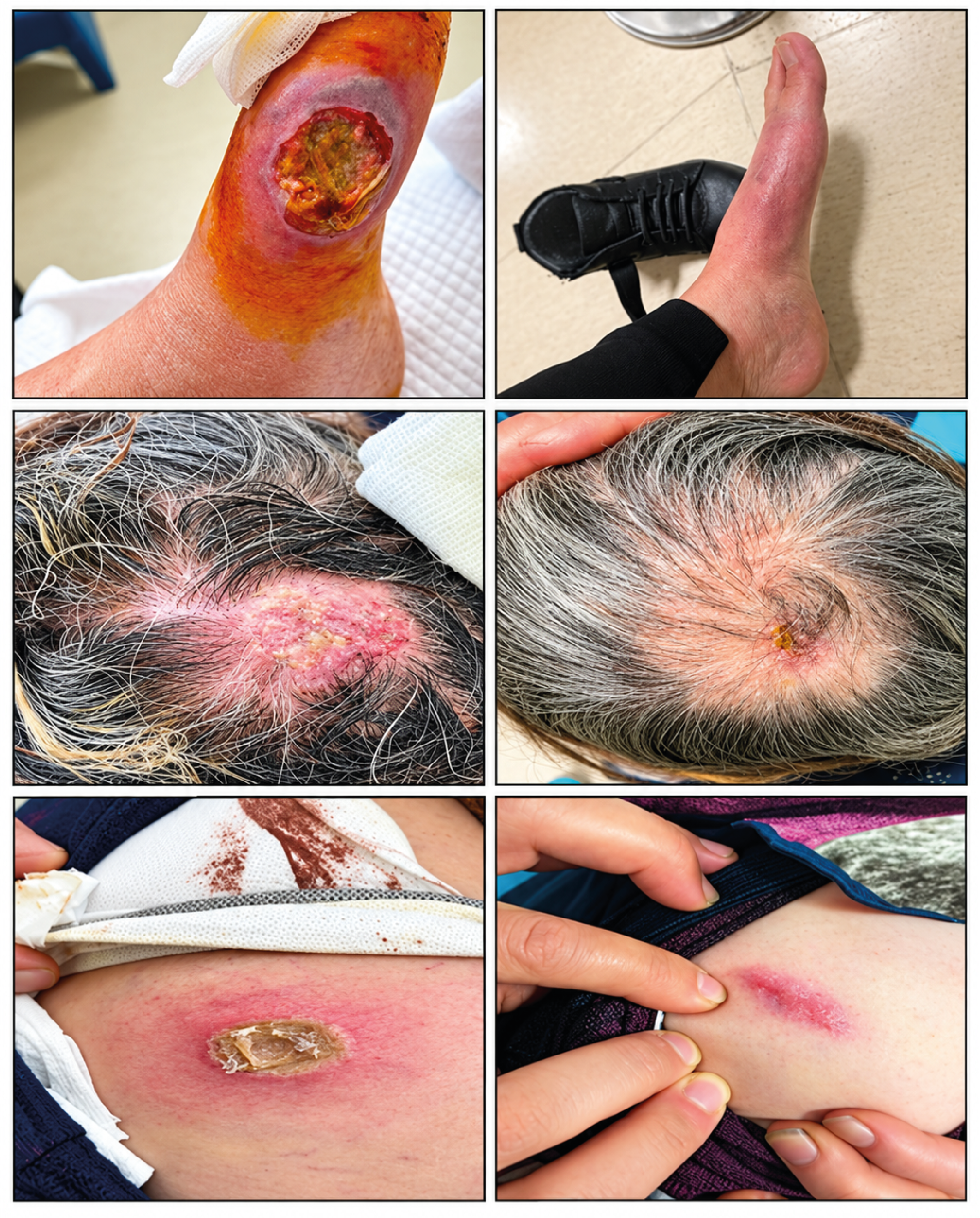
Figure 1. Pyoderma Gangrenosum Ulcerated Skin Lesions Before and After Treatment (On the Left and Right, Respectively) Appear to Have Completely Healed
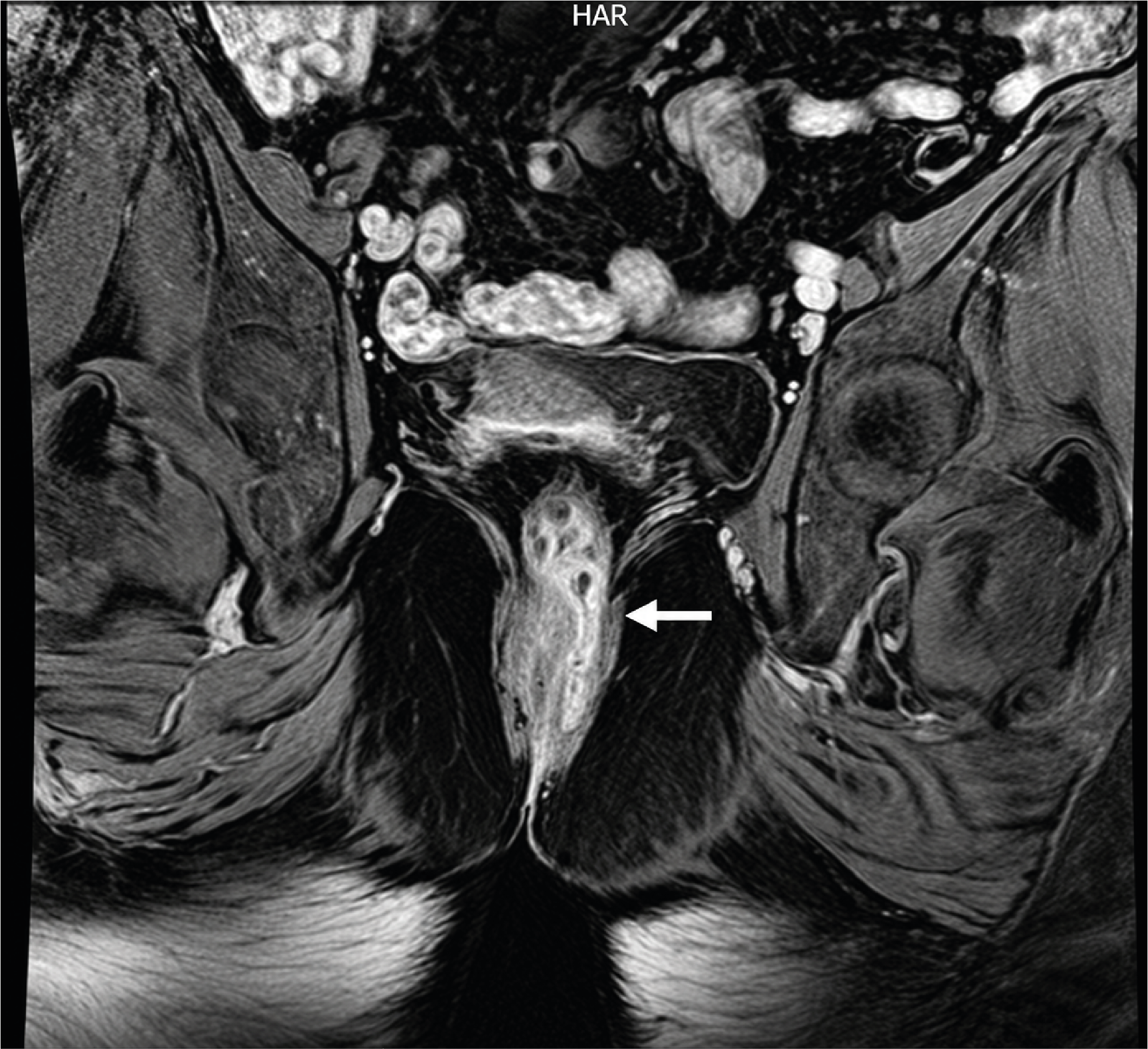
Figure 2. MRI Imaging Reveals the Presence of a Perianal Fistula Tract
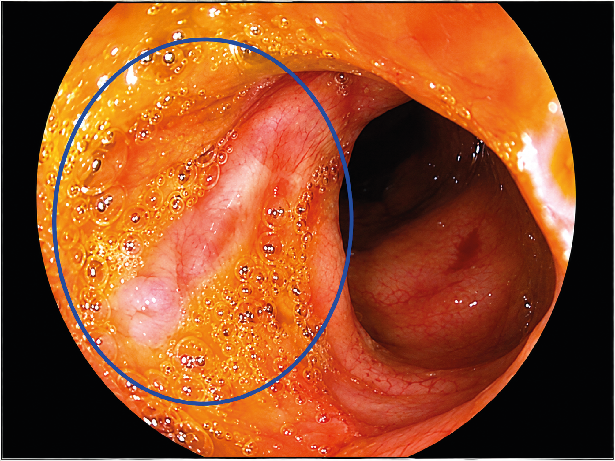
Figure 3. Colonoscopy Demonstrates the Presence of an Ulcer Adjacent to the Ileocecal Valve
Additional Information
Publisher’s Note
Bayrakol MP remains neutral with regard to jurisdictional and institutional claims.
Rights and Permissions

This work is licensed under a Creative Commons Attribution-NonCommercial 4.0 International License (CC BY-NC 4.0). To view a copy of the license, visit https://creativecommons.org/licenses/by-nc/4.0/
About This Article
How to Cite This Article
İdris Kurt. Geometric phagedenism: A Case Report. Ann Clin Anal Med 2024;15(Suppl 2):93-95. doi:10.4328/ACAM.22228
Publication History
- Received:
- 21.04.2025
- Accepted:
- 27.05.2025
- Published Online:
- 03.07.2024
- Printed:
- 20.10.2024