
Abstract
AimWe aimed to examine the relationship between OCT findings and oxidation and antioxidation balance, which is thought to have a role in the etiopathogenesis of the disease in patients with fibromyalgia.MethodsA total of 39 female patients with FMS and 41 healthy volunteers were included in the study. Plasma native thiol (NT), total thiol (TT), disulfide (DS), NT/TT, DS/NT, DS/TT, TOS, TAS and OSI values, and eye OCT measurements were evaluated. Regarding OCT measurements, the subfoveal choroid layer, retinal nerve fiber layer, ganglion cell layer, and macular thickness were assessed.ResultsWhen compared to the healthy control group, TAS, TOS, NT, TT, DS levels were high in the FMS patient group (all P < .05), whereas other measurements were found to be similar (all P > .05). The increase in TOS levels (P < .001) in the FMS group was much more significant than the increase in TAS levels (P = .030). In terms of OCT findings, there was no significant difference between the right and left eye subfoveal choroid thicknesses between the two groups (both <0.001). ConclusionThe increase in TOS levels suggests that the level of antioxidants for compensation of increased oxidant status also increases because it is much higher than TAS increase. Since the OSI values are similar, we can state that we did not detect an increase in oxidative stress. We think that FMS patients with high FIQ scores, symptoms, and complaints should be evaluated in terms of oxidative stress and related possible complications.
Keywords
Introduction
Fibromyalgia syndrome (FMS) is one of the most common causes of chronic widespread musculoskeletal pain, the etiopathogenesis of which has not yet been determined, and systemic symptoms are often accompanied. Autonomic nervous system dysregulation and increased oxidative stress are two of the many factors implicated in the etiopathogenesis.1
During oxidative phosphorylation, where most of the body’s energy is produced, free oxygen radicals are formed. In the normal state, there is a balance between reactive oxygen species (ROS) and antioxidants in the intracellular, membranes and extracellular spaces. The increase in oxidative stress is due to the imbalance between oxidation products and antioxidant defense products. Parameters such as thiol/disulfide balance, total oxidant status (TOS), total antioxidant status (TAS) and oxidative stress index (OSI) are used to calculate the balance between oxidation and antioxidation. Thiols are important antioxidants and form reversible disulfide bonds with reactive oxygen radicals in order to balance oxidative stress in the body. It has an important role in many cellular activities such as thiol/disulfide homeostasis, maintenance of oxidant/antioxidant balance, detoxification, cellular signal transduction, transcription, cell growth and apoptosis. The etiopathogenetic role of changes in thiol/disulfide homeostasis in hypertension, preeclampsia, many autoimmune diseases, gastrointestinal and cardiovascular system diseases has been investigated previously. It is thought that imbalances in thiol/disulfide homeostasis cause an increase in oxidative stress products, leading to the emergence of diseases through inflammation.2,3 Although there are a limited number of studies in the literature examining the thiol/disulfide balance or TAS, TOS values in FMS, we found only one study in which both parameters were studied together.4
Optical coherence tomography (OCT) devices can be used in the diagnosis and follow-up of diseases by obtaining high-resolution tomographic images in biological tissues, especially in the eye, without using any contrast agent or radiation.5,6 The number of studies evaluating the OCT findings of patients with FMS is very limited.7
In our study, we aimed to examine the relationship between OCT findings and oxidation and antioxidation balance, which is thought to have a role in the etiopathogenesis of the disease in patients with FMS. Despite our extensive literature review, our study is a first in this regard, as we have not found any publication examining the OCT findings of patients with FMS and the oxidant/antioxidant balance relationship.
Materials and Methods
Female patients with FMS, aged 20-50 years, with old and new diagnoses, who applied to the Physical Medicine and Rehabilitation outpatient clinic between March 2019 and March 2020, and healthy volunteer women in the same age group were included in the study.
The participants in our study were divided into two groups as those with FMS and healthy volunteers. Demographic data of both groups were recorded. In addition, venous blood samples were taken from the forearm of the two groups and intraocular pressure was measured before the OCT procedure. The 2010 ACR diagnostic criteria, Fibromyalgia Impact Questionnaire (FIQ) and Visual Pain Scale (VAS) were filled in the FMS group.
A 5 cc tube blood sample of both groups included in the study was taken and stored at –80°C until the study time. Serum total oxidant, total antioxidant parameters and thiol/disulfite levels were studied considering the manufacturer’s recommendations and expiration dates.
In addition to the characteristics of all participants such as age, BMI, smoking, occupation, marital status, number of children, sports/exercise habits, duration of diagnosis and medical treatments of FMS patients were recorded.
In order to evaluate the central macular, optic nerve and choroidal thicknesses of all participants, SD-OCT recordings were made with the Heidelberg spectralis OCT device (Heidelberg Engineering, Heidelberg, Germany) in our hospital. All results were evaluated by the same ophthalmologist. In this evaluation, retinal nerve fiber thickness, ganglion cell layer thickness, subfoveal choroidal layer thickness and macular thickness were evaluated. In addition, in order to exclude retinal nerve fiber thickness and eye hypertension affecting other structures, eye pressures of all groups were measured before extraction and those less than 21 mmHg were included in the study. This study was produced in the specialization thesis.Ethical ApprovalThis study was approved by the Ethics Committee of Harran University, Faculty of Medicine (Date: 11.03.2019, Decision No: HRÜ/19.03.03).Statistical AnalysisStatistical Package for the Social Sciences (SPSS) 20 for Windows package program (SPSS Inc., Armonk, NY USA) was used for data analysis. The Shapiro Wilk Test and histogram graphs were used to assess whether the data conformed to the normal distribution. Descriptive statistics were given as mean ± standard deviation, median (minimum-maximum), or number and percentage. Numerical data between groups were compared using Student’s t test or Mann Whitney U test. Categorical data were compared using Chi-Square or Fischer’s Exact test. Pearson or Spearman correlation tests were used for correlation analysis. The relationship between the parameters that were significant between the groups was determined by multiple linear regression analysis. P < .05 was considered statistically significant.
Reporting GuidelinesThis study was reported in accordance with the STROBE guideline.
Results
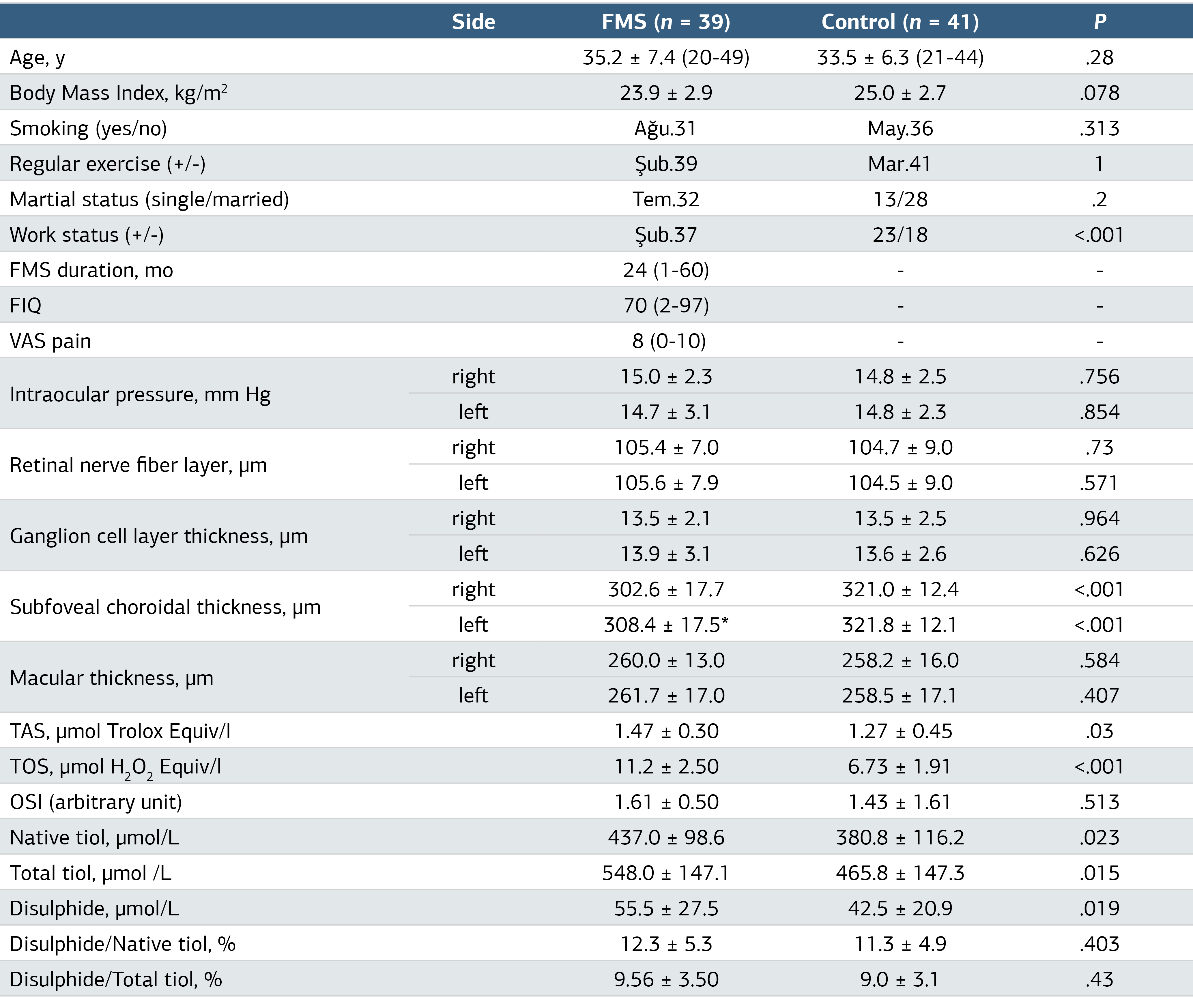
39 women with FMS and 41 healthy volunteer women aged 20-50 years were included in the study. The mean age of all participants was 34.4 ± 6.9 years, and their mean BMI was 24.5 ± 2.8 kg/m2. The mean values of the right and left intraocular pressures of the participants were measured as 14.9 ± 2.4mmHg and 14.7 ± 2.7mmHg, respectively. While 82.1% (n = 32) of FMS patients were married, 68.3% (n = 28) of healthy volunteers were married. While 94.9% (n = 37) of those with FMS were housewives, this rate was 43.9% in the other group. While there was no participant with a history of alcohol use, the rate of smoking was 20.5% (n = 8) in the group with FMS, while this rate was 12.2% (n = 5) in the healthy group.
The comparison of demographic and OCT measurement values of the group with FMS and the group consisting of healthy volunteers is shown in Table 1. In the OCT findings of both groups, only the right and left eye subfoveal choroidal thickness measurements were significantly different, and these two values were lower in the FMS group.
Comparative blood tests for the examination of oxidative stress parameters and thiol/difulfide balance of the two groups are summarized in Table 1. TOS and TAS values were found to be significantly higher in the group with FMS.
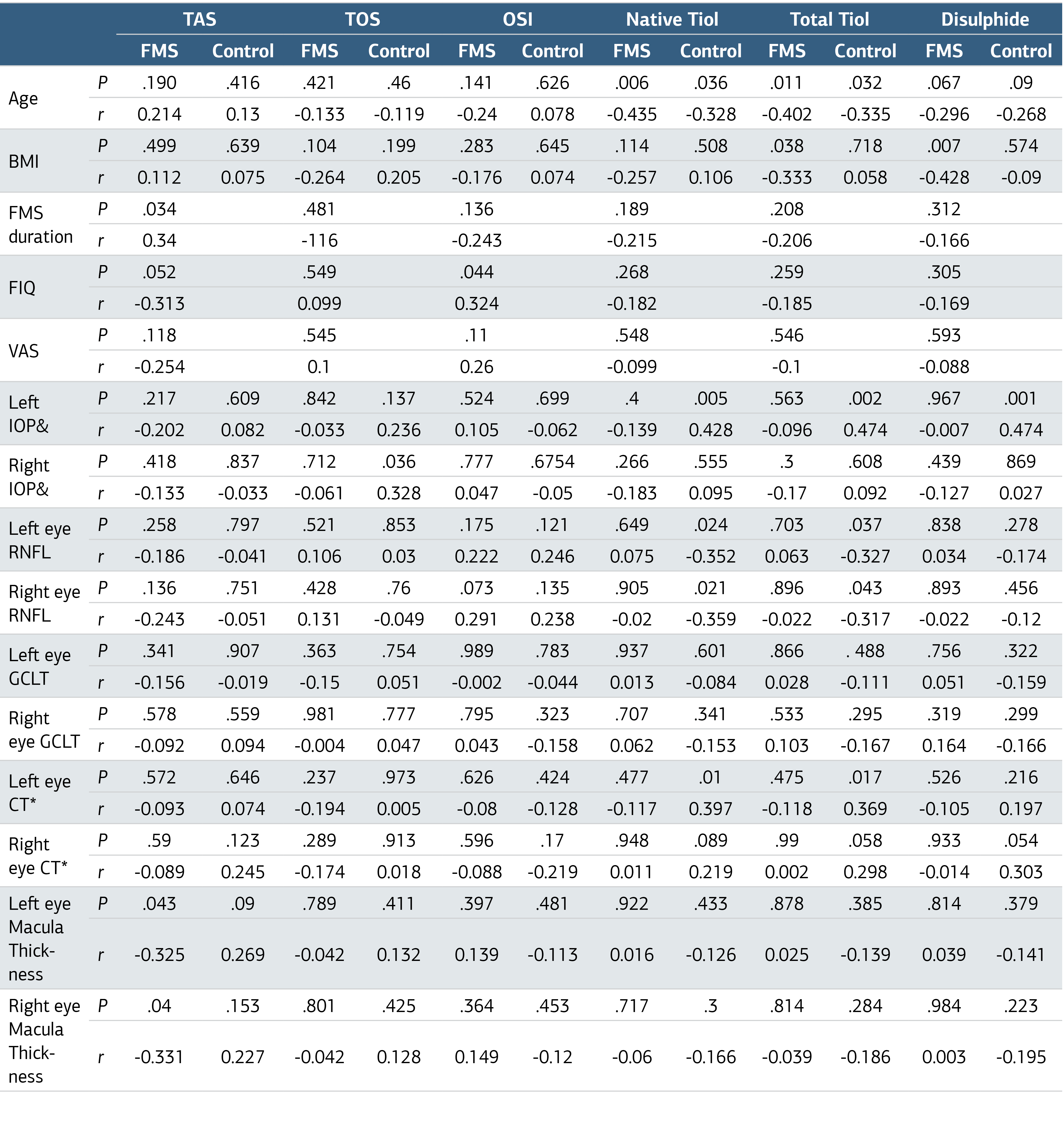
The correlations between demographic and clinical characteristics of FMS patients and healthy control group are summarized in Table 2. There was a negative correlation between the ages of all participants and their native and total Thiol values. FMS diagnosis time was not correlated with any blood or OCT finding, except for a positive correlation with TAS level. While there were no findings correlated with VAS, FIQ was positively correlated with OSI. Right and left eye RNFL measurements were negatively correlated with native thiol and total thiol only in the healthy group. It was determined that the macular thickness of the right and left eyes were negatively correlated with TAS levels only in the FMS group.
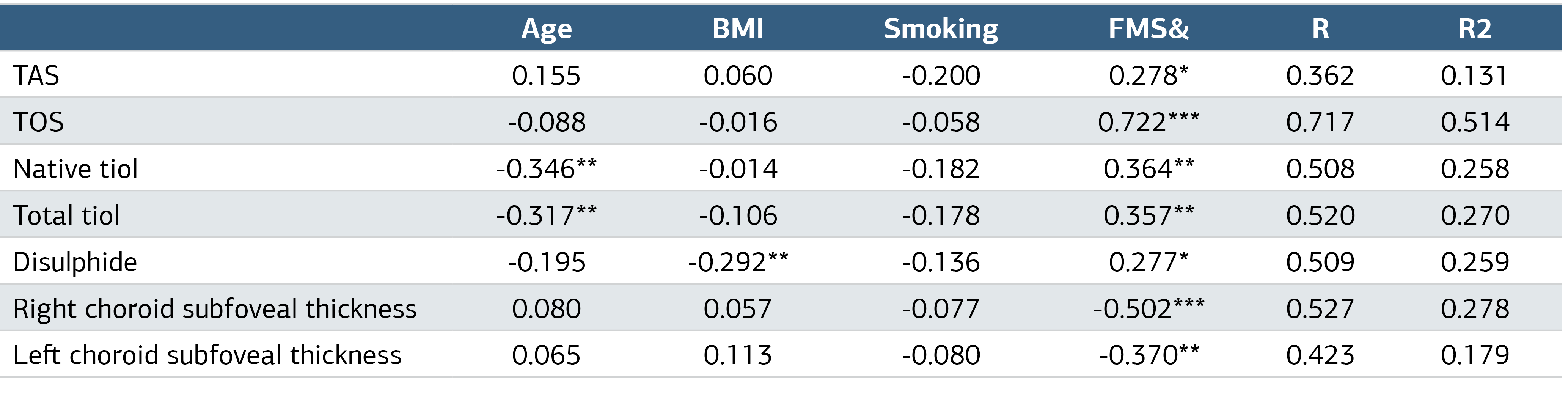
The relationship between oxidant-antioxidant results and eye findings, which differed significantly between the FMS group and the healthy control group, and FMS after excluding (normalizing) the effect of age, BMI, and smoking by regression analysis is summarized in Table 3.
Discussion
There are studies suggesting that oxidative stress disorder may play a role in the pathogenesis of fibromyalgia syndrome.4 Increased oxidative stress results from an imbalance between oxidation products and antioxidant defense products. It has been stated that increased oxidative stress may lead to some inflammatory clinical conditions. Recent data suggest an association between oxidative stress and pain. Lipid peroxidation resulting from increased oxidative stress has been described in patients suffering from depression and fatigue, and these two typical symptoms are also frequently found in FMS patients.1,4,8
In our study, in the OCT evaluation of the patients; Subfoveal choroidal thickness measurements were found to be significantly lower in both eyes of the FMS group compared to the healthy control group. However, the retinal nerve fiber layer, ganglion cell layer thickness and macular thickness were similar in both groups.
Compared with the healthy control group, total oxidant status and total antioxidant status were significantly higher in the group with FMS, but there was no significant difference in the oxidative stress index. In addition, while native thiol, total thiol and disulfide levels were high in patients with FMS, there was no significant difference between disulfide/native thiol, disulfide/total thiol, and native thiol/total thiol ratios.
No correlation was observed between subfoveal choroidal thicknesses and age, disease duration, FIQ, VAS and oxidative stress parameters in the group with FMS. A positive correlation was detected with native thiol and total thiol only in the left eyes of the healthy group. Native thiol and total thiol levels were negatively correlated with age in both groups. No correlation was found between TAS, TOS and OSI values and thiol/disulfide homeostasis parameters in both groups.
Ulusoy et al. He measured the eye choroidal thickness of patients with FMS with OCT.5 In addition to the subfoveal region, which was our measurement site, he also examined the nasal and temporal areas. The finding of significant thinning in the choroidal thickness of the eyes in all three regions of the patients with FMS was considered as a result consistent with our study.
Endothelial dysfunction may occur in patients with FMS due to an overactive sympathetic nervous system. Choroidal layer thickness may be affected in these patients, since the control of choroidal blood flow is carried out by the autonomic nervous system and autonomic nervous system changes are observed in FMS. Sympathetic stimulation causes vasoconstriction and decreases choroidal blood flow, while parasympathetic stimulation increases blood flow by causing vasodilation via nitrous oxide. It has been shown that there is also a decrease in optic disc perfusion in patients with FMS. In patients with FMS, worsening of endothelial functions and arterial elasticity features was detected with the increase in FIQ score.9,10
Endothelial dysfunction may have resulted in choroidal ischemia, resulting in thinning of the choroidal layer with an extensive vascular network. Thinning of the choroidal layer has also been detected in diseases that cause high sympathetic activity, such as chronic heart failure and coronary artery disease.11,12 We believe that measuring the thickness of the choroidal layer of the eye with OCT, which is a non-invasive method in FMS patients, may be a stimulant for both the diagnosis of the disease and the prevention of future complications secondary to autonomic nervous system dysfunction.
In our study, there was no significant difference between the FMS and healthy groups in the retinal nerve fiber layer, ganglion cell layer and macular thickness. Garcia et al. OCT measurements of patients with FMS detected a significant thinning in the retinal nerve fiber layer, inner plexiform layer, and ganglion cell layers compared to the healthy group.6 As a result of this study, in which the thickness of the choroid and macula layers was not evaluated, they mentioned the presence of axonal damage in the optic nerve due to thinning of the RNFL, and stated that it suggests the presence of neurodegenerative processes in FMS.6 The fact that current OCT findings are not similar to our study may be related to patient selection.
Aktekin et al. reported that there was no significant difference in native thiol, total thiol, disulfide levels, disulfite/native thiol, disulfide/total thiol and native thiol/total thiol ratios, TAS, TOS, and OSI values between FMS patients and the control group.9 As a result of their studies, they stated that they found that oxidative stress did not increase in FMS patients. In our study, we did not find a significant difference in OSI values, DS/NT, DS/TT and NT/TT ratios between FMS patients and healthy controls. Again, similar to the aforementioned study, we did not detect any correlation between TAS, TOS, OSI and thiol/disulfide balance parameters. Bozkurt et al. found a positive correlation between the FIQ values and TOS and OSI values of patients with FMS in their studies, but they did not detect a relationship with TAS.13 In our study, the OSI value of the group with a FIQ value of >50 was significantly higher.
Unlike Bozkurt et al., there was no difference between the high FIQ group and the low group in terms of TOS, while the TAS level of the high group was low. Two studies show that there is a relationship between the severity of the disease and the oxidative stress state in FMS.
Antioxidants may be increased to prevent increased oxidative load in mitochondria from causing oxidative damage and deterioration.14 Although the TOS level was significantly higher in the group with FMS in our study, we believe that the less significantly higher TAS level can be considered as an increased antioxidant status in response to the increased oxidant status. Since the patient group included in the study was relatively young, the OSI value may have been normal by providing compensation for the oxidation response.
Perhaps, if the older patient group had been chosen, antioxidant compensation would not have been sufficient and the OSI value would have been high. Fidan et al. and Karataş et al. found similar results in their studies. They showed that the native thiol level and native thiol/total thiol ratio increased, while the disulfide level and disulfide/native thiol and disulfide/total thiol ratios decreased in those with FMS.15,16 In both studies, the mean age and BMI of patients with FMS were considerably higher than in our study. While Fidan et al. did not include any information about smoking in their study, Karataş et al. They stated that smoking status of FMS and the healthy group was similar. Fidan et al. Considering the thiol/disulfide balance, they stated that their results are compatible with the views supporting that FMS is more suitable for proliferative diseases than degenerative diseases and that oxidative stress does not increase.
As can be seen from these results, the results of the few studies examining the thiol/disulfide balance in FMS are contradictory.17 and it is clear that new studies with a high number of participants are needed.
Limitations
The main limitation of our study was the relatively small number of participants. In addition, although smoking status was similar in the healthy and FMS group, the age, duration and amount of starting smoking were not evaluated. Another limitation of the study was the possibility that most of the patients in the FMS group were using drugs, which might affect the results. In addition, the lack of similar studies caused difficulties in comparing the results.
Conclusion
In our study, for the first time, eye OCT findings of patients with FMS and blood oxidant-antioxidant parameters were examined together. Subfoveal choroidal thickness measurements were significantly lower in the patient group with FMS. In addition, we found that there was no significant difference in the retinal nerve fiber layer, ganglion cell layer thickness and macular thickness.
Declarations
Animal and Human Rights Statement
All procedures performed in this study were in accordance with the ethical standards of the institutional and/or national research committee and with the 1964 Helsinki Declaration and its later amendments or comparable ethical standards.
Informed Consent
Informed consent was waived due to the retrospective nature of the study.
Data Availability
The datasets used and/or analyzed during the current study are not publicly available due to patient privacy reasons but are available from the corresponding author on reasonable request.
Conflict of Interest
The authors declare that there is no conflict of interest.
Funding
None.
Abbreviations
ACR: American College of Rheumatology
BMI: Body mass index
CT: Choroid thickness
DS: Disulfide
FIQ: Fibromyalgia Impact Questionnaire
FMS: Fibromyalgia syndrome
IOP: Intraocular pressure
NT: Native thiol
OCT: Optical coherence tomography
OSI: Oxidative stress index
RNFL: Retinal nerve fiber layer
ROS: Reactive oxygen species
SD-OCT: Spectral-domain optical coherence tomography
SPSS: Statistical Package for the Social Sciences
TAS: Total antioxidant status
TOS: Total oxidant status
TT: Total thiol
References
- Clauw DJ. Fibromyalgia: a clinical review. JAMA. 2014;311(15):1547-1555. doi:10.1001/jama.2014.3266
- Erel O, Neselioglu S. A novel and automated assay for thiol/disulphide homeostasis. Clin Biochem. 2014;47(18):326-332. doi:10.1016/j.clinbiochem.2014.09.026
- Ates I, Altay M, Yilmaz FM, et al. Dynamic thiol/disulfide homeostasis in patients with autoimmune subclinical hypothyroidism. Endocr Res. 2016;41(4):343-349. doi:10.3109/07435800.2016.1156124
- Beyaztaş H, Aktaş S, Güler EM, Ata E. Oksidatif stres fibromiyalji hastalarının ağrı mekanizmalarında katkıda bulunan bir faktör olabilir. Reumatismo. 2023;75(3):121-127.
- Ulusoy MO, Kal A, Işik-Ulusoy S, Kal Ö. Choroidal thickness in patients with fibromyalgia and correlation with disease severity. Indian J Ophthalmol. 2018;66(3):428-432. doi:10.4103/ijo.ijo_877_17
- Garcia-Martin E, Garcia-Campayo J, Puebla-Guedea M, et al. Fibromyalgia is correlated with retinal nerve fiber layer thinning. PLoS One. 2016;11(9). doi:10.1371/journal.pone.0161574
- Pilar Bambo M, Garcia-Martin E, Gutierrez-Ruiz F, et al. Study of perfusion changes in the optic disc of patients with fibromyalgia syndrome using new colorimetric analysis software. J Fr Ophtalmol. 2015;38(7):580-587. doi:10.1016/j.jfo.2015.01.010
- İnanici F. Fibromiyalji sendromu [Fibromyalgia syndrome]. In: Beyazova M, Gökçe Kutsal Y, eds. Ankara: Güneş Kitapevi; 2011:2365-2377.
- Ekinci B, Baran P, Bicer CK, Haliloglu S, Uzkeser H, Carlioglu A. The relationship between thiol/disulphide homeostasis and fibromyalgia. Ann Med Res. 2020;27(2):688-694. doi:10.5455/annalsmedres.2019.12.860
- Ozcan O, Sezgin Ozcan D, Candemir B, Aras M, Koseoglu B, Erol C. Evaluation of aortic elastic properties in patients with fibromyalgia. Int Angiol. 2016;35(1):84-89.
- Ahmad M, Kaszubski PA, Cobbs L, Reynolds H, Smith RT. Choroidal thickness in patients with coronary artery disease. PLoS One. 2017;12(6). doi:10.1371/journal.pone.0175691
- Teberik K, Eski MT, Doğan S, Pehlivan M, Kaya M. Ocular abnormalities in morbid obesity. Arq Bras Oftalmol. 2019;82(1):6-11. doi:10.5935/0004-2749.20190007
- Bozkurt M, Caglayan M, Oktayoglu P, et al. Serum prolidase enzyme activity and oxidative status in patients with fibromyalgia. Redox Rep. 2014;19(4):148-153. doi:10.1179/1351000213y.0000000079
- Shen X, Zheng S, Metreveli NS, Epstein PN. Protection of cardiac mitochondria by overexpression of MnSOD reduces diabetic cardiomyopathy. Diabetes. 2006;55(3):798-805. doi:10.2337/diabetes.55.03.06.db05-1039
- Karatas G, Gunduz R, Haskul I, et al. Dynamic thiol and disulphide homoeostasis in fibromyalgia. Arch Med Sci. 2020;16(3):597-602. doi:10.5114/aoms.2019.87052
- Fidan F, Alkan BM, Uğurlu FG, et al. Dynamic thiol/disulphide homeostasis in patients with fibromyalgia. Arch Rheumatol. 2017;32(2):112.
- Fatima G, Das SK, Mahdi AA. Some oxidative and antioxidative parameters and their relationship with clinical symptoms in women with fibromyalgia syndrome. Int J Rheum Dis. 2017;20(1):39-45. doi:10.1111/1756-185x.12550
Tables
Table 1. Comparison of General Demographic and Eye Findings of FMS Patients and Healthy Controls
FMS, Fibromiyalgia Syndrome; *, P < .001 between left and right eye; FMS, Fibromiyalgia Syndrome; TAS, Total antioxidant status; TOS, Total oxidant status; OSI, Oxidative stress index.
Table 2. Correlations of FMS and Healthy Control Group Demographic and Laboratory Examinations
FMS, Fibromyalgia Syndrome; BMI, Body Mass Index; IOP, Intraocular Pressure; RNFL, Retinal Nerve Fiber Layer; GCLT, Ganglion Cell Layer Thickness; *, Subfoveal region measurement; CT, Choroid Thickness.
Table 3. The Relationship of FMS Presence with Oxidant and Antioxidant Parameters and Subfoveal Choroidal Thickness After Excluding the Effects of Age, BMI, Smoking Status
FMS, Fibromyalgia Syndrome & FMS compared to healthy control; BMI, Body mass index; TAS, Total antioxidant status; TOS, Total oxidant status; OSI, Oxidative stress index; * P < .05, ** P < .01, ***P < .001.
Additional Information
Publisher’s Note
Bayrakol MP remains neutral with regard to jurisdictional and institutional claims.
Rights and Permissions

This work is licensed under a Creative Commons Attribution-NonCommercial 4.0 International License (CC BY-NC 4.0). To view a copy of the license, visit https://creativecommons.org/licenses/by-nc/4.0/
About This Article
How to Cite This Article
Mustafa Tuna, Alparslan Yetişgin, Müslüm Toptan, Adnan Kirmit. Evaluation of plasma total antioxidant, total oxidant, thiol-disulphide and optical coherence tomography data in fibromyalgia patients. Ann Clin Anal Med 2024;15(11):768-773. doi:10.4328/ACAM.22247
Publication History
- Received:
- 04.05.2024
- Accepted:
- 29.07.2024
- Published Online:
- 11.09.2024
- Printed:
- 01.11.2024