
Abstract
AimCalcaneal fractures pose challenges in orthopaedic surgery due to their intricate anatomy and potential impact on the function of the feet. This study compares the outcomes of two surgical approaches, the Lateral Extensile approach (LE) and the Sinus Tarsi approach (ST) and aims to resolve the ongoing debate on optimal strategies in the management of the calcaneal fractures.MethodsWe analyzed a total of 131 eligible patients. Of these, 60 patients (63 feet) underwent ST procedures, and 71 patients (74 feet) underwent LE procedures.ResultsThe ST approach demonstrated significantly shorter operation times and fewer complications compared to the LE approach. Patients in the LE group had longer periods of hospital stays and prolonged union duration. The ST approach was also superior regarding postoperative talocalcaneal angle and talocalcaneal height. Postoperative AOFAS hindfoot and VAS scores were significantly better in the ST group. The mean AFOAS score was 85.6 for the ST group and 79.6 for the LE group. Complications including infections and implant-related issues were notably lower in the ST group when compared to the LE group.ConclusionThe ST approach demonstrated advantages in terms of efficiency, lower complication rates, and improved postoperative functionality which supports the idea of it being the preferred method in the management of the calcaneal fractures for appropriate cases.
Keywords
Introduction
Calcaneal fractures pose a significant challenge to orthopaedic surgeons due to their complex anatomy and potential impact on the functioning of the foot. Various surgical approaches have been employed to repair these fractures, such as two prominent techniques, lateral extensile (LE) approach and sinus tarsi (ST) approach.1 The selection of a surgical approach for calcaneus fractures is a critical decision that involves the fracture pattern, associated injuries, soft tissue status, the surgeon’s preference, and experience. The LE approach, which involves a more extensive exposure of the calcaneus, allows a more direct visualization of the fracture site for precise reduction and fixation.2 Also, the ST approach is a more minimally invasive technique that preserves soft tissues and reduces surgical morbidity.3 However, the choice of these approaches remains a subject of ongoing debate within the orthopaedic community.
This retrospective study aims to compare the outcomes of these two approaches and focuses on factors such as operation times, complication rates, and overall efficacy. Our hypothesis is that the ST approach will demonstrate better results in terms of shorter operation times and fewer complication rates compared to the LE approach. This hypothesis assumes that the ST approach might reduce postoperative complications because it is less invasive than the LE approach. Moreover, fixation with headless screws in the ST approach causes less irritation than fixation with the plate, and the need for autogenous graft use is also less in the ST approach.
Materials and Methods
Patients ≥16 years of age with calcaneal fractures, operated using LE or ST approach, and patients with necessary medical records including appropriate X-rays and having at least 12 months of follow-up data were included in the study. Patients who did not have adequate medical records, any pathological or open fractures, accompanying additional lower extremity injuries, congenital lower extremity pathologies, new or previous injuries in the same extremity, underwent a previous foot surgery, or had not completed 12 months of follow-up were excluded from the study.
Many clinical and radiologic parameters were evaluated. None of the patients were operated with ST approach until 2016. The ST approach was employed and preferred after this date. Patients with fracture or displacement of the subtalar articular facet and patients with a change in Gissane and Böhler angles were more appropriate for ST incision. Patients with a previous history of foot/ankle fractures and patients with tongue-type fractures were not appropriate for this type of incision. The skin condition is critical in patients who will undergo planned LE surgery, and the predictive wrinkle test is considered for these patients. Regardless of the technique used in the patient’s surgery, all patients had post-op short leg brace and were followed up for 3 weeks and mobilized with support afterwards.Lateral Extensile ApproachAn incision is made from 5–6 cm proximal to the lateral malleolus and also between Achilles tendon and malleolus. L incision is extended to the base of the 5th metatarsal bone. The corner (middle) of the incision is especially given close attention in order not to cause postoperative wound problems. In order to obtain a flap with full thickness, the bone is reached. Calcaneofibular and talocalcaneal ligaments are separated from the bone. Peroneal tendons and sural nerve are left within the flap and are not exposed. Flap is widened upwards and subtalar joint and sinus tarsi are exposed. After that, the flap is temporarily fixated with K-wires. After the joint restoration and fixation, the site is checked for bleeding and a hemovac drain is placed. Skin saturation is provided with Allgöwer-Donati technique, and attention is paid not to cause any tightness.4Sinus Tarsi ApproachA 4–5 cm incision is made starting from 1 cm inferior and posterior of the tip of the lateral malleolus towards the base of the 4th metatarsal bone. Peroneal tendons and sural nerves are protected, and calcaneofibular ligament is cut to reach the joint. The subtalar joint is visible proximally and the anterior process is visible distally. In order to obtain a clear view of the subtalar joint reduction, peroneal tendons are proximally loosened and a Hintermann retractor is placed. The collapsed posterior facet fragment is corrected with the help of an elevator and reduced after the sustentaculum level is taken as a reference and temporarily fixated with K-wires. Fractures usually tend to extend to varus with the pulling power of Achilles tendon, and to fix this malposition, a transcalcaneal thick K-wire was inserted from the middle calcaneus, and varus deformity is corrected, and fixation is achieved with temporary K-wires in this position. After the subtalar joint reduction and calcaneal height are provided, first of all, 2 or 3 headless cannulated screws are placed from lateral to medial and subchondral to the posterior facet. Secondly, to prevent the collapse of the posterior facet, two 5–6 mm cannulated headless screws are passed through right under the first screws from the lateral and medial of the Achilles tendon and from the posterior to the anterior of the calcaneus. Thirdly, to prevent varus deformity and to achieve the length of the calcaneus, one more 5–6 mm headless screw is placed from the calcaneal tubercle along the long axis of the calcaneus and fixation is achieved. The site is checked for bleeding, calcaneofibular ligament is repaired, and the skin layers are appropriately closed.5Ethical ApprovalThis study was approved by the Ethics Committee of Ümraniye Training and Research Hospital (Date: 16.12.2021, Decision No: 00154840608).Statistical AnalysisData were analyzed using IBM SPSS Statistics 22.0. Interobserver reliability for qualitative data was assessed with Cohen’s kappa coefficient. Nonparametric variables were analyzed using the Mann–Whitney U test, and parametric variables were analyzed using the Student t-test and ANOVA. Categorical variables were compared using the Pearson chi-square test and Monte Carlo simulations with the Fisher exact test. Qualitative variables were expressed as frequencies or ratios. A P value <0.05 was considered statistically significant.
Reporting GuidelinesThis study was reported in accordance with the STROBE guideline.
Results
A total of 159 patients were operated on for calcaneus fractures, and 28 of them were found to be ineligible for inclusion in the study. Of the remaining 131 patients, 60 (63 feet) and 71 (74 feet) underwent ST approach and LE approach, respectively. The mean age, gender, trauma pattern, and other demographic characteristics of the patients were similar between groups (Table 1). Until 2016, all patients operated in our clinic were treated using LE approach, and ST approach was employed after this date and was used in most cases (Figure 1). Therefore, the duration of follow-up was significantly longer for patients operated with the LE approach (Figure 2).
The mean time to surgery was 3.1 ( ± 1.1) days and 4.7 ( ± 2.3) days for the ST approach and the LE approach groups, respectively. Patients in the LE group had waited for surgery longer than the ST group (P = .001). Moreover, both postoperative and total hospital stays were longer in the LE group (Table 1). The duration of surgery was significantly longer in the LE group than in the ST group, and union was achieved after a significantly longer period in LE patients compared to ST patients (P = .001) (Table 1).
There was good to excellent agreement between the two observers for quantitative variables with ICC values ranging from 0.78 to 0.90. Substantial to almost perfect agreement was observed for categorical variables between the two observers with κ values ranging from 0.75 to 0.96.
Preoperative Gissane angles were significantly increased in the LE group (P = .012). However, postoperative values were similar between groups (P = .88) (Table 3). Preoperative and postoperative Böhler angles and calcaneal inclination angles measured at the last follow-up were similar between groups. Talocalcaneal angle measured at the last follow-up was significantly increased in the ST group (P = .001). Also, talocalcaneal height was longer in the ST group than in the LE group (P = .02).
Mean AOFAS hindfoot scores and VAS scores were significantly better in the ST group compared to the LE group (P = .001 for both) (Table 2). The mean AOFAS score was 85.6 ( ± 70) (7.23–96) and 79.6 ( ± 65) (6.76–95) for the ST and LE groups, respectively.
Complications were statistically more common in the LE group than in the ST group (P = .001). Complications occurred only in 4 (6.7%) patients in the ST group. Of these, 2 were superficial infections treated with oral antibiotics, and 1 was a Sudeck atrophy (Figure 3). A squamous cell tumour developed in a patient at the incision site although there was no infection. However, there were 26 (36.6%) complications in the LE group. Of these, 10 were due to implant irritation symptoms. The plate failed in one patient. Three (4.2%) patients developed osteomyelitis, which was treated with implant removal and several rounds of debridement. Two (2.8%) patients had superficial infections along with necrosis at the incision site that required debridement and oral antibiotic use. Four (5.6%) patients had contracture of the subtalar joint, which led to stiffness and ongoing pain until the last follow-up (Table 3). Moreover, the need for secondary surgeries, including implant removal with permanent deformity, was significantly more common among patients operated with the LE approach compared to patients operated with the ST approach (Table 3).
Discussion
In this retrospective study, we found distinct advantages associated with each technique. The ST approach demonstrated superior results, shorter operation times, fewer complications, and enhanced postoperative functionality. Notably, this minimally invasive method exhibited a lower rate of complications including superficial infections and implant-related issues, contributing to a more favourable overall patient experience. Conversely, the LE approach was linked to prolonged operation times, increased complications, and a higher need for secondary surgeries.
Timing of surgical intervention is critical in calcaneal fractures. Wrinkle sign is a common parameter used in deciding on the timing of surgery. In the literature, patients who underwent LE approach had a longer waiting time until surgery compared to patients who had ST approach. In a study by Li et al. published in 2016, time to surgery in LE group was found to be 6.9 and 4.6 days in LE and ST groups, respectively.6 A similar study demonstrated comparable outcomes.7 In our study, we observed that the time to surgery was shorter in the ST group. We believe the reason is that surgeons wait longer when planning to use LE approach due to concerns about postoperative complications.
In the literature, many articles comparing the two groups show a longer period of hospital stay for LE patients.8,9 We also found a similar result in our study, and we believe this is because LE group is more susceptible to surgical site infection.
Operation time was significantly higher in LE group than in ST group. This leads to an increase in tourniquet application time and associated morbidities. Xie et al. found a mean operation time of 129.0 min and 108.5 min for LE and ST, respectively, and indicated a significant difference between the two groups.8 Similar results were found in another randomized controlled study carried out between 2019 and 2021.10,11 According to the outcomes of our patient groups, we support the findings in the literature.
In our study, mean union time was 9 and 10 weeks for ST and LE groups, respectively, and nonunion was not observed in any patient. Park et al. did not observe any nonunion in patients they operated with ST incision for displaced intraarticular calcaneus fracture, and the mean union time was 3.2 months for all patients.12
There are various results in the literature regarding radiological parameters.13,14 Joseph et al. detected a significant difference between the two groups regarding Gissane and Böhler angle, calcaneal length, and width.15 In another study, a better postoperative improvement was found regarding Böhler angle in LE group, but no significant difference was found regarding Gissane angle. In the same study, calcaneal height was significantly higher in LE than ST, but calcaneal width and length were similar for both approaches.3 In our study, we observed a significant difference in talocalcaneal angle and length in ST group. No difference was found between the groups regarding other radiological parameters, although a significant increase was observed in both groups. In this context, we believe both approaches are generally successful and applicable.
Patients are clinically evaluated with AOFAS ankle-hindfoot score, which assesses parameters like pain, hindfoot motion, gait distance, and they can also be assessed according to their VAS score, another scale for pain evaluation. Zhang et al. found better AOFAS scores in ST patients operated for calcaneus fractures compared to patients who underwent minimally invasive longitudinal surgery.16 In another study, ST patients demonstrated significant improvement and difference in postoperative AOFAS and VAS scores. In another randomized controlled study carried out with 64 patients, no difference was observed in both parameters.6 In the literature, there are also studies showing no radiological and clinical difference between the two groups.17 Both our study and a meta-analysis published in 2018 indicated that ST approach was more effective than LE approach in terms of AOFAS and VAS scores.18 The main reason for this result is the early wound healing, earlier ankle movement, and earlier confidence development observed in ST patients.
Literature data is consistent regarding the complications that may occur after both approaches. According to many articles, surgical site problems mostly occur after LE approach.6,19 We observed that the incidence of incision site problems in LE patients was higher than in ST patients.20,21 In randomized controlled studies and meta-analyses comparing the two groups, surgical site problems were found to be much higher in LE group, which is also consistent with the findings of our study.6,11,13,14,16,22
After union, deformity developed in 6 (8.5%) out of 71 patients operated with LE. Zhou et al. reported varus deformity in 4 (10.8%) out of 37 patients, which indicated a significant difference; however, patients did not need revision surgery.23 Similar to the findings in the literature, our study also reported deformity in 6 patients, which was significantly different.
Limitations
While this study provides valuable insights regarding the outcomes of the LE and ST approaches for calcaneal fractures, there are several limitations. Firstly, the retrospective nature of the study introduces inherent biases and limitations associated with data collection from medical records. Selection bias may also have influenced the results, as the choice of surgical approach was at the discretion of the operating surgeon. The relatively small sample size and single-center design restricted the generalizability of our findings. Despite these limitations, our study contributes to the ongoing discourse regarding surgical approaches for calcaneal fractures.
Conclusion
Our study revealed the distinct advantages of the ST approach over the LE method for calcaneal fractures. The aforementioned superior outcomes were shorter operation times, fewer complications, and improved postoperative functionality. These findings support a preference for the ST technique when it comes to expecting a more favorable outcome and efficiency while also ensuring the patient’s well-being.
Declarations
Animal and Human Rights Statement
All procedures performed in this study were in accordance with the ethical standards of the institutional and/or national research committee and with the 1964 Helsinki Declaration and its later amendments or comparable ethical standards.
Informed Consent
Informed consent was waived due to the retrospective nature of the study.
Data Availability
The datasets used and/or analyzed during the current study are not publicly available due to patient privacy reasons but are available from the corresponding author on reasonable request.
Conflict of Interest
The authors declare that there is no conflict of interest.
Funding
None.
Abbreviations
ANOVA: Analysis of variance
AOFAS: American Orthopaedic Foot & Ankle Society
ICC: Intraclass correlation coefficient
LE: Lateral extensile
ST: Sinus tarsi
VAS: Visual analog scale
References
- Xia S, Lu Y, Wang H, Wu Z, Wang Z. Open reduction and internal fixation with conventional plate via L-shaped lateral approach versus internal fixation with percutaneous plate via a sinus tarsi approach for calcaneal fractures: a randomized controlled trial. Int J Surg. 2014;12(5):475-480. doi:10.1016/j.ijsu.2014.03.001
- Jain V, Kumar R, Mandal DK. Osteosynthesis for intra-articular calcaneal fractures. J Orthop Surg (Hong Kong). 2007;15(2):144-148. doi:10.1177/230949900701500203
- Ersin M, Demirel M, Ekinci M, Sungur İ, Yilmaz M. Deplase eklem içi kalkaneal kırıkların tedavisinde sinüs tarsi yaklaşımı ile ekstansil lateral yaklaşımın karşılaştırılması: tek merkezli çalışma [Comparison of sinus tarsi approach versus extensile lateral approach in the management of displaced intra-articular calcaneal fractures: a single-center study]. Ulus Travma Acil Cerrahi Derg. 2023;29(9):1061-1067. doi:10.14744/tjtes.2023.13642
- Palmer I. The mechanism and treatment of fractures of the calcaneus: open reduction with the use of cancellous grafts. J Bone Joint Surg Am. 1948;30-A(1):2-8. doi:10.2106/00004623-194830010-00002
- Sclamberg EL, Davenport K. Operative treatment of displaced intra-articular fractures of the calcaneus. J Trauma. 1988;28(4):510-516. doi:10.1097/00005373-198804000-00016
- Li LH, Guo YZ, Wang H, et al. Less wound complications of a sinus tarsi approach compared with an extended lateral approach for the treatment of displaced intra-articular calcaneal fracture: a randomized clinical trial in 64 patients. Medicine (Baltimore). 2016;95(36). doi:10.1097/md.0000000000004628
- Schepers T, Backes M, Dingemans SA, de Jong VM, Luitse JSK. Similar anatomical reduction and lower complication rates with the sinus tarsi approach compared with the extended lateral approach in displaced intra-articular calcaneal fractures. J Orthop Trauma. 2017;31(6):293-298. doi:10.1097/bot.0000000000000819
- Xie W, Cui X, Zhang C, Chen X, Rui Y, Chen H. Modified sinus tarsi approach with a variable-angle locking anterolateral plate for Sanders type II and III calcaneal fractures. Foot Ankle Surg. 2022;28(7):872-878. doi:10.1016/j.fas.2021.11.012
- Dai F, Xu YF, Yu ZH, Liu JT, Zhang ZG. Percutaneous prodding reduction and K-wire fixation via sinus tarsi approach versus ORIF for Sanders type III calcaneal fractures: a prospective case-controlled trial. J Foot Ankle Surg. 2022;61(1):37-42. doi:10.1053/j.jfas.2021.06.005
- Zeng Z, Yuan L, Zheng S, Sun Y, Huang F. Minimally invasive versus extensile lateral approach for Sanders type II and III calcaneal fractures: a meta-analysis of randomized controlled trials. Int J Surg. 2018;50:146-153. doi:10.1016/j.ijsu.2017.12.034
- Park CH, Yan H, Park J. Randomized comparative study between extensile lateral and sinus tarsi approaches for the treatment of Sanders type 2 calcaneal fracture. Bone Joint J. 2021;103-B(2):286-293. doi:10.1302/0301-620x.103b.bjj-2020-1313.r1
- Park J, Che JH. The sinus tarsi approach in displaced intra-articular calcaneal fractures. Arch Orthop Trauma Surg. 2017;137(8):1055-1065. doi:10.1007/s00402-017-2714-y
- Ma C, Zhao J, Zhang Y, et al. Comparison of the modified sinus tarsi approach versus the extensile lateral approach for displaced intra-articular calcaneal fractures. Ann Transl Med. 2021;9(8):695. doi:10.21037/atm-21-1226
- Lin J, Xie C, Chen K, et al. Comparison of sinus tarsi approach versus extensile lateral approach for displaced intra-articular calcaneal fractures Sanders type IV. Int Orthop. 2019;43(9):2141-2149. doi:10.1007/s00264-019-04318-w
- Joseph NM, Sinkler MA, Magister S, et al. Fixation of displaced intra-articular calcaneus fractures is safe using either sinus tarsi or extensile lateral approach. Injury. 2023;23(2). doi:10.1016/j.injury.2023.02.036
- Zhang T, Su Y, Chen W, Zhang Q, Wu Z, Zhang Y. Displaced intra-articular calcaneal fractures treated in a minimally invasive fashion: longitudinal approach versus sinus tarsi approach. J Bone Joint Surg Am. 2014;96(4):302-309. doi:10.2106/jbjs.l.01215
- Yeo JH, Cho HJ, Lee KB. Comparison of two surgical approaches for displaced intra-articular calcaneal fractures: sinus tarsi versus extensile lateral approach. BMC Musculoskelet Disord. 2015;16:63. doi:10.1186/s12891-015-0519-0
- Bai L, Hou YL, Lin GH, Zhang X, Liu GQ, Yu B. Sinus tarsi approach versus extensile lateral approach for treatment of closed displaced intra-articular calcaneal fractures: a meta-analysis. Orthop Traumatol Surg Res. 2018;104(2):239-244. doi:10.1016/j.otsr.2017.12.015
- Seat A, Seat C. Lateral extensile approach versus minimal incision approach for open reduction and internal fixation of displaced intra-articular calcaneal fractures: a meta-analysis. J Foot Ankle Surg. 2020;59(2):356-366. doi:10.1053/j.jfas.2019.08.007
- Ebraheim NA, Elgafy H, Sabry FF, Freih M, Abou-Chakra IS. Sinus tarsi approach with transarticular fixation for displaced intra-articular fractures of the calcaneus. Foot Ankle Int. 2000;21(2):105-113. doi:10.1177/107110070002100203
- Harvey EJ, Grujic L, Early JS, Benirschke SK, Sangeorzan BJ. Morbidity associated with ORIF of intra-articular calcaneus fractures using a lateral approach. Foot Ankle Int. 2001;22(11):868-873. doi:10.1177/107110070102201102
- Bandyopadhyay A, Kumar S, Mandal P. Calcaneal fractures management, change of clinical practice in recent years from ELA to STA: a systematic review and meta-analysis. Indian J Orthop. 2023;57(6):800-817. doi:10.1007/s43465-023-00871-0
- Zhou HC, Yu T, Ren HY, et al. Clinical comparison of extensile lateral approach and sinus tarsi approach combined with medial distraction technique for intra-articular calcaneal fractures. Orthop Surg. 2017;9(1):77-85. doi:10.1111/os.12310
Tables
Table 1. Perioperative and Postoperative Follow-Up Characteristics of the Patients
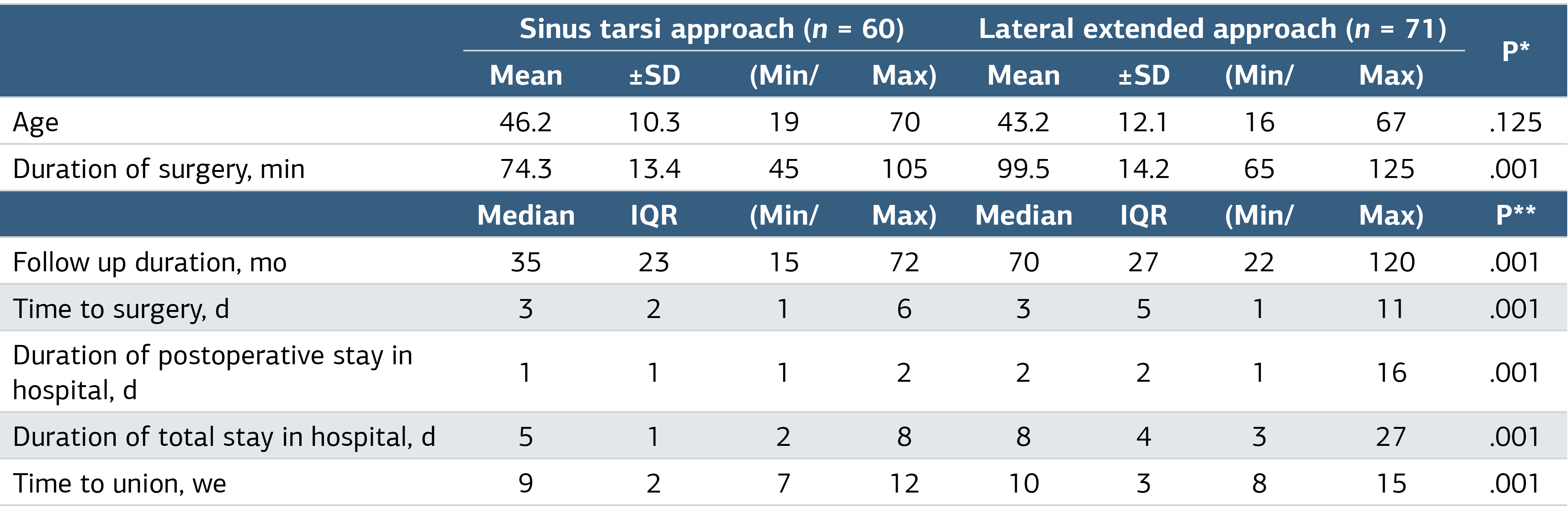
SD, Standart deviation; *,One Way ANOVA; **, Mann Whitney U test; Min, minimum; Max, maximum; Bold P values represent statistical significance.
Table 2. Change in Perioperative and Postoperative Anatomical and Clinical Measurements of Calcaneus
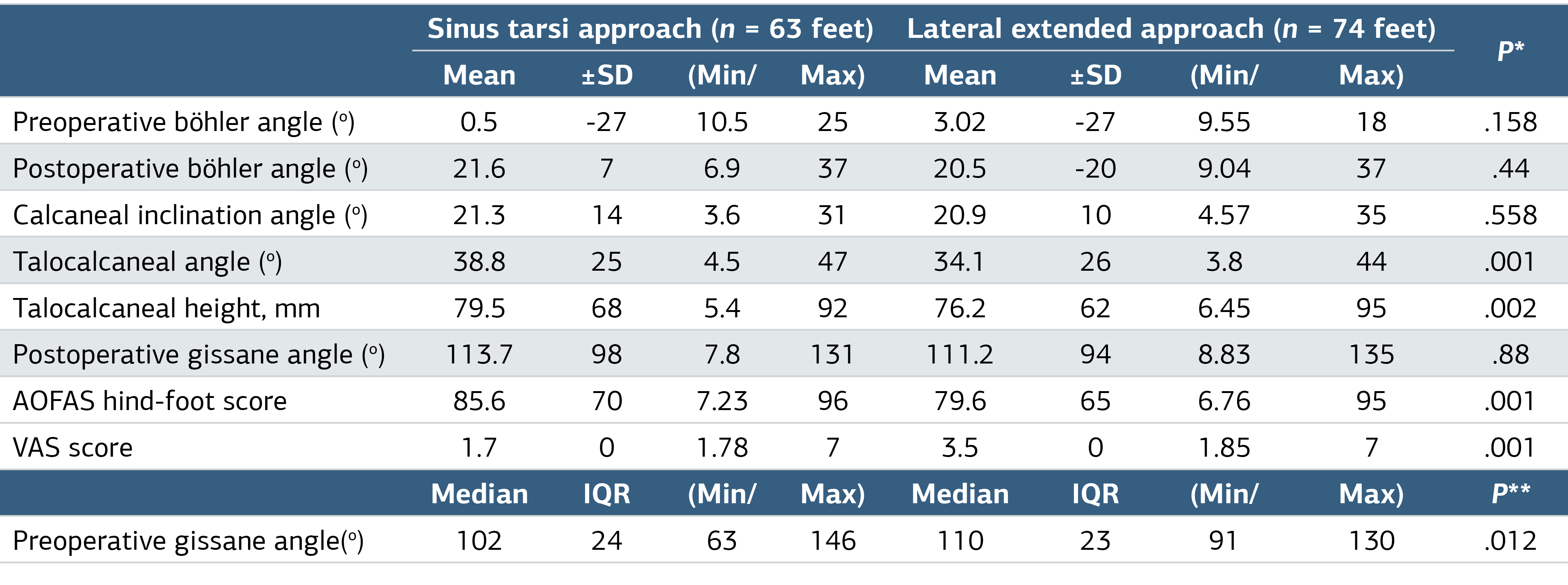
SD, Standart deviation; *, One Way ANOVA; **, Mann Whitney U test; Min, minimum; Max, maximum; Bold P values represent statistical significance.
Table 3. Comparison of the Groups for Revision Surgery, Complication and Patient Related Outcome Measures
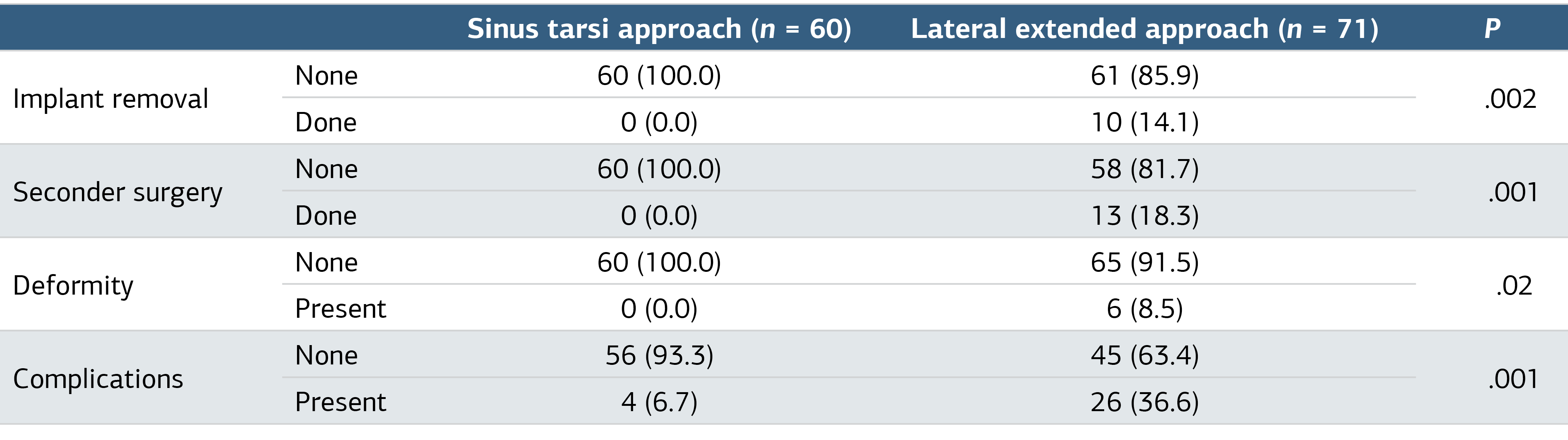
Pearson chi-square.
Figures
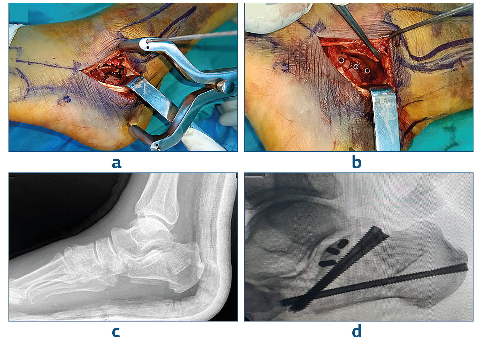
Figure 1. Perop Images of a Patient Operated by ST Approach
Preop and postop radiographs of a patient operated by ST approach; a, Hintermann retractor placement and displaced joint fragment; b, After reduction and cannulated headless screw fixation; c, Preop radiographs; d, Postop scopy images.
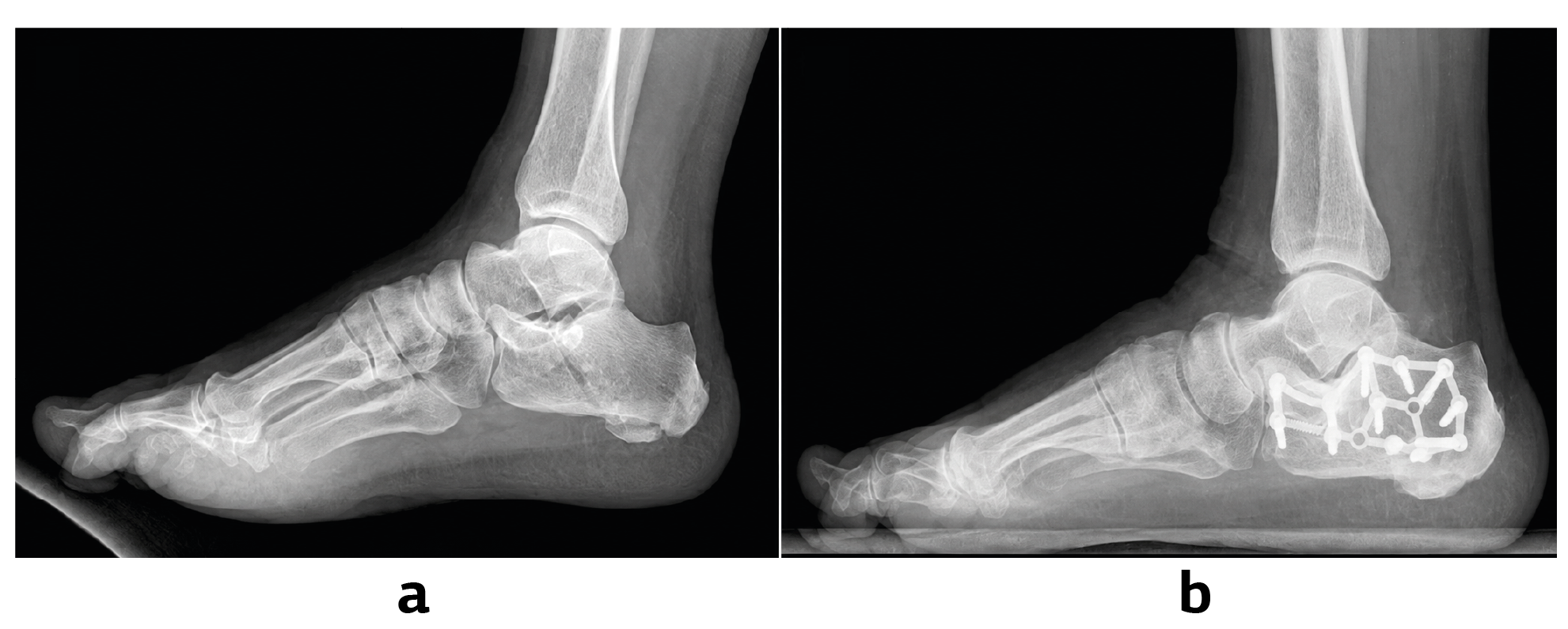
Figure 2. Patient Operated with LE Approach
a, Preop radiograph of the patient; b, Postop radiography.
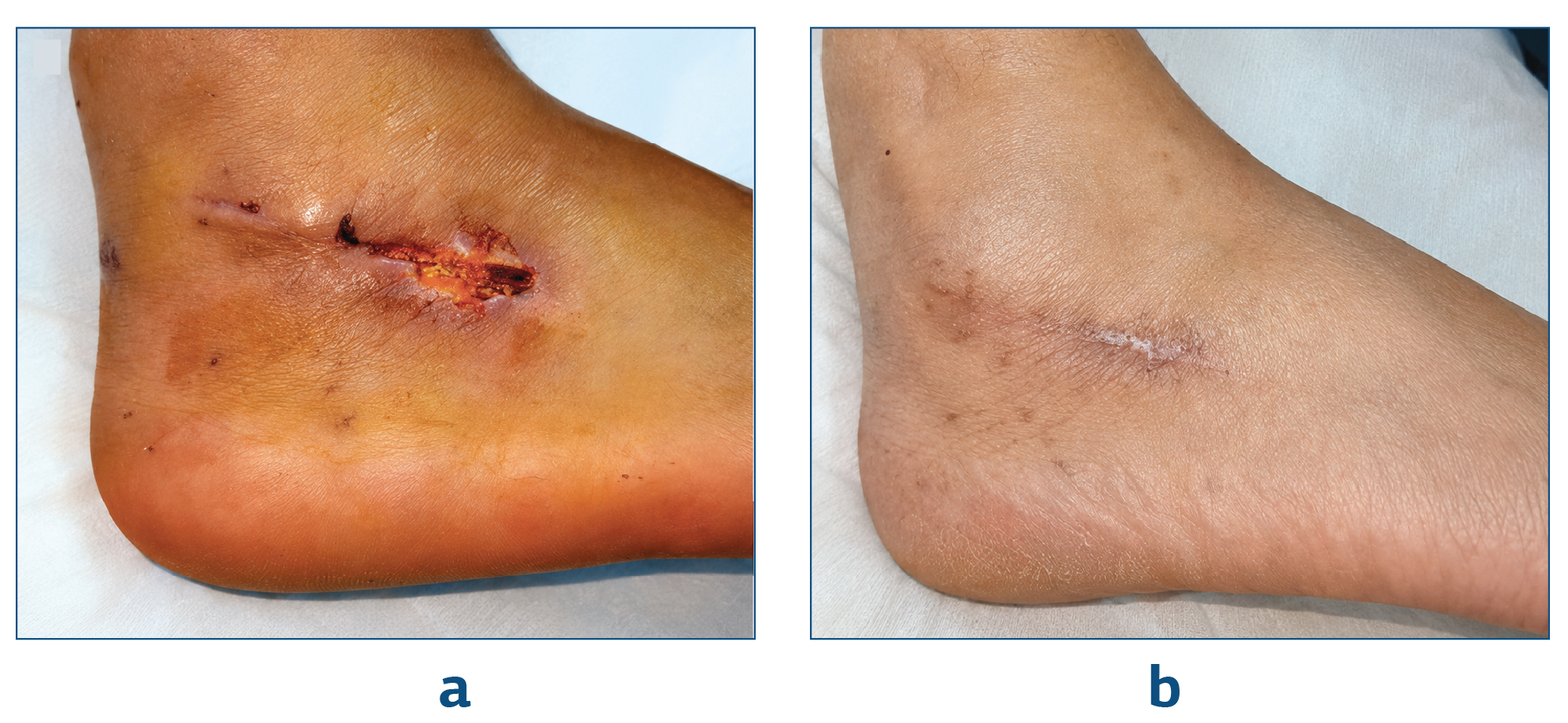
Figure 3. Patient with Superficial Wound Site Problem Operated Through ST Approach
a, Detachment present at the incision site; b, Wound healed after oral antibiotics.
Additional Information
Publisher’s Note
Bayrakol MP remains neutral with regard to jurisdictional and institutional claims.
Rights and Permissions

This work is licensed under a Creative Commons Attribution-NonCommercial 4.0 International License (CC BY-NC 4.0). To view a copy of the license, visit https://creativecommons.org/licenses/by-nc/4.0/
About This Article
How to Cite This Article
Suat Batar, Muhammed Enes Karataş, Yusuf Kaya, Serdar Kamil Çepni, Bahattin Kemah, Mehmet Salih Söylemez, Necdet Sağlam. Comparative analysis of lateral extensile approach and sinus tarsi approach in the surgical treatment of calcaneal fractures: a retrospective study. Ann Clin Anal Med 2024;15(9):650-655. doi:10.4328/ACAM.22318
Publication History
- Received:
- 28.06.2024
- Accepted:
- 12.08.2024
- Published Online:
- 27.08.2024
- Printed:
- 01.09.2024