
Abstract
AimThis study aimed to examine the nasolacrimal canal both proximally and distally in patients with septum deviation, in order to investigate the relationship between septum deviation and nasolacrimal canal diameter and to explore its relationship with maxillary sinus diameter.MethodsPreoperative paranasal sinus computed tomography images of 50 male and 50 female adult patients who underwent septoplasty were examined, and the deviated side (ipsilateral) and opposite side (contralateral) nasolacrimal canal diameters were measured at the proximal and distal parts. Moreover, bilateral maxillary sinus height, anteroposterior, and horizontal diameters were also measured.ResultsNo significant difference was found in the nasolacrimal canal diameter between the ipsilateral and contralateral sides, whether measured proximally or distally (p>0.05). As a result of the measurements, it was observed that the nasolacrimal canal was wider in the proximal part than in the distal part without being affected by the deviation side (p<0.05). Likewise, it was observed that the nasolacrimal canal was wider in males than in females without being affected by the deviation side (p<0.05). Our study revealed no statistically significant differences between the ipsilateral and contralateral MS dimensions (p>0.05).ConclusionThe nasolacrimal canal in men is typically wider than in women; and it tends to be broader at the proximal end compared to the distal end. This anatomical difference may explain why Primary Acquired Nasolacrimal Duct Obstruction is more prevalent in women and why obstruction is frequently observed in the distal part of the canal. Whether septum deviation is one of the etiological factors causing Primary Acquired Nasolacrimal Duct Obstruction is still a controversial issue in the medical community.
Keywords
Introduction
Nasal septal deviation is one of the most common anatomical variations observed in the nose.1 The nasal septum is a structure located in the inner part of the nose, which plays an important physiological role by regulating and supporting the airway. Asymmetrical sloping of the nasal septum to either side is called nasal septal deviation.2 Possible risk factors for asymmetric growth of the maxillary bone include childhood traumas, as well as genetic and environmental influences.3 The nasolacrimal canal; is mainly formed by the nasolacrimal sulcus of the maxillary bone. Its cranial and lateral ends are bounded by the lacrimal bone, while its inferomedial end is formed by the lacrimal process of the inferior turbinate.4
The nasolacrimal canal (NLC) is composed of two parts. The intraosseous superior part is approximately 12 mm long, while the membranous lower part is around 5 mm long. The intraosseous part starts from the floor of the lacrimal fossa. It is bounded laterally by “the maxillary bone, medially by the lacrimal bone, and inferior turbinate”.5 It opens into the lateral nasal wall, terminating at the Hasner valve in the inferior meatus. In women, the NLC becomes narrower towards the valve of Hasner. Therefore, Primary Acquired Nasolacrimal Duct Obstruction (PANCO) is more common in women.6
Thin-section axial computed tomography (CT), is an exceptionally effective imaging method for evaluating the lacrimal drainage system and its relationship with surrounding tissues, primarily due to its ability to clearly distinguish contrast differences between soft tissue and bone.4 This study was undertaken due to the limited number of studies in the literature that examine the relationship between nasal septum deviation, the NLC, and other paranasal anatomical structures.
The NLC was examined both proximally and distally in patients with septum deviation to investigate the relationship between septum deviation and NLC diameter and to reveal its relationship with maxillary sinus (MS) diameter.
Materials and Methods
SubjectsThis study, conducted retrospectively, involved 50 male and 50 female patients who underwent septoplasty following a diagnosis of septum deviation, and who applied to Kırıkkale University Faculty of Medicine, Kırıkkale Clinic between August 2015 and July 2019. The material of our study consisted of PNS CT images of these patients taken before the septoplasty operation between August 2015 and July 2019 at Kırıkkale University Medical Faculty Hospital, Department of Kırıkkale The individuals within the scope of the study were between the ages of 19-62, and the mean age was found to be 33.86±11.26. There are 32 people in the ≤25 age group, and 68 people in the >25 age group. The side where the septum deviates is referred to as ipsilateral, while the opposite side of the deviation is called contralateral.Inclusion CriteriaThe study included patients aged 18 to 75 who had PNS CT images taken before the septoplasty operation with the diagnosis of septum deviation, and there were no signs of nasal polyposis, acute or chronic sinusitis in the CT images examined. PNS CT images of 100 patients in the CT archive were analyzed retrospectively.Inclusion Criteria When Selecting Images• The patient is18 years old or older
• Bilateral nasolacrimal canals are totally in the field of view
• Full inclusion of bilateral maxillary sinuses in the field of view
• Presence of septum deviation
• No nasolacrimal canal obstruction symptomsExclusion Criteria When Selecting Images• The patient is younger than 18 years old
• Bilateral nasolacrimal ducts or maxillary sinuses are not totally in the field of view
• Image quality does not allow for inspection
• Patients have had a previous operation on the nasolacrimal duct or paranasal sinuses
• Patients with previous septoplasty
• Patients with allergic rhinitisPNS CT Imaging and AnalysisAll scans were obtained using routine PNS CT imaging in the supine position, without the use of contrast or sedation. The images were captured with a 64-slice CT scanner (MSCT; Brilliance 64, Philips Medical System, Best, the Netherlands) using the following parameters: tube voltage = 120 kV, effective mAs = 350, slice thickness = 1.00 mm, the field of view (FOV) = 180 mm, and image matrix = 768 × 768. The evaluation was performed on the coronal, axial, and sagittal planes in the workstation by a single radiologist.Measurements1. Measurement of NLC diameters: The transverse diameters of the NLC were measured by identifying the sections where the canal is the widest, both proximally and distally on both sides (Figure 1).
2. Measurement of MS diameters: The widest section of the MS was identified on PNS CT images, and the maximum anteroposterior and mediolateral dimensions were measured (Figure 2). The height of the MS was assessed from the sections in the coronal plane (Figure 3).Ethical ApprovalEthics committee approval was obtained from the Kirikkale University Non-invasive Research Ethics Committee (Date: 07.08.2019, Decision No: 2019/12).Statistical AnalysisThe data collected in the study were analyzed using SPSS for Windows version 20.0 software (SPSS, INC, an IBM Company, Chicago, Illinois). The paired t-test, independent sample t-test, and Pearson correlation test were used. A p-value of less than 0.005 was considered to indicate statistical significance.
Results
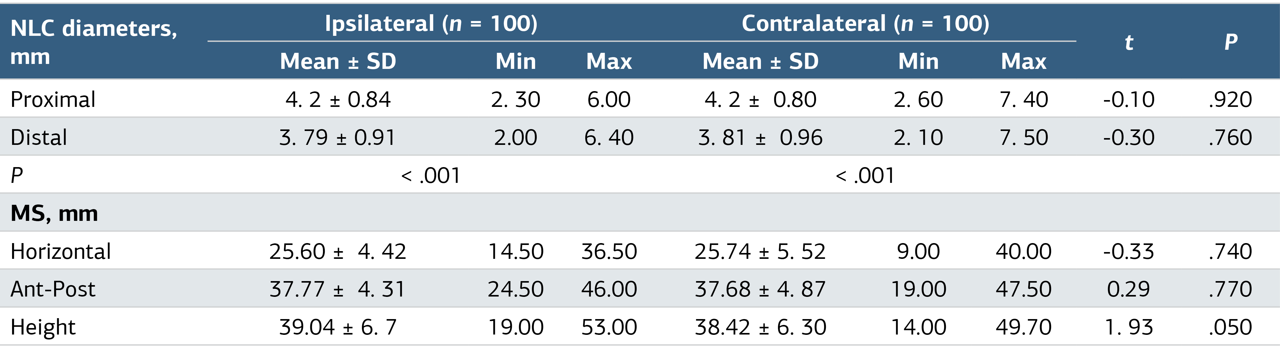
Nasolacrimal Canal Proximal and Distal MeasurementsNo significant differences were found between ipsilateral and contralateral NLC proximal; and distal diameters (p>0.05) (Table 1). In each of the ipsilateral, and contralateral groups separately, proximal NLC diameters were significantly higher than that of the distal NLC diameters (p<0.001) (Table 1).
Maxillary Sinus MeasurementsNo significant differences were found between ipsilateral and contralateral MS measurement values (horizontal, anterior-
posterior dimensions and length) (p>0.05) (Table 1).
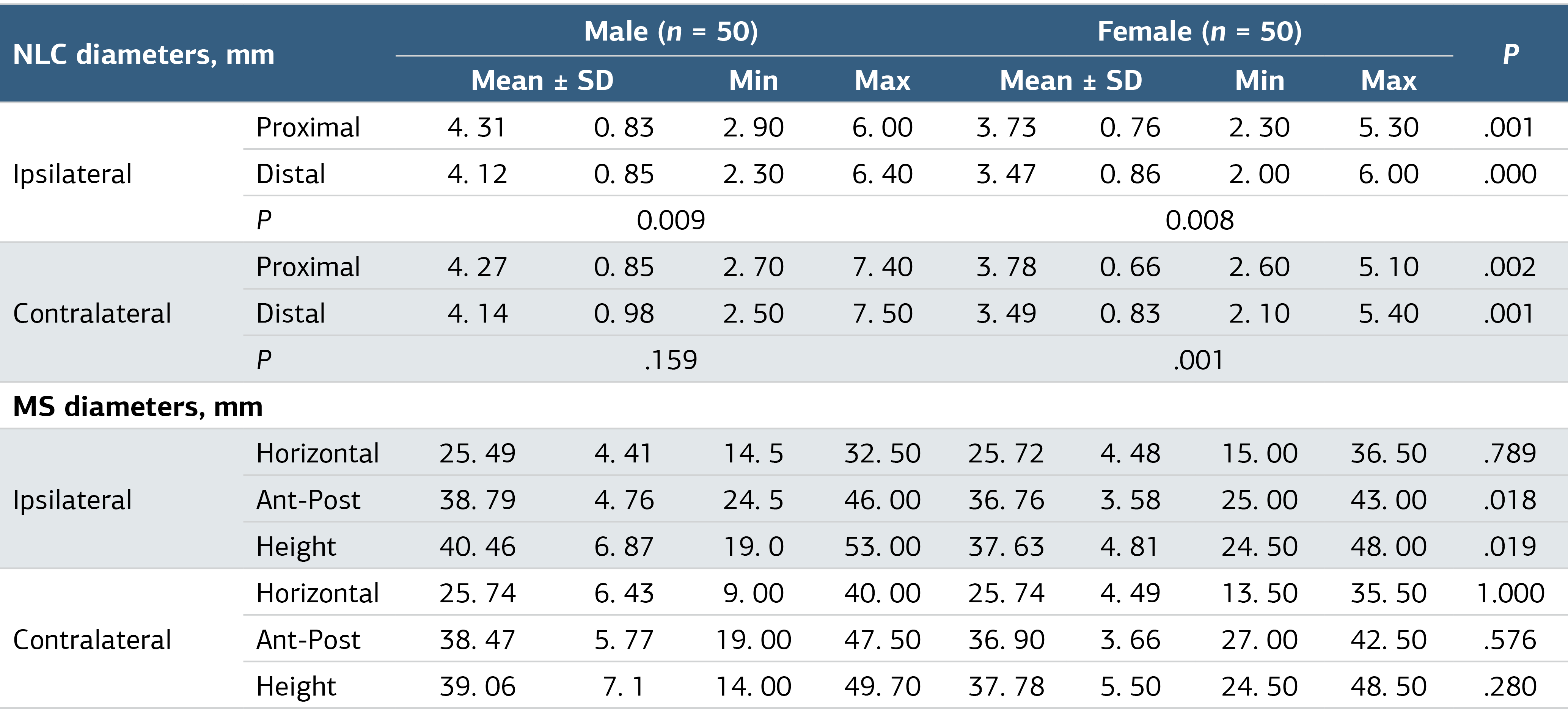
The NLC and MS measurements by gender are presented in Table 2.Nasolacrimal Canal Proximal and Distal Measurementsİpsilateral and contralateral NLC proximal and distal diameters were significantly higher in males compared to females (p<0.05) (Table 2). In both males and females, the ipsilateral proximal NLC diameters were significantly higher than the distal NLC (p<0.05) (Table 2). In females only, contralateral proximal NLC diameters were significantly higher than the distal NLC (p<0.05) (Table 2).
Maxillary Sinus MeasurementsIn females, the anterior-posterior dimension and height of the ipsilateral MS were significantly lower than those in the males (p<0.05) (Table 2). No significant differences were detected in the contralateral MS measurements (horizontal, anterior-posterior dimensions, and height) between male and female patients (p>0.05) (Table 2).
Nasolacrimal Canal Proximal and Distal Measurements
No significant differences were found between ipsilateral proximal, distal, contralateral proximal, and distal NLC dimensions of the subjects aged ≤25 years and those aged >25 years (p>0.05).
In the ≤25 years age group, contralateral proximal NLC diameters were significantly higher than the distal NLC (p<0.05) (Table 3). In the >25 years age group, both ipsilateral and contralateral proximal NLC diameters were significantly higher than the distal NLC (p<0.05).
Maxillary Sinus MeasurementsNo significant differences were found in the ipsilateral and contralateral MS measurements (horizontal, anterior-posterior dimensions, and height) between subjects aged 25 years or younger and those older than 25 years (p>0.05).
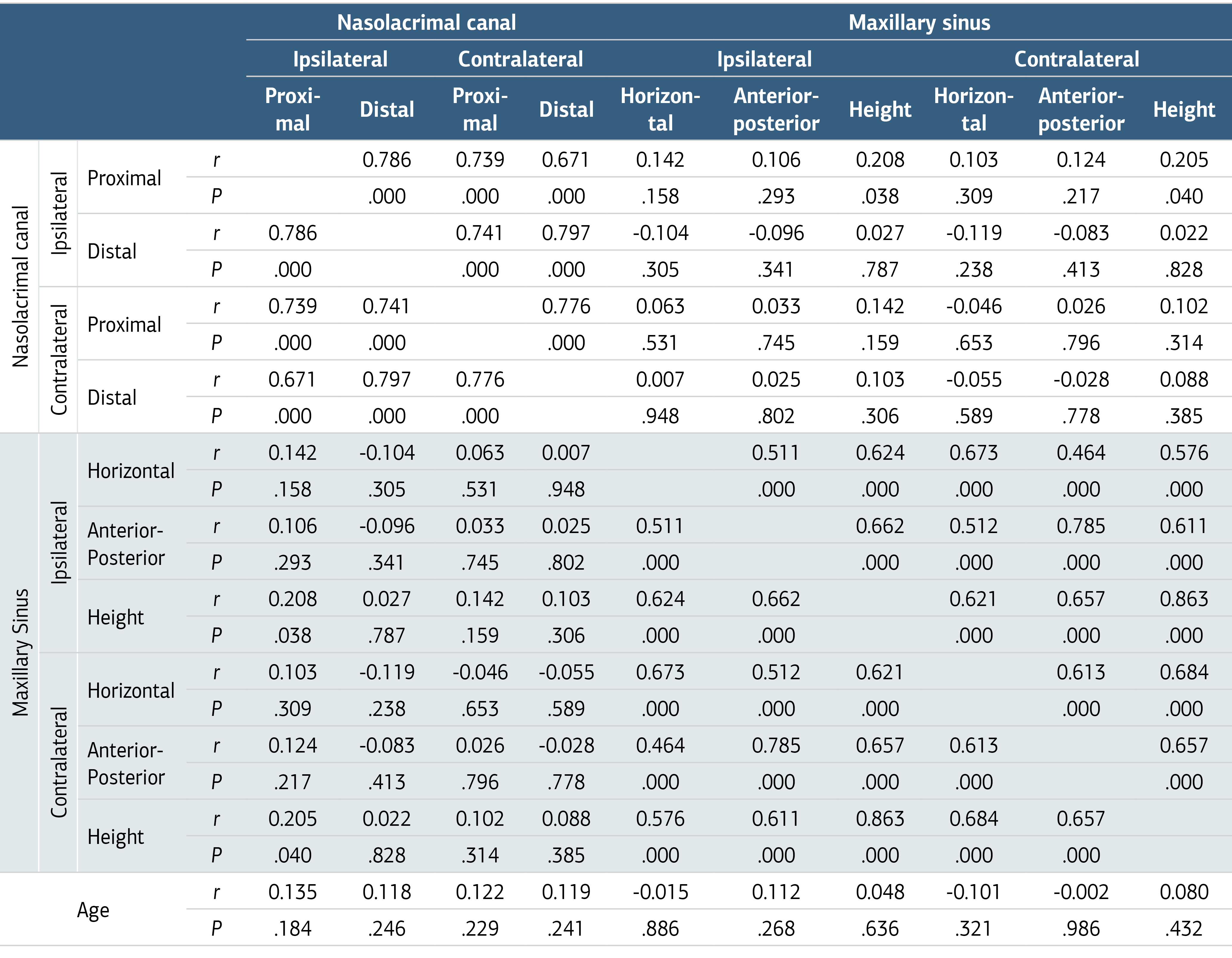
Correlation test results are presented in Table 3:
-A positive correlation was identified between the ipsilateral proximal NLC diameters and ipsilateral MS heights (p=0.038) (Table 3)
-A positive correlation was found between the ipsilateral proximal NLC diameters and contralateral MS heights. (p=0.040) (Table 3)
-A strong positive correlation exists among all NLC diameter measurements (p<0.05) (Table 3)
-A strong positive correlation was observed among all MS diameter measurements (p<0.05) (Table 3).
-No significant correlation was detected between age and any of the measurement parameters (p>0.05) (Table 3).
Discussion
The ipsilateral and contralateral NLC were evaluated independently. The ipsilateral proximal NLC diameters of the males (4.31±0.83 mm) were significantly wider than females (3.73±0.76 mm), and distal NLC diameters of the males (4.12±0.85 mm) were significantly wider than females (3.47±0.86 mm). Upon examining the contralateral side, the proximal diameter of NLC was 4.27±0.85 mm in men and 3.78±0.66 mm in women (p=0.002). When we examined the contralateral distal part, it was observed as 4.14±0.98 mm in males and 3.49±0.83 mm in females (p=0.001). These findings, consistent with the studies of Sirik et al.7 and Alfred et al.8 indicate that the nasolacrimal duct is wider in males than in females, both proximally and distally, and that this difference is not influenced by septum deviation. This could account for the higher prevalence of acquired nasolacrimal duct obstruction in women.9-10
MS development is proportional to facial bone development.11-12 In our study, the positive correlation between the proximal diameter of the ipsilateral nasolacrimal duct and the height of MS supports this information. In the study of Whyte et al.13 it was stated that the volume of fully developed MS is higher in men than in women. In our study, the anteroposterior and horizontal diameters of MS on the ipsilateral side were higher in men than in women (p<0.05). However, there is no difference in heights of MS between men and women (p>0.05).
In our study, septum deviation and MS diameters were assessed. It was found that there were no statistically significant differences between ipsilateral and contralateral side diameters (p>0.05). Although we believe that the dimensions of the MS are not affected by the deviation of the septum, it will be beneficial to conduct prospective studies examining whether the deviation occurs during development or in adulthood.
In the study of Cervelli et al.14 involving adult patients, it was reported that nasolacrimal canal obstruction is more affected by turbinate hypertrophy than by septum deviation.14 Our findings, which show that NLC diameter measurements were unaffected by septum deviation, support this conclusion. Additionally, many publications in the literature that NLC obstruction frequently occurs at the distal, internal ostium level. In our study, it was observed that the proximal part of the NLC was wider than the distal part on both the ipsilateral and contralateral sides, and this difference was statistically significant (p<0.05). The higher frequency of NLC obstruction in the distal region may be attributed to the narrower canal in that area.
In the study of Dikici et al.15 37 patients with primary acquired nasolacrimal canal obstruction (PANCO) in 48 eyes were included, along with 37 patients in the control group. A positive correlation was found between right and left, proximal and distal diameters. There was no significant difference between the groups when the diameter lengths were compared. There was no significant difference between the groups according to the direction and location of the deviation and Mladina classification (p>0.05).15 Similar to the findings of Dikici et al.15 our study also identified a positive correlation between the ipsilateral and contralateral diameters, both proximally and distally. However, no significant difference was observed between the ipsilateral and contralateral groups.
In the studies of Janssen.8 Groel.16 and Lee.17 it was concluded that having a small nasolacrimal canal diameter is one of the etiological factors causing nasolacrimal canal obstruction. In our results, the diameter of the proximal part of the canal was larger than the diameter of the distal part in all groups. Therefore, it can be inferred that the pathologies in the distal part of the canal are more likely to cause obstruction. The fact that lower turbinate hypertrophy causes more obstruction in the studies of Habesoglu.18 Cervelli.14 and Dikici.15 supports this.
In the study of Habeşoğlu et al.19 involving 41 patients with PANCO, osteomeatal complex disease was observed on the occluded side in 19.5% of the patients and on the healthy side in 5% of the patients. Maxillary sinusitis was observed on the occluded side in 24.4% of the cases, while it was observed on the healthy side in 7.3% of the cases (p<0.05).19 Based on the study of Habeşoğlu et al.19 it can be suggested that there is a relationship between the NLC and MS physiology. In our study, a positive correlation was found between the proximal diameter of the ipsilateral NLC and the height of both the ipsilateral and contralateral MS (p<0.05). Additional research is required to elucidate the developmental connection between the nasolacrimal canal and the maxillary sinus. In the study of Habeşoğlu et al.19 found no statistically significant association between septum deviation and nasolacrimal duct obstruction in their study.19
Sirik et al.7 examined the NLC diameters between the groups with and without deviation, no statistically significant difference was detected.7 Although the proximal and distal parts were not assessed separately in this study, our analysis showed no statistically significant differences when these parts were evaluated separately. This suggests that there is no relationship between septum deviation and nasolacrimal canal diameter. However, our study did not consider the timing of septal deviation during the developmental process and its potential relationship with NLC diameter.
In Wang et al.’s study.19 PNS CT images of 126 patients with PANCO and a control group consisting of 76 people were examined. The narrowest diameter of the NLC was significantly smaller in the PANCO patient group (p<0.01). The angle between the inferior turbinate and the medial wall of the MS was significantly narrower in the PANCO patient group than in the control group. Given that the distal part of the nasolacrimal canal is narrower, it supports our perspective that pathologies in this region have a greater impact. Wang et al.19 found no significant difference in the rate of septal deviation and the side of occlusion between PANCO and the control group, similar to our findings. This supports our view that nasal septal deviation does not affect the diameter of the NLC or the development of PANCO.
In the measurements taken, a positive correlation was found between the proximal diameter of the NLC and bilateral MS heights on the side of the deviation (p<0,005). This suggests that in cases where MS is higher, the proximal diameter of the nasolacrimal duct might be wider, as observed in coronal PNS CT or Waaters X-ray. Particularly in individuals with MS atrophy, there might be issues with the nasolacrimal duct on the same side. Future studies are crucial to further explore and clarify this relationship.
Conclusion
In conclusion, the NLC is wider in men than in women, and it is also wider proximally than distally. This may explain why PANCO is more common in women and why obstruction is frequently observed in the distal part of the canal. The role of septum deviation as an etiological factor in PANCO remains a controversial issue in the medical community, and future studies are important to clarify this issue.
Declarations
Animal and Human Rights Statement
All procedures performed in this study were in accordance with the ethical standards of the institutional and/or national research committee and with the 1964 Helsinki Declaration and its later amendments or comparable ethical standards.
Data Availability
The datasets used and/or analyzed during the current study are not publicly available due to patient privacy reasons but are available from the corresponding author on reasonable request.
Conflict of Interest
The authors declare that there is no conflict of interest.
Funding
None.
References
- Paksoy M, Sanli A, Evren C. Konka bullozanın nazal patolojilerdeki rolü [The role of concha bullosa in nasal pathologies]. Kulak Burun Bogaz Ihtis Derg. 2008;8(4):41-238.
- Smith K, Edwards P, Saini T. The prevalence of concha bullosa and nasal septal deviation and their relationship to maxillary sinusitis by volumetric tomography. Int J Dent. 2010;2010:404982. doi:10.1155/2010/404982
- Arslan E, Tulaci KG, Canakci H, Arslan S, Yazici H. Effects of nasal septum deviation and concha bullosa surgery on the frequency and financial burden of acute rhinosinusitis. Ir J Med Sci. 2023;192(1):341-347. doi:10.1007/s11845-022-03156-x
- Nair JR, Syed R, Chan IYM, Gorelik N, Chankowsky J, Del Carpio-O’Donovan R. The forgotten lacrimal gland and lacrimal drainage apparatus: pictorial review of CT and MRI findings and differential diagnosis. Br J Radiol. 2022;95(1135):20211333. doi:10.1259/bjr.20211333
- Unlu HH, Toprak B, Aslan A, Guler C. Comparison of surgical outcomes in primary endoscopic dacryocystorhinostomy with and without silicone intubation. Ann Otol Rhinol Laryngol. 2002;111(8):9-704. doi:10.1177/000348940211100809
- Çukurova I, ed. Inferior lacrimal system obstructions. In: Endoscopic Dacryocystorhinostomy Diagnosis to Treatment. 2018:10-30.
- Sirik M, Inan I. Evaluation of the relationship between concha bullosa and nasal septum deviation with diameter of nasolacrimal duct. Ann Med Res. 2019;26(2):12-209.
- Janssen AG, Mansour K, Bos JJ. Diameter of the bony lacrimal canal: normal values and values related to nasolacrimal duct obstruction: assessment with CT. AJNR Am J Neuroradiol. 2001;22:845-850.
- Ivanišević M, Bojić L, Lešin M, Žuljan I, Buan K, Kovačić Z. Primary acquired nasolacrimal duct obstruction: epidemiological analysis of 91 patients. Med Jadertina. 2007;37(1-2):37-41.
- Woog JJ. The incidence of symptomatic acquired lacrimal outflow obstruction among residents of Olmsted County, Minnesota, 1976-2000: an American Ophthalmological Society thesis. Trans Am Ophthalmol Soc. 2007;105:66-649.
- Lorkiewicz-Muszyńska D, Kociemba W, Rewekant A, et al. Development of the maxillary sinus from birth to age 18: postnatal growth pattern. Int J Pediatr Otorhinolaryngol. 2015;79(9):400-1393. doi:10.1016/j.ijporl.2015.05.032
- Scuderi AJ, Harnsberger HR, Boyer RS. Pneumatization of the paranasal sinuses: normal features of importance to the accurate interpretation of CT scans and MR images. AJR Am J Roentgenol. 1993;160(5):4-1101. doi:10.2214/ajr.160.5.8470585
- Whyte A, Boeddinghaus R. The maxillary sinus: physiology, development, and imaging anatomy. Dentomaxillofac Radiol. 2019;48(8). doi:10.1259/dmfr.20190205
- Cervelli V, Gravante G, Colicchia GM, et al. Asymptomatic lacrimal flow abnormalities in patients with septal deviations and turbinate hypertrophy. Aesthetic Plast Surg. 2008;32(1):6-72. doi:10.1007/s00266-007-9009-6
- Dikici O, Ulutaş HG. Relationship between primary acquired nasolacrimal duct obstruction, paranasal abnormalities, and nasal septal deviation. J Craniofac Surg. 2020;31(3):782-786. doi:10.1097/scs.0000000000006108
- Groell R, Schaffler GJ, Uggowitzer M. CT anatomy of the nasolacrimal sac and duct. Surg Radiol Anat. 1997;19:189-191. doi:10.1007/bf01627974
- Lee H, Ha S, Lee Y, Park M, Baek S. Anatomical and morphometric study of the bony nasolacrimal canal using computed tomography. Ophthalmologica. 2012;227:153-159. doi:10.1159/000331986
- Habesoglu M, Eriman M, Habesoglu TE, Kinis V, Surmeli M, Deveci I. Co-occurrence and possible role of sinonasal anomalies in primary acquired nasolacrimal duct obstruction. J Craniofac Surg. 2013;24(2):497-500. doi:10.1097/scs.0b013e31827c83ea
- Wang W, Gong L, Wang Y. Anatomic characteristics of primary acquired nasolacrimal duct obstruction: a comparative computed tomography study. Quant Imaging Med Surg. 2022;12(11):5068-5079. doi:10.21037/qims-22-170
Tables
Table 1. Nasolacrimal Canal and Maxillary Sinus Measurements of the Ipsilateral and Contralateral Sides
*NLC, Nasolacrimal canal; MS, Maxillary sinus; Ant-Post, Anterior-Posterior.
Table 2. Nasolacrimal Canal and Maxillary Sinus Measurements According to Gender
*NLC, Nasolacrimal canal; MS, Maxillary sinus; Ant-Post, Anterior-Posterior.
Table 3. Correlation Test Results
Figures
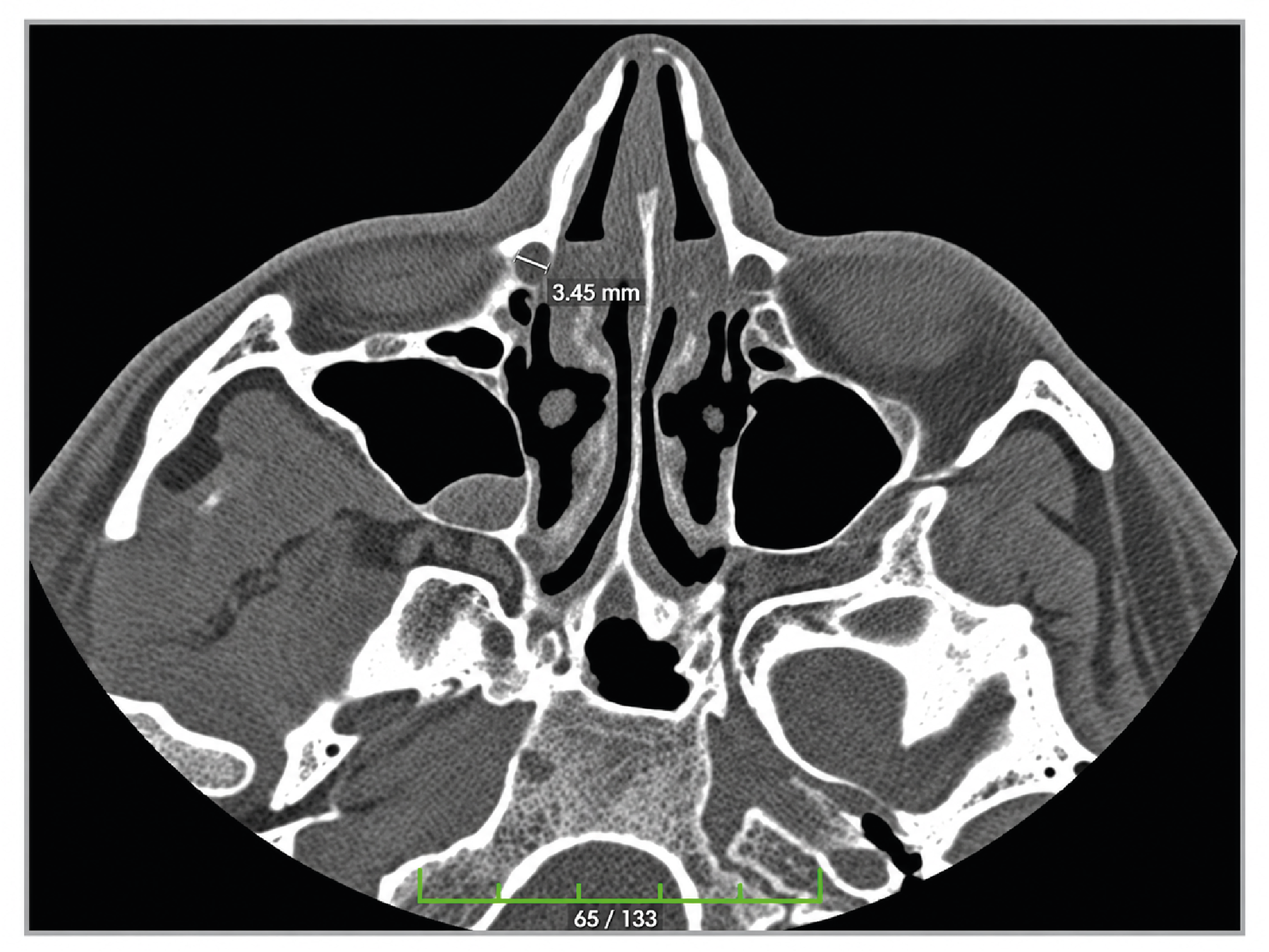
Figure 1. Axial Plane Ipsilateral Nasolacrimal Canal Transverse Diameter Measurements
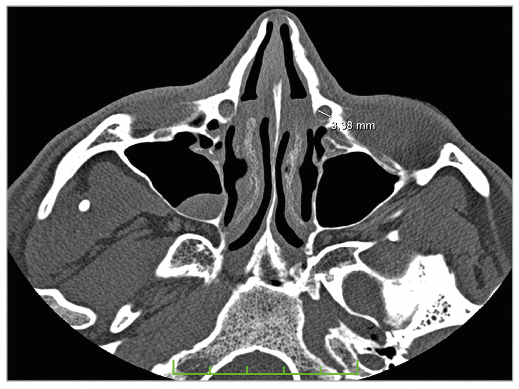
Figure 2. Contralateral Nasolacrimal Canal Transverse Diameter Measurement in the Axial Plane
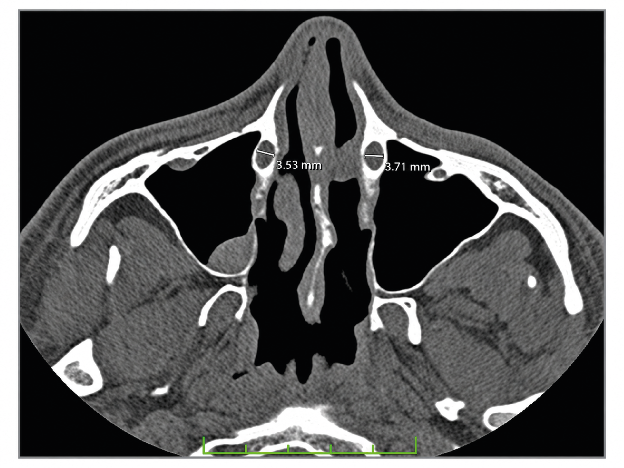
Figure 3. Ipsilateral and Contralateral Nasolacrimal Canal Transverse Diameter Measurement in the Distal Section
Additional Information
Publisher’s Note
Bayrakol MP remains neutral with regard to jurisdictional and institutional claims.
Rights and Permissions

This work is licensed under a Creative Commons Attribution-NonCommercial 4.0 International License (CC BY-NC 4.0). To view a copy of the license, visit https://creativecommons.org/licenses/by-nc/4.0/
About This Article
How to Cite This Article
Furkan Melih Koçak, Nuray Bayar Muluk, Mikail İnal, Selçuk Başer, Ziya Şencan, Ela Cömert, Gökçe Özel. Evaluation of the nasolacrimal canal by computed tomography in patientswith septal deviation. Ann Clin Anal Med 2024;15(Suppl 2):127-132. doi:10.4328/ACAM.22319
Publication History
- Received:
- 30.06.2024
- Accepted:
- 26.08.2024
- Published Online:
- 09.10.2024
- Printed:
- 20.10.2024