
Push-out bond strength of bioactive repair materials exposed to endodonticirrigants
Bond strength of bioactive repair materials
Authors
Abstract
AimThis study aimed to evaluate the push-out bond strength (PBS) of three endodontic repair materials (ProRoot MTA, NeoPutty, and Endocem MTA Premixed) to root dentin, assess the impact of different irrigation solutions on PBS, and classify the post-testing failure modes.
MethodsA total of 180 root slices were randomly divided into three groups based on the repair material (n=60). After repair materials were inserted, the samples were randomly subdivided into four subgroups (n=15) based on the irrigation solution exposure: saline, sodium hypochlorite (NaOCl), ethylenediaminetetraacetic acid (EDTA), and unexposed. An Instron Universal Testing Machine measured the PBS of the three study materials, and failure modes post-PBS testing were identified using a stereomicroscope.
ResultsPBS assessments revealed that ProRoot MTA and Endocem MTA Premixed exhibited significantly superior bond strengths compared to NeoPutty. Irrigation with saline reduced the PBS of ProRoot MTA relative to NaOCl and the control group. In contrast, NaOCl irrigation significantly diminished the PBS of NeoPutty when compared to control group, saline, and EDTA. All three solutions did not affect the PBS of Endocem MTA Premixed. Failure mode analysis indicated a predominance of cohesive failure in ProRoot MTA (71.7%) and Endocem MTA Premixed (66.7%), whereas NeoPutty mainly showed mixed failure (65%).
ConclusionThe results of the present study indicate that Endocem MTA Premixed presents a robust bioactive cement alternative for perforation repair, withstanding various irrigation solutions.
Keywords
Introduction
Furcal root perforations, defined as iatrogenic or pathological communications between the root canal system and the surrounding periodontal tissues, most commonly occur during access cavity preparation in teeth exhibiting canal calcification.1 Prompt furcal root perforation repair is necessary to achieve an effective seal preserving dentin and restorative material interface integrity.2 Ideal repair materials should demonstrate biocompatibility, dimensional stability, antibacterial properties, radiopacity, and ease of manipulation. Additionally, they require strong adhesion to root dentin and resistance to dislodgement forces.3
Mineral trioxide aggregate (MTA), which has a wide range of uses in endodontics such as apexification, apexogenesis, pulp capping, apical plug, and revascularization.4,5,6,7 is a bioactive material composed of tricalcium silicate, dicalcium silicate, tricalcium aluminate, and bismuth oxide.8 First developed in the 1990s and later approved by the United States Food and Drug Administration, MTA became commercially available under the trade name ProRoot MTA (PR-MTA) (Dentsply, Tulsa, OK, USA). Despite advantageous properties including biocompatibility, superior sealing ability, bioactivity, and antibacterial effects, MTA has drawbacks such as difficult handling, prolonged setting times, tooth discoloration, and the need for multiple treatment visits.8 As such, there is a recognized need to refine the chemical composition of MTA to improve its clinical utility. To address inconsistent mixtures seen with conventional powder-liquid formulations, premixed tricalcium silicate repair materials have emerged. One example is NeoPutty (NuSmil Ltd., Houston, TX, USA), a bioactive bioceramic incorporating calcium aluminate, an inorganic tricalcium/dicalcium silicate blend, a water-free organic liquid, and tantalum oxide for radioopacity. As a user-friendly, pre-mixed material requiring direct application, NeoPutty leverages inherent dentinal moisture as a natural curing agent.9 Recently, another injectable hydraulic bioceramic cement was introduced, Endocem MTA Premixed (ECM-Premixed) (Maruchi, Wonju, South Korea). Its composition of calcium sulfate, dimethyl sulfoxide (DMSO), hydroxypropyl methylcellulose, and zirconium oxide aims to enhance biocompatibility and physicochemical properties.10 In clinical practice, prompt furcal perforation repair using suitable materials is necessary to mitigate microbial infiltration and subsequent periodontal tissue infection.1 Following perforation repair, endodontic therapy proceeds with various irrigation solutions. However, contact with these solutions during endodontic therapy can affect the sealing capacity and physical/chemical characteristics of repair materials.3 While previous investigations have extensively assessed the push-out bond strength (PBS) of materials to root dentin, few studies exist evaluating the displacement resistance of novel bioactive cements employed for perforation repair, especially after contact with different irrigation solutions. Therefore, this study was designed to evaluate the PBS of PR-MTA, NeoPutty, and ECM-Premixed after irrigations with saline, sodium hypochlorite (NaOCl), and ethylenediaminetetraacetic acid (EDTA). The study tested two null hypotheses: 1) there would be no difference in PBS between PR-MTA, NeoPutty, and ECM-Premixed, and 2) irrigation solutions would not affect the post-exposure PBS of the three materials.
Materials and Methods
Sample Selection and PreparationA power analysis was conducted to determine the minimum sample size required to achieve sufficient statistical power.11 Based on a significance level set at 0.05 and a 95% power, results indicated that a minimum of 144 dentin slices (12 slices per group) would be needed. To meet this requirement, 60 intact, recently extracted mature human maxillary central incisors were obtained. All teeth were examined under a microscope at 10× magnification and teeth with calcified canals, radicular caries, external resorption, or visible cracks were excluded from the study. The included teeth were cleaned of residual soft and hard tissues using Gracey curettes and subsequently immersed in NaOCl solution for 10 min. Afterward, they were stored in distilled water at 9°C until sample preparation. Dentin slices were acquired from the middle third of each tooth using a water-cooled precision diamond saw (Metkon Instruments Inc., Bursa, Türkiye). In total, 180 slices with a thickness of 1 ± 0.1 mm were sectioned, and verified with a digital caliper. A centered circular perforation measuring 1.4 mm in diameter was created in each dentin slice using a cylindrical diamond bur. All samples were immersed in 2.5% NaOCl solution for 5 min, thoroughly rinsed with distilled water, and dried with absorbent paper points. Subsequently, the sections were randomly assigned into three experimental groups (n=60) based on the repair material: PR-MTA; NeoPutty; ECM-Premixed. Each repair material was placed into the artificial perforations, and incubated at 37°C with 100% humidity for 10 min to allow initial setting. Then, the samples in each material group were randomly divided into four subgroups (n =15) for irrigant exposure (saline, 5.25% NaOCl, 17% EDTA, control). The saline and 5.25% NaOCl subgroups were immersed for 30 min.12 with renewal of the solutions every 5 min. Conversely, the 17% EDTA subgroup was immersed for 5 min.11 An unexposed control subgroup from each material group was retained without solution immersion. Following exposure to the designated solution, all experimental samples were rinsed to remove any residue and then incubated for 48 h at 37°C and 100% humidity to allow the materials to set completely.
PBS TestPBS testing was performed using an Instron Universal Testing Machine (Elista, Istanbul, Türkiye). A uniaxial compressive force was applied in a corona-apical direction to each sample at a crosshead speed of 1 mm/min using a 1.2-mm diameter stainless steel cylindrical plunger. The peak debonding force required to displace the material was measured in Newtons (N). To calculate the PBS (MPa), the recorded peak force value was divided by the adhesive area of root canal filling: PBS=F/ (2×π×r×h). F is the maximum load (N), π is a constant (3.14), r is the canal radius (mm), and h is the slice thickness (mm). Samples that exhibited fractures during PBS testing were excluded and new dentin sections were prepared from the remaining eligible teeth following the same experimental protocol.
Stereomicroscope AnalysisA stereomicroscope with 40× magnification was utilized to visually examine and categorize the failure modes following PBS testing. Adhesive failure was determined when separation was observed at the interface between the dentin and restorative material, denoting inadequate adhesion between the filling and tooth structure. Cohesive failure occurred when fracture transpired within the body of the restorative material itself. Finally, mixed failure was characterized by the concurrent presence of aspects indicative of both adhesive failure and cohesive failure.
Ethical ApprovalThis study was approved by the Ethics Committee of Recep Tayyip Erdoğan University (Date: 29.04.2021, Decision No: 2021/87).
Statistical AnalysisData normality was evaluated utilizing the Shapiro-Wilk test, which revealed skewed distributions. Consequently, the nonparametric Kruskal-Wallis H test was conducted to compare PBS values between repair materials. When significant differences were noted, post hoc Bonferroni’s tests were performed. One-way ANOVA was applied to analyze PBS across different irrigation solutions for each repair material, contingent on normality adherence per Shapiro-Wilk tests. Homogeneity of variances between ANOVA groups was verified through Levene’s test. Where equal variances were established, Tukey post hoc examinations were applied. Alternatively, Tamhane’s T2 analysis was implemented for unequal variances, when group variance heterogeneity was disclosed per Levene’s test. Analyses were performed using SPSS, version 22.0, with all tests two-sided at a 5% level of significance.
Reporting GuidelinesThe study was reported in accordance with STROBE guidelines.
Results
Both PR-MTA (9.74 ± 3.59 MPa, p<0.001) and ECM-Premixed (11.80 ± 4.33 MPa, p<0.001) displayed significantly superior PBS than NeoPutty (5.08 ± 2.09 MPa) (Figure 1). However, the PBS values of PR-MTA and ECM-Premixed did not differ significantly (p=0.130).
Table 1 summarizes the PBS data obtained for each repair material following exposure to different irrigation solutions. Statistically significant differences in PBS were noted within both the PR-MTA (p=0.016) and NeoPutty (p<0.001) subgroups.
For PR-MTA, saline irrigation resulted in inferior PBS relative to both the control (p=0.021) and NaOCl (p=0.036). Regarding NeoPutty, NaOCl irrigation yielded significantly reduced PBS compared to the negative control (p=0.001), saline (p=0.003), and EDTA (p=0.015).
Across all irrigants, ECM-Premixed displayed significantly superior PBS over NeoPutty (p<0.05). Specifically, in the saline (p=0.032) and EDTA (p=0.041) subgroups, ECM-Premixed achieved significantly higher PBS compared to PR-MTA. And, with NaOCl and EDTA irrigation, NeoPutty exhibited markedly lower PBS versus PR-MTA (p<0.001 for both) (Figure 2). Stereomicroscopic images depicting failure modes are presented in Figure 3. PR-MTA and ECM-Premixed predominantly displayed cohesive failures (71.7% and 66.7%, respectively), while NeoPutty exhibited mixed-type failures (65%).
Discussion
The integrity of the interface and bond strength between repair materials and root dentin are critical factors influencing the success of perforation repair procedures.13 In this study, we examined the bond strength of different repair materials exposed to routinely used endodontic irrigants. In accordance with previous investigations, PR-MTA was utilized as the control material in this study. Its popularity stems from favorable properties including expansion during setting to optimize adaptation to root dentin and the release of calcium ions that promote the deposition of apatite-like crystals at the material–dentin interface. These interfacial crystalline structures have been associated with reduced microleakage and enhanced bond strength.14 In the present study, no statistically significant difference in PBS was detected between ECM-Premixed and PR-MTA. This finding is in agreement with the study conducted by Park et al.10 and can be attributed to comparable capacities for interfacial apatite crystal formation and analogous calcium/ phosphorus ratios of the two materials that serve to strengthen the material-dentin interface.
Unlike ECM-Premixed, NeoPutty exhibited significantly lower bond strength compared to other materials, and the first null hypothesis was rejected. Reduced calcium ion release from perforation repair materials can negatively impact dentin bond strength. This phenomenon may be attributable to the compositional characteristics of the material in question.14 As reported by Ipek et al.15 the calcium ion elution observed with NeoPutty was significantly lower than that of Biodentine, potentially explaining the inferior PBS of NeoPutty detected in the present study. In contrast to conventional calcium silicate formulations, ECM-Premixed uniquely contains DMSO to enhance dentin wettability and hydroxypropyl methylcellulose to improve flowability.10 The incorporation of these agents may facilitate material adaptation and bonding at the perforation site and root dentin interface.16 Thus, the significantly lower PBS of NeoPutty compared to ECMPremixed could result from differing capacities for calcium ion-mediated biomineralization, as well as specific chemical compositions. However, additional research is warranted to further characterize the biomineralization potential of these perforation repair materials.
Root canal irrigants can alter the surface properties of repair materials and negatively impact their dislodgement resistance.12,13 In a previous study.12 PR-MTA immersed in saline demonstrated larger surface crystals and greater PBS compared to non-treated controls. However, Asgary et al.17 observed an absence of hydroxyapatite crystal formation on the surfaces of PR-MTA samples soaked in saline, resulting in deficient interfacial bonding with dentin. In line with those prior findings, we found that saline irrigation significantly reduced the PBS of PR-MTA, while not affecting the other tested materials. Additionally, after saline treatment, PR-MTA and NeoPutty showed lower PBS values compared to Endocem MTA. The reduced performance of PR-MTA with saline exposure corroborates previous observations.17 potentially attributable to inhibited interfacial crystallization. Thus, the second null hypothesis that irrigants would not impact PBS was rejected. The published literature presents conflicting findings regarding the effects of NaOCl irrigation on the PBS of repair materials. Some studies.3,12 have shown that NaOCl exposure does not impact the PBS of calcium silicate formulations, whereas other investigations.18,19 have demonstrated significant effects. Yan et al. reported that NaOCl exposure did not influence the PBS of MTA, as the interfacial dentin layers appeared microstructurally similar between NaOCl-treated and control groups.20 Aligning with those results, herein we found no significant differences in the PBS of PR-MTA or ECM-Premixed between NaOCl-irrigated and control specimens. The comparable bond strength responses may stem from analogous NaOCl-induced physicochemical alterations occurring on the surfaces of both materials. However, as no studies have specifically examined the microstructural changes elicited by NaOCl treatment on ECM-Premixed, additional investigations in this area are warranted.
NeoPutty is a novel premixed, putty-consistent material with limited available data regarding the effects of irrigants on its PBS. Ilısulu et al.21 have previously shown that NaOCl exposure did not influence the bond strength of NeoPutty. However, in the present study, immersion in NaOCl resulted in a significant reduction in the PBS of NeoPutty compared to non-treated controls. Additionally, after NaOCl treatment, NeoPutty demonstrated lower bond strength than both PR-MTA and ECMPremixed. Similarly, Alamoudi et al.19 found that exposure to 5.25% NaOCl compromised the PBS of another high-viscosity putty formulation, which they attributed to dissolution of organic material components leading to depletion of surface minerals and impaired fracture resistance. Thus, based on the current findings, NaOCl may induce alterations in the surface microstructure of some repair materials, including NeoPutty, that disrupt bonding capacity. Further investigations are warranted to confirm this possibility.
EDTA can chelate calcium ions by interacting with the hydrated phases of MTA, and may thereby impact the physical properties of repair materials. While Prasanthi et al.22 has shown diminished PBS of PR-MTA after EDTA exposure, others have demonstrated no effects on the PBS of PR-MTA or NeoPutty specifically.11,21 Aligning with those latter reports, in the present work, EDTA irrigation did not influence the PBS of any tested material. Notably, ECM-Premixed exhibited higher bond strength than both PR-MTA and NeoPutty in the context of EDTA treatment. Variability in these findings may stem from differences in the materials’ capacity for biomineralization, chemical composition, and irrigation protocols across studies, including EDTA concentration and duration of exposure.
The formation of tag-like extensions into the dentinal tubules may promote robust bonding of repair materials to root dentin, predisposing fractures to propagate within the material rather than at the bonded interface (cohesive-type failures).13 In the present study, PR-MTA and ECM-Premixed predominantly displayed cohesive failures, potentially attributable to pronounced tubule penetration and resin-dentin adhesion, by previous reports.11,18 Moreover, materials with high solubility are prone to suboptimal bonding with root dentin, often due to unfavorable composition and setting reactions.23 The low solubility of PR-MTA and ECM-Premixed have been demonstrated in previous studies.24,25 which may be indicative of high adaptation and may further underlie the high incidence of cohesive failures observed in our investigation. The literature presents inconsistent findings regarding the predominant failure modes observed with NeoPutty. Ilısulu et al.21 reported primarily adhesive failures, whereas Ipek et al.15 found that mixed and adhesive failures predominated. The latter was attributed to the high viscosity of this material, limiting penetration into dentinal tubules. Mirroring those results.15 mixed failures were most common among NeoPutty specimens in the current work.
Limitations
Simulating furcal perforations using the canal lumens of maxillary central incisors may not precisely replicate the complexities of the in vivo environment. Regional variations in dentinal tubule density and structure are also expected to influence PBS. Furthermore, vital clinical factors including physiologic temperature, pH, and contaminants such as blood, which may impact material setting and dentin bonding, could not be incorporated into the experimental model. Though the controlled nature of in vitro studies imparts advantages, additional investigations are also needed to substantiate these results.
Conclusion
The results of our study indicate that ECM-Premixed exhibited PBS on par with the extensively utilized PR-MTA. We also observed significantly superior PBS compared to NeoPutty when subjected to immersion in a range of endodontic irrigants. Collectively, our findings suggest that ECM-Premixed should be considered a viable bioactive cement option for perforation repair.
Declarations
Animal and Human Rights Statement
All procedures performed in this study were in accordance with the ethical standards of the institutional and/or national research committee and with the 1964 Helsinki Declaration and its later amendments or comparable ethical standards.
Informed Consent
Written informed consent was waived due to the use of extracted teeth and the in vitro design of the study.
Data Availability
The datasets used and/or analyzed during the current study are not publicly available due to patient privacy reasons but are available from the corresponding author on reasonable request.
Conflict of Interest
The authors declare that there is no conflict of interest.
Funding
None.
Abbreviations
ANOVA: Analysis of variance
DMSO: Dimethyl sulfoxide
ECM: Endocem MTA Premixed
EDTA: Ethylenediaminetetraacetic acid
MTA: Mineral trioxide aggregate
NaOCl: Sodium hypochlorite
PBS: Push-out bond strength
PR-MTA: ProRoot mineral trioxide aggregate
References
-
Tsesis I, Fuss Z. Diagnosis and treatment of accidental root perforations. Endod Topics. 2006;13(1):95-107. doi:10.1111/j.1601-1546.2006.00213.x
-
Akbulut MB, Bozkurt DA, Terlemez A, Akman M. The push-out bond strength of BIOfactor mineral trioxide aggregate, a novel root repair material. Restor Dent Endod. 2019;44(1):e5. doi:10.5395/rde.2019.44.e5
-
Elnaghy AM. Influence of QMix irrigant on the micropush-out bond strength of Biodentine and white mineral trioxide aggregate. J Adhes Dent. 2014;16(3):277.
-
Hatipoğlu FP, Hatipoğlu Ö, Taha N, et al. Attitude and practice of regenerative endodontic procedures among endodontists and paediatric dentists: a multinational survey from 13 countries. Int J Paediatr Dent. 2023;33(5):521-534. doi:10.1111/ipd.13101
-
Hatipoğlu Ö, Hatipoğlu FP, Javed MQ, et al. Factors affecting the decision-making of direct pulp capping procedures among dental practitioners: a multinational survey from 16 countries with meta-analysis. J Endod. 2023;49(6):675-685. doi:10.1016/j.joen.2023.04.005
-
Hatipoğlu Ö. Factors affecting the decision-making of direct pulp capping procedures amongst Turkish dental practitioners. Eur Endod J. 2018;3(3):167. doi:10.14744/eej.2018.09609
-
Aricioglu B, Gunacar DN, Kosar T, Sanal Cikman A, Pertek Hatipoglu F, Maras E. Assessment of bone quality of apical periodontitis treated with MTA plug and regenerative endodontic techniques. Aust Endod J. 2022;48(3):444-450. doi:10.1111/aej.12697
-
Parirokh M, Torabinejad M. Mineral trioxide aggregate: a comprehensive literature review—part I: chemical, physical, and antibacterial properties. J Endod. 2010;36(1):16-27. doi:10.1016/j.joen.2009.09.006
-
Sun Q, Meng M, Steed JN, et al. Manoeuvrability and biocompatibility of endodontic tricalcium silicate-based putties. J Dent. 2021;104:103530. doi:10.1016/j.jdent.2020.103530
-
Park JH, Kim HJ, Lee KW, Yu MK, Min KS. Push-out bond strength and intratubular biomineralization of a hydraulic root-end filling material premixed with dimethyl sulfoxide as a vehicle. Restor Dent Endod. 2023;48(1). doi:10.5395/rde.2023.48.e8
-
Rebolloso de Barrio E, Gancedo-Caravia L, García-Barbero E, Pérez-Higueras JJ. Effect of exposure to root canal irrigants on the push-out bond strength of calcium silicate-based cements. Clin Oral Investig. 2021;25:3267-3274. doi:10.1007/s00784-020-03658-x
-
Guneser MB, Akbulut MB, Eldeniz AU. Effect of various endodontic irrigants on the push-out bond strength of Biodentine and conventional root perforation repair materials. J Endod. 2013;39(3):380-384. doi:10.1016/j.joen.2012.11.033
-
Reyes-Carmona JF, Felippe MS, Felippe WT. The biomineralization ability of mineral trioxide aggregate and Portland cement on dentin enhances the push-out strength. J Endod. 2010;36(2):286-291. doi:10.1016/j.joen.2009.10.009
-
Han L, Kodama S, Okiji T. Evaluation of calcium-releasing and apatite-forming abilities of fast-setting calcium silicate-based endodontic materials. Int Endod J. 2015;48(2):124-130. doi:10.1111/iej.12290
-
İpek İ, Ünal M, Güner A, Candan M. Push-out bond strength of Biodentine, MTA Repair HP, and a new premixed NeoPutty bioactive cement: scanning electron microscopy energy dispersive X-ray spectroscopy analysis. J Aust Ceram Soc. 2022;58:171-179.
-
Jang YJ, Kim YJ, Vu HT, et al. Physicochemical, biological, and antibacterial properties of four bioactive calcium silicate-based cements. Pharmaceutics. 2023;15(6):1701. doi:10.3390/pharmaceutics15061701
-
Asgary S, Eghbal MJ, Parirokh M, Ghoddusi J. Effect of two storage solutions on surface topography of two root-end fillings. Aust Endod J. 2009;35(3):147-152. doi:10.1111/j.1747-4477.2008.00137.x
-
Alsubait SA. Effect of sodium hypochlorite on push-out bond strength of four calcium silicate-based endodontic materials when used for repairing perforations on human dentin: an in vitro evaluation. J Contemp Dent Pract. 2017;18(4):289-294. doi:10.5005/jp-journals-10024-2033
-
Alamoudi RA, Abu Zeid ST. Effect of irrigants on the push-out bond strength of two bioceramic root repair materials. Materials (Basel). 2019;12(12):1921. doi:10.3390/ma12121921
-
Yan P, Peng B, Fan B, Fan M, Bian Z. The effects of sodium hypochlorite, chlorhexidine, and Glyde File Prep on the bond strength of MTA-dentin. J Endod. 2006;32(1):58-60. doi:10.1016/j.joen.2005.10.016
-
İlisulu SC, Gürcan AT, Sismanoglu S. Effects of different irrigation protocols on push-out bond strength of premixed calcium silicate-based cements. J Aust Ceram Soc. 2023;59(5):1381-1388. doi:10.1007/s41779-023-00920-9
-
Prasanthi P, Garlapati R, Nagesh B, Sujana V, Kiran Naik KM, Yamini B. Effect of 17% ethylenediaminetetraacetic acid and 0.2% chitosan on push-out bond strength of Biodentine and ProRoot mineral trioxide aggregate: an in vitro study. J Conserv Dent. 2019;22(4):387-390. doi:10.4103/jcd.jcd_56_19
-
Back S, Jang Y, Lee J, et al. pH, ion release capability, and solubility value of premixed mineral trioxide aggregates. J Korean Acad Pediatr Dent. 2022;49(4):379-391. doi:10.5933/jkapd.2022.49.4.379
-
Kaup M, Schäfer E, Dammaschke T. An in vitro study of different material properties of Biodentine compared to ProRoot MTA. Head Face Med. 2015;11:1-8.
-
Jang Y, Kim Y, Lee J, Kim J, Lee J, Han MR, et al. Evaluation of setting time, solubility, and compressive strength of four calcium silicate-based cements. J Korean Acad Pediatr Dent. 2023;50(2):217-228. doi:10.5933/jkapd.2023.50.2.217
Figures

Figure 1. Comparisons of push-out bond strength (PBS) values between the groups were presented as the mean ± SD. The PBS values of ProRoot MTA, Endocem MTA Premixed, and NeoPutty were 9.74±3.59 MPa, 11.80±4.33 MPa, and 5.08±2.09 MPa, respectively

Figure 2. Graphical representation of the push-out bond strength (PBS) values of the materials in each solution group

Figure 3. Stereomicroscope images (40× magnification) showing failure modes after push-out bond strength testing: (a) Adhesive failure at the interface between filling material and dentin; (b) Cohesive failure within the filling material; (c) Mixed failure showing a combination of adhesive and cohesive bond failures
Tables
Table 1. Push-out bond strength values of repair materials after immersion in irrigation solutions
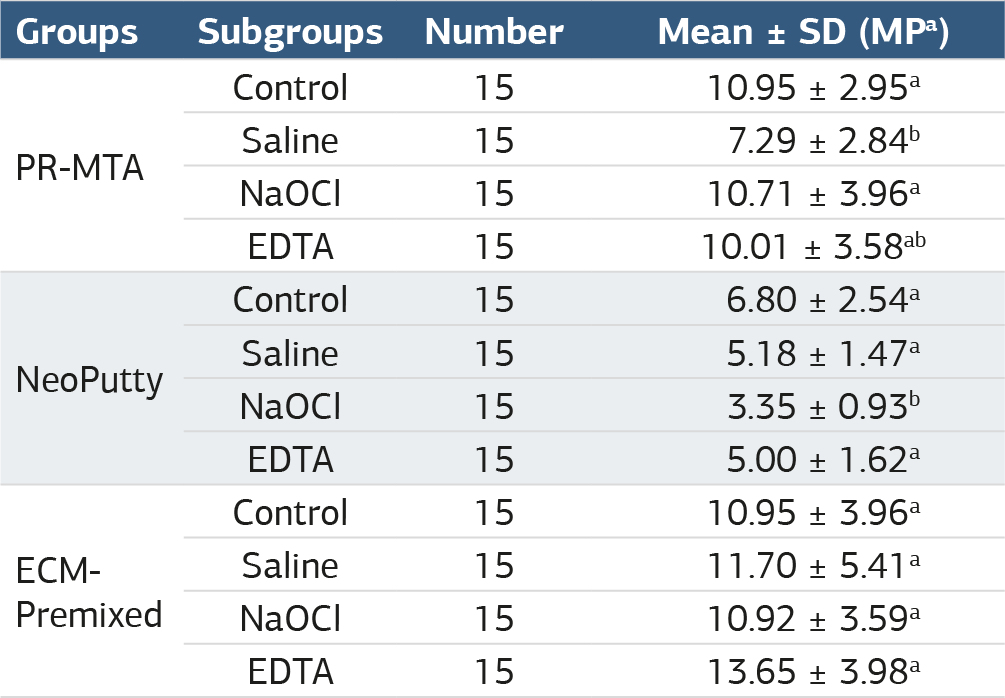
Lowercase superscript letters compare the effects of different irrigation protocols within the same material group. Mean values accompanied by the same superscript letter were not significantly different in pairwise comparisons
Additional Information
Publisher’s Note
Bayrakol MP remains neutral with regard to jurisdictional and institutional claims.
Rights and Permissions

This work is licensed under a Creative Commons Attribution-NonCommercial 4.0 International License (CC BY-NC 4.0). To view a copy of the license, visit https://creativecommons.org/licenses/by-nc/4.0/
About This Article
How to Cite This Article
Nihan Çelik Uzun, Ahter Şanal Çıkman, Banu Arıcıoğlu. Push-out bond strength of bioactive repair materials exposed to endodonticirrigants. Ann Clin Anal Med 2025;16(1):25-29. doi:10.4328/ACAM.22332
- Received:
- July 19, 2024
- Accepted:
- August 26, 2024
- Published Online:
- November 2, 2024
- Printed:
- January 1, 2025