
Abstract
IntroductionThis case report details the diagnosis and treatment of Langerhans Cell Histiocytosis (LCH) in the frontal bone of a 14-year-old male.Case PresentationThe patient presented with a palpable swelling on the forehead, and subsequent imaging studies revealed significant bony destruction. Histopathological examination confirmed the diagnosis of LCH. A surgical resection was successfully performed, resulting in symptom resolution without recurrence at a six-month follow-up.ConclusionThis case points to the importance of considering LCH in differential diagnoses of cranial lesions in children, highlighting the successful role of surgical approaches in managing localized LCH. Multidisciplinary collaboration was crucial for early diagnosis and optimal outcomes in this uncommon pediatric condition.
Keywords
Introduction
Langerhans Cell Histiocytosis (LCH) is a medical condition that was first identified in the early 1900s as case reports and is defined as the accumulation of aberrant proliferation of Langerhans cells within several tissues and organs and is characterized by granulomatous lesions.1,2 Kapukaya et al. proposes that LCH acts as a localized form of eosinophilic granuloma in the bone tissue. No genetic research proves that LCH may be a genetic disorder. Since some types of LCH have a terrible prognosis, it is classified as a neoplasm. However, current ideas focus on whether LCH is immunologic.2 It is possible for the disease to affect one organ or multiple systems. LCH is sporadic, with an incidence ranging from 2 to 9 cases per million children under the age of 15, with this being the most common age group, while the peak incidence belongs to children aged 1 to 3 years. In adults, LCH is extremely rare.3 This case report illustrates a rare manifestation of LCH in the frontal bone of a 14-year-old male, a presentation seldom reported in medical literature, thereby offering valuable insights focusing on the diagnostic obstacles and efficient management methods for pediatric LCH cases, especially those involving atypical cranial sites.
Case Presentation
A 14-year-old male patient applied to our outpatient clinic complaining of “swelling on the right side of the forehead and pain” that had been present for one month. There was nothing special in his medical history other than inguinal hernia and tonsillectomy surgeries. On physical examination, there was a soft swelling on the forehead’s skin, positioned slightly offcenter from the midline (Figure 1).
No pathological findings were detected in the neurological examination. In the cranial MRI and CT examinations performed, significant destruction is caused in the external table just to the right of the midline in the frontal bone, which is assumed to mainly come from the diploe distance. Its dimensions were measured as 19 x 11 mm in the axial plane, showing iso-hyperintense in the T2-weighted series (Figure 2a) and heterogeneous contrast enhancement in the post-contrast series (Figure 2b). The lesion was initially evaluated as Langerhans cell histiocytosis. A surgical resection under general anesthesia via a linear incision was planned and carried out (Figures 3a, 3b, 3c, and 3d). The differential diagnosis for cranial lesions in pediatric patients includes a variety of conditions, each with distinct imaging and histopathological features. In this case, the differential included osteomyelitis, and metastatic disease, among others.
Osteomyelitis was initially considered due to a bone lesion with associated swelling. However, this was ruled out based on the absence of elevated inflammatory markers typically seen in infection, such as C-reactive protein and erythrocyte sedimentation rate, and the lack of fever or other symptoms indicating systemic infection.
Metastatic disease was also considered due to the destructive nature of the lesion. The lack of other systemic symptoms often associated with cancer, such as weight loss or night sweats, along with a normal complete blood count, helped to exclude this diagnosis.
The final diagnosis of Langerhans Cell Histiocytosis was supported by the radiologic finding of a lesion with mixed lytic and sclerotic features, predominantly arising from the diploic space—typical of LCH. Histopathological findings confirmed the existence of Langerhans cells with characteristic CD1a and S-100 protein-positive staining, which are definitive for LCH and not observed in the other conditions considered.
The pathological examination was compatible with Langerhans cell histiocytosis. A follow-up MRI in the sixth month showed no residual or recurrent lesion (Figure 2c), and the patient was free of any symptoms.Ethical ApprovalNot required.
This case report was prepared in accordance with the CARE guidelines.
Discussion
The skull is the most affected part of the skeleton in LCH cases. In our case, the frontal bone, which is one of the most frequent involvements in LCH cases, was affected. The parietal and temporal bones constitute the rest of the majority. In Türkiye, one of the most detailed analyses of LCH cases is an evaluation of 217 patients presented by Yağcı et al. In this retrospective analysis, the most frequently affected bone was the skull, as expected based on other involvements reported so far.4 A typical LCH lesion presents as a soft palpable cyst and manifests with pain.3 Similarly, in our case, the patient’s main complaints were swelling and pain.
Besides the clinical presentation, histological assessments are needed for a differential diagnosis.5 Further, radiological investigations have an essential role in LCH cases, particularly in bones, as in this case.6 However, to diagnose LCH, radiological imaging may be insufficient since LCH may show features similar to other etiologies, such as multiple myeloma. Therefore, a biopsy is essential to diagnose specific lesions.7
The LCH cases with frontal bone involvement do not carry a risk for central nervous system involvement, thus surgical treatment is generally preferable.3 Curettage operations might be performed for lesions smaller than 2 cm.8 Treatment planning depends on the disease’s severity and the organ involved.7 Because of these factors and the possibility of spontaneous remission, there is currently no specific or standardized treatment protocol for this disease.2
In our case, the lesion’s dimensions were measured as 19 x 11 mm. There was a one-month continuance, and as a result of the cranial MRI and CT examinations, a surgical resection under general anesthesia was performed. Then, the lesion was initially evaluated as LCH, and this evaluation became absolute with the biopsy.
Limitations
This report is limited by its single-case design.
Conclusion
This case report highlights a presentation of Langerhans Cell Histiocytosis in the frontal bone of a young patient, emphasizing the challenges associated with diagnosing and treating this uncommon disease. The successful surgical resection of the lesion, without recurrence at a six-month follow-up, demonstrates the effectiveness of prompt and precise surgical intervention in localized LCH cases. Furthermore, this case underscores the importance of considering LCH in diagnosing bone lesions in children and adolescents, mainly when presenting as palpable cyst-like swellings in the skull.
Multidisciplinary collaboration and a high index of suspicion are crucial for early diagnosis and optimal management of LCH, aiming to prevent potential complications and improve patient outcomes. Our experience suggests that surgical intervention, complemented by thorough radiological and histopathological evaluation, remains a cornerstone in managing localised LCH, contributing positively to patient prognosis. Future studies and case reports are essential to enhance our understanding of LCH, refine diagnostic criteria, and improve therapeutic strategies for this complex condition.
Declarations
Animal and Human Rights Statement
All procedures performed in this study were in accordance with the ethical standards of the institutional and/or national research committee and with the 1964 Helsinki Declaration and its later amendments or comparable ethical standards.
Informed Consent
Informed consent was obtained from patient.
Data Availability
The datasets used and/or analyzed during the current study are not publicly available due to patient privacy reasons but are available from the corresponding author on reasonable request.
Conflict of Interest
The authors declare that there is no conflict of interest.
Funding
None.
Abbreviations
LCH: Langerhans cell histiocytosis
MRI: Magnetic resonance imaging
References
- Allen CE, Merad M, McClain KL. Langerhans-cell histiocytosis. N Engl J Med. 2018;379(9):856-868. doi:10.1056/nejmra1607548
- Kapukaya A. Langerhans-hücreli histiyositoz [Langerhans-cell histiocytosis]. TOTBID Derg. 2013;12(6):547-556. doi:10.14292/totbid.dergisi.2013.64
- Leung AK, Lam JM, Leong KF. Childhood Langerhans cell histiocytosis: a disease with many faces. World J Pediatr. 2019;15(6):536-545. doi:10.1007/s12519-019-00304-9
- Yağcı B, Varan A, Çağlar M, et al. Langerhans cell histiocytosis: retrospective analysis of 217 cases in a single center. Pediatr Hematol Oncol. 2008;25(5):399-408. doi:10.1080/08880010802107356
- Krooks J, Minkov M, Weatherall AG. Langerhans cell histiocytosis in children. J Am Acad Dermatol. 2018;78(6):1047-1056. doi:10.1016/j.jaad.2017.05.060
- Tawashi K, Khattab K. Langerhans cell histiocytosis of the frontal bone with unexpected manifestations: rare case report. Int J Surg Case Rep. 2023;109:108580. doi:10.1016/j.ijscr.2023.108580
- Mo JT, Darrow MA, Sharma JD. Langerhans cell histiocytosis with aneurysmal bone cyst-like changes: a case-based literature review. Childs Nerv Syst. 2023;39(11):3057-3064. doi:10.1007/s00381-023-06108-7
- Haupt R, Minkov M, Astigarraga I, et al. Langerhans cell histiocytosis: guidelines for diagnosis, clinical work-up, and treatment for patients till the age of 18 years. Pediatr Blood Cancer. 2013;60(2):175-184. doi:10.1002/pbc.24367
Figures
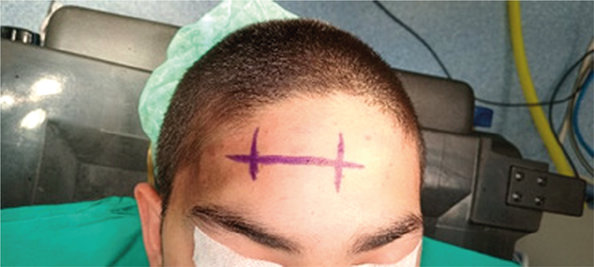
Figure 1. Localization of the soft tissue swelling on the skin of the forehead
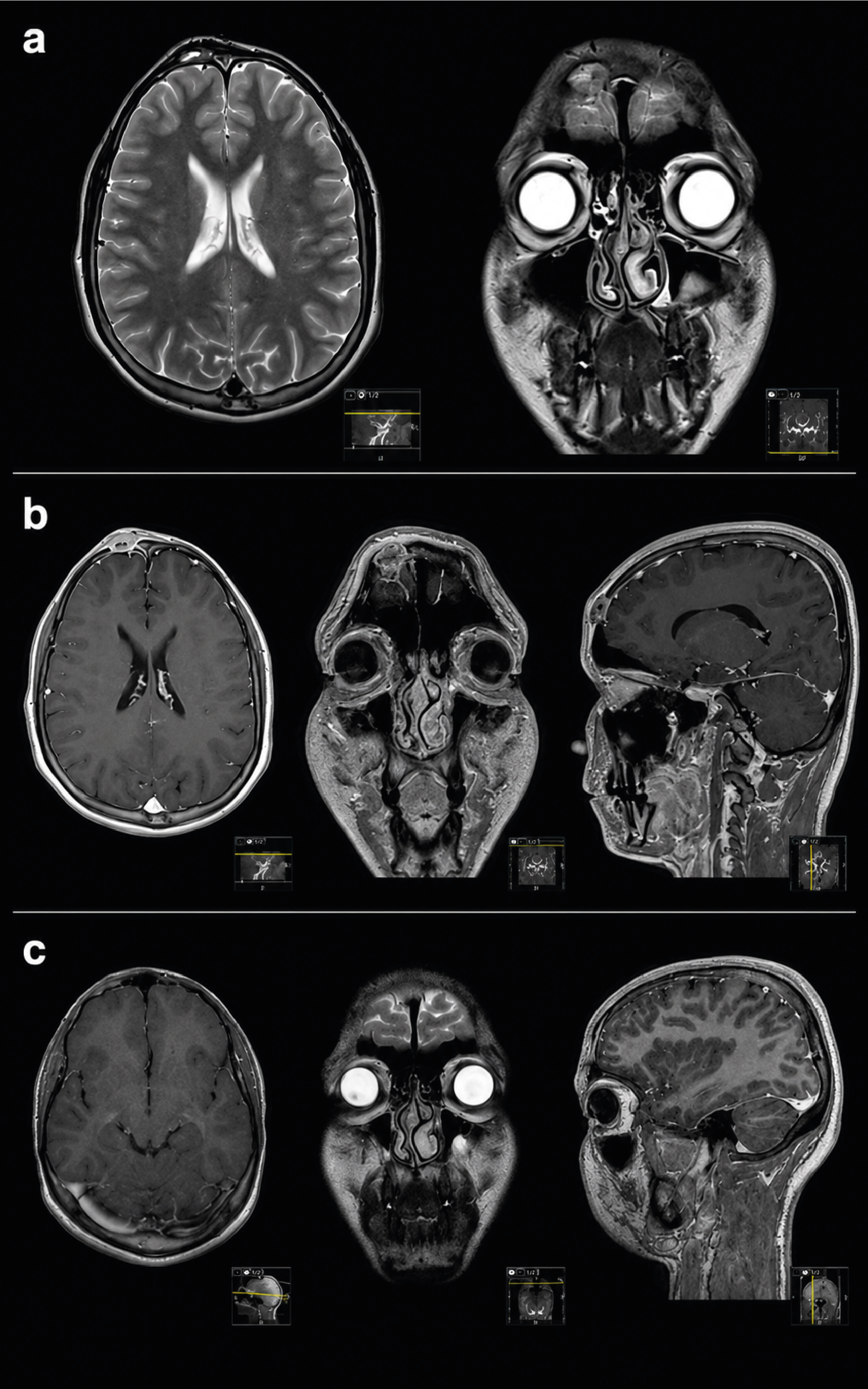
Figure 2. Pre-operational and Post-operational radiological assessments
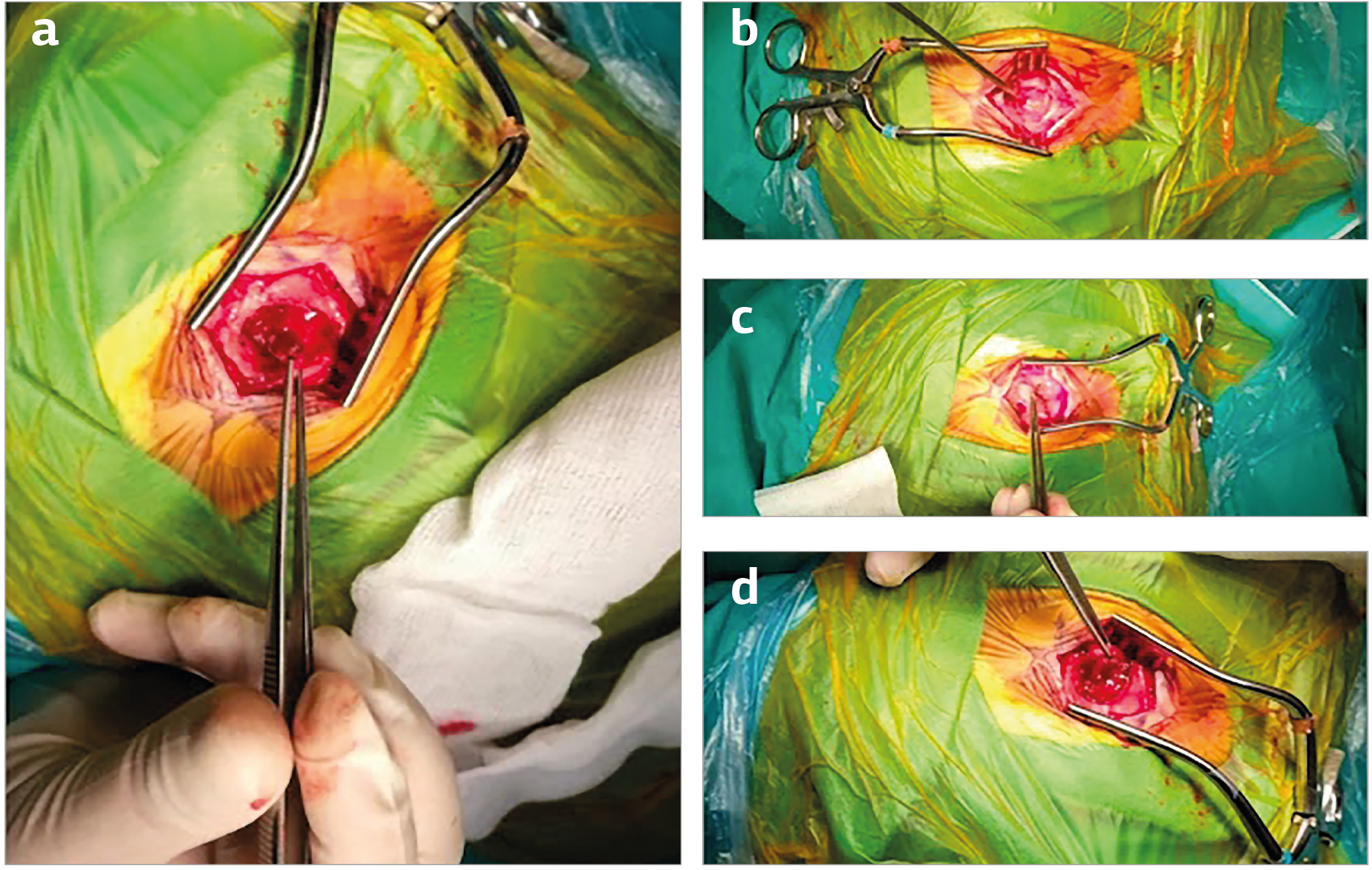
Figure 3. Surgical resection
Additional Information
Publisher’s Note
Bayrakol MP remains neutral with regard to jurisdictional and institutional claims.
Rights and Permissions

This work is licensed under a Creative Commons Attribution-NonCommercial 4.0 International License (CC BY-NC 4.0). To view a copy of the license, visit https://creativecommons.org/licenses/by-nc/4.0/
About This Article
How to Cite This Article
Ekin Lal Akat, Beyza Öztürk, Gözde Erçetin, Ahmet Tolgay Akıncı. A case of langerhans cell histiocytosis in the frontal bone: surgical and clinical perspectives. Ann Clin Anal Med 2025;16(Suppl 2):108-111. doi:10.4328/ACAM.22344
Publication History
- Received:
- 29.07.2024
- Accepted:
- 03.10.2024
- Published Online:
- 04.11.2024
- Printed:
- 25.05.2025