
Abstract
AimAs a result of the developments in breast cancer treatment, the life expectancy of patients has been extended. This fact has brought the morbidity of breast cancer surgery to the fore. Axillary dissection is the major factor responsible for morbidity. In the present study, we aimed to determine the safety of sentinel node biopsy in patients without pathological lymph node involvement using breast MRI taken after neoadjuvant chemotherapy.MethodsThe study was carried out in the General Surgery Department of Bagcilar Training and Research Hospital between 15.02.2021 and 15.07.2021 on patients planned to undergo surgery after neoadjuvant chemotherapy. Postoperative axillary pathology findings were compared with patients without pathological axillary lymphadenopathy in breast MRI results after neoadjuvant chemotherapy.ResultsNinety patients were operated. After the treatment, 57 of them became cN0 on breast MRI. When ≥ 3 negative SLNB was removed in the group of patients who were cN0 after the treatment, no pathological lymph involvement was observed in the axilla.ConclusionThe gold standard for evaluating chemotherapy efficacy is pathologic assessment, the residual cancer burden score (RCB) and the Miller-Payne score are used for this purpose. However, these evaluations are not practical since they need the patient to undergo surgery. For this reason, imaging methods such as mammography, US, MRI and PET-CT are used in the follow-up of NACT treatment response. WHO criteria and RECIST are used to standardize treatment response values with imaging methods.
Keywords
Introduction
Breast cancer is the most common type of cancer which causes the most deaths among women in the world and in Turkey.1 According to the 2020 data of the World Health Organization (WHO), breast cancer patients accounted for 11.7% of newly diagnosed cancer types in all ages and genders, and this number is 2.261.419 people annually. The widespread use of screening methods in developed countries has allowed breast cancer to be diagnosed at an early stage and potential treatment for the disease. In addition, continuously developing medical treatments reduce mortality due to breast cancer. The prolonged life expectancy has led to the questioning of the morbidities experienced by patients in the postoperative period and the improvement of quality of life has become increasingly prominent. The most important morbidity of breast cancer surgery is associated with surgical intervention on the axilla. In the current surgical approach, in patients who have not received preoperative chemotherapy and whose axilla is clinically and radiologically negative, sentinel lymph node biopsy (SLNB) instead of axillary dissection is unquestionably accepted. In patients undergoing neoadjuvant chemotherapy (NACT), the gold standard is still axillary dissection. Nevertheless, studies on the adaptation of SLNB to patients who have received NACT have been started, and many such studies have been published on how to evaluate the axilla accurately and reliably using various methods and lymph node detection agents.
This study was planned to investigate the answer to the question of whether the SLNB method with breast MRI taken after NACT could fully reflect the surgical staging of the axilla (low false negativity rate and high true positivity rate) in patients with locally advanced breast cancer.
Materials and Methods
This study was conducted retrospectively with the participation of 90 patients who were diagnosed with locally advanced breast cancer between 15.02.2021 and 15.07.2021 at the General Surgery Clinic of Bagcilar Training and Research Hospital. The patients had been given neoadjuvant chemotherapy and subjected to re-staging with radiological controls based on breast MRI after the treatment. In all patients diagnosed with locally advanced breast cancer, breast staging is performed with MRI before and after NACT in our clinic in accordance with the guidelines. The age, demographic data, the Tru-cut pathology result of the breast and/or axillary mass before NACT, the number and the size (mm) of masses in the breast in the breast MRI taken after NACT and the status of the axilla (presence/absence of pathological LAP) were recorded by obtaining them from patient files in electronic environment. The cases that responded completely to the treatment based on physical examination and breast MRI performed after NACT were included in the study.Inclusion Criteria1. Female patients older than 18 years of age who were T0-4, N1-3 and M0 according to the TNM system after clinical and radiological evaluation before NACT and who were diagnosed with primary invasive breast cancer based on Tru-cut biopsy.
2. The patients with a performance status of 0-1 according to Eastern Cooperative Oncology Group (ECOG).
3. Patients who received NACT treatment.
4. The patients with complete axillary response in the physical examination performed after NACT and on control breast MRI and who underwent surgical treatment.Exclusion Criteria1. The patients who had previously undergone surgical treatment to the ipsilateral axillary area for any reason.
2. The patients with pathological axillary LAP or metastatic disease were observed in clinical examination and imaging methods after NACT.
3. The patients with a history of allergy to isosulfan blue and/or indocyanine green.
Procedure
A dual method (isosulfan blue (ISB) and indocyanine green (ICG)) was applied to determine sentinel lymph node (SLNB) in patients participating in the study. Axillary dissection, which is the standard treatment, was performed in patients who completed SLNB sampling, and the dissection material was sent in a separate container for pathological examination. It was aimed to remove at least two SLNBs with ISB and ICG. Both SLNB and axillary dissection material were sent to permanent pathology and the false negativity, false positivity, true negativity and true positivity rates of SLNB were calculated from the obtained data.Ethical ApprovalThis study was approved by the Ethics Committee of Bağcılar Training and Research Hospital Clinical Research Ethics Committee (Date: 02.03.2021, Decision No: 2021.01.1.04.004).Statistical AnalysisFrequency and percentage values were presented for categorical variables. Mean, standard deviation, median, minimum and maximum values were presented for continuous variables. Chi-square analysis was performed for relationships between categorical variables. When appropriate, a single sample Chi-square was evaluated by Fisher’s exact and Fisher Freeman Halton Test. The normality of the distribution was determined by Kolmogorov Smirnov Test. For variables that did not satisfy the normal distribution assumption, the Mann Whitney U test was used for two independent group comparisons and the Kruskal Wallis H test was used for more than two group comparisons. The Bonferroni-corrected Dunn Multiple Comparison test was used to determine the source of the significant difference in the Kruskal Wallis H test. The analyses were performed with SPSS 23 (Statistical Package for the Social Sciences) software. p<0.05 was considered significant.Reporting GuidelinesThis study was reported according to the STROBE guideline.
Results
Ninety patients with breast cancer who met the study criteria and received preoperative NACT were operated. SLN was not found in 10 patients. Thus, 80 patients were included in the study. The median age of the patients was 48 years (range: 29-79). After physical examination and radiological imaging, it was found that the primary tumor was in the upper outer quadrant in 35 patients, in the upper inner quadrant in 15 patients, in the lower outer quadrant in 13 patients, in the lower inner quadrant in 7 patients, and in the retroareolar region in 10 patients. Tumor was single-focused in 58 patients, multicentric in 14 patients and multifocal in 8 patients. The mean size of primary masses on MRI was 21.8 ± 12.05 mm, and the mean size of satellite lesion was 17.45 ± 6.95 mm in 22 patients with satellite lesions. In the pre-NACT Tru-cut biopsy evaluation of the patients, diagnosis was invasive ductal carcinoma in 75 patients, invasive lobular carcinoma in 4 patients, and invasive ductal carcinoma + invasive lobular carcinoma in 1 patient.
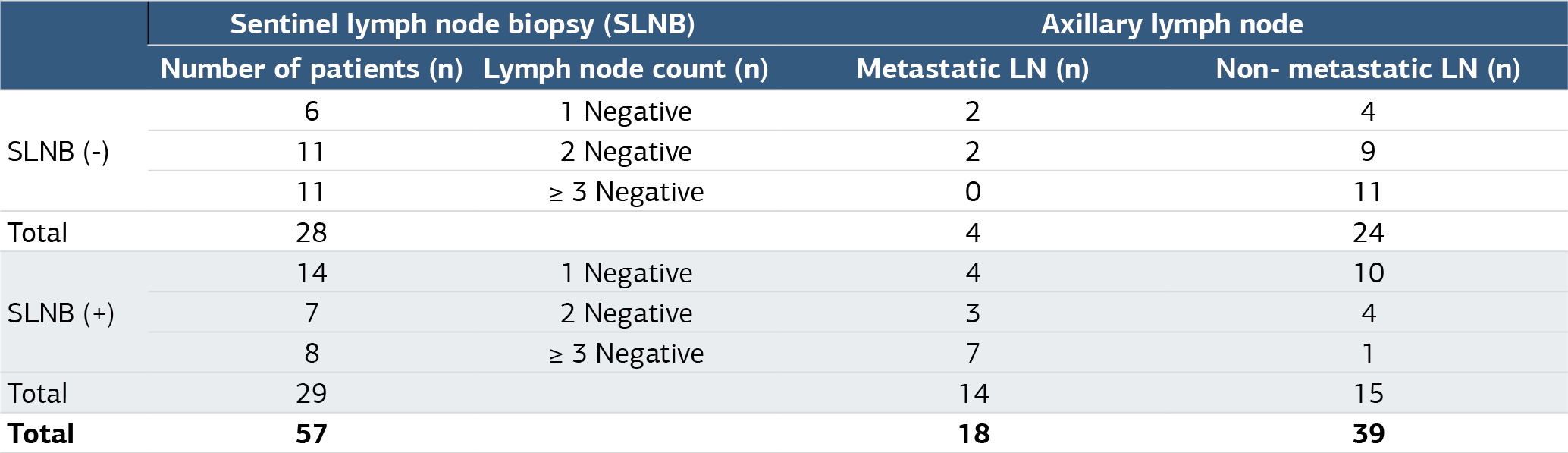
There were 57 patients who were evaluated as cN0 and had SLNB in radiological (breast MRI) evaluations after NACT. Dual method (ICG + ISB) was used to determine sentinel lymph nodes. All patients underwent axillary dissection after sentinel lymph node removal. SLNB was positive in 29 patients and negative in 28 patients. In the evaluation of SLNB negative patients, 2 of the 6 patients with one SLNB negative had metastatic nodule in axillary area and 2 of the 11 patients who had two SLNBs removed had additional metastatic lymph node while none of the 11 patients who had three or more SLNBs removed had additional metastatic lymph node. According to the results of this evaluation, when axillary cN0 was taken into account in breast MRI, the presence of ≥ 3 negative SLNB had a positive predictive value of 100% (Table 1).
Based on the evaluations, the sensitivity of the breast MRI taken after NACT for metastatic lymph node determination in the axillary region was 41.67% while the specificity was 95.45% and the accuracy was 71.25%.
Discussion
Breast cancer is the most common cancer with the highest death toll among women throughout the world.1 Due to the advances in the diagnosis and treatment of breast cancer, its mortality has decreased considerably with a concomitant longer life expectancy.2 This has brought to the fore the quality of life in patients after breast cancer surgery. The most important cause of morbidity after breast cancer surgery is the interventions made to axilla. Axillary involvement is the most important prognostic factor in determining the prognosis and treatment of breast cancer and recurrences occur most frequently.3 For this reason, the question of how to evaluate and treat the axilla with the least intervention that would treat the axilla correctly without compromising the principles of oncological surgery has gained importance. In the 1990s, the lymph node in which cancer first metastasized in lymphatic spread was identified and it was shown that if there was no involvement in this lymph node, the tumor had not yet metastasized.4 The lymph node where the tumor first metastasizes is called the “sentinel lymph node”. Clinically, SLNB has been shown to be equivalent to axillary dissection in patients who are axillary lymph node negative. Thus, morbidities such as lymphedema, seroma, numbness, wound infection, limitation of movement and chronic pain caused by axillary dissection could be minimized.5 Therefore, NACT regimens were started to be applied to patients who were clinically axillary lymph node positive. As in early stage breast cancers, it was hypothesized that the axilla could be evaluated with SLNB in patients whose axilla became clinically negative, and studies were started on this issue. In the present study, it was aimed to find SLNB with the dual method (ICG + ISB) for the evaluation of the axilla in patients who did not have axillary pathological lymph nodes clinically and radiologically after NACT. According to the results of our study, it was observed that the negative SLNB ≥ 3 lymph node in patients who did not have pathological LAP in the axilla clinically and radiologically determined the absence of metastatic lymph nodes in the axillary area with a 100% accuracy. This is an important consideration for the evaluation of patients receiving NACT using SLNB instead of axillary dissection.
In breast cancer, NACT is preferred to treat nodal involvement in the axilla in order to both reduce and destroy the mass in the breast. If the chemotherapy regimen given is ineffective, the patient would be treated with an ineffective regimen again because the same regimen would be used both in breast cancer treatment and after surgery. Therefore, follow-up of the response after the chemotherapy regimen used has gained importance. The gold standard for evaluating chemotherapy efficacy is pathologic assessment, the residual cancer burden score (RCB) and the Miller-Payne score are used for this purpose.6 However, these evaluations are not practical since they need the patient to undergo surgery. For this reason, imaging methods such as mammography, US, MRI and PET-CT are used in the follow-up of NACT treatment response. WHO criteria and RECIST are used to standardize treatment response values with imaging methods.7 In our study, we used the Miller-Payne scoring system to evaluate the pathology response according to the RECIST criteria and to evaluate the radiological response. US is used in the evaluation of treatment response after NACT due to its advantages such as low cost, no radiation exposure, and ability to be performed on point of care basis. Conventional US has difficulty separating fibrotic and tumor tissues. Another disadvantage of the US in assessing the treatment response is that it depends on the experience of the person who performs NACT. Vriens et al. evaluated the response to NACT in 172 breast cancer patients and showed that US measurements of the tumor tissue size were on the average 20% larger than they actually are.8 Many studies showed that MRI exhibits higher accuracy in predicting the effectiveness of NACT.9 Although conventional US has low sensitivity in assessing the treatment response of NACT, new techniques such as electrography, CEUS (Contrast Enhanced Ultrasound using microbubbles) can improve US prediction accuracy.10 In our study, we used breast MRI to evaluate the treatment response of NACT because of the low sensitivity of traditional US. Fibrous tissue has no vascularization whereas the tumor tissue has good vascularization. Since MRI is an imaging method that differentiates these two tissues efficiently, it is used to determine the size of fibrous tissue that develops in response to NACT and remaining tumor tissue. The NACT regimen may affect the evaluation of the MRI because drugs containing taxane and anthracycline have a greater antiangiogenic effect, which negatively affects the evaluation of MRI by disrupting vascularization.11 In the presence of fibrosis and reactive inflammation around the tumor bed, MRI shows the residual disease larger size than it is. The presence of residual DCIS causes false positive diagnosis, while small residual lesion leads to a false negative diagnosis.12 De Los Santos et al. found the accuracy of MRI to be 74% in predicting pCR (pathological complete response).13 Choi et al. found that in the evaluation of NACT treatment response, MRI had a sensitivity of 88.2%, a specificity of 62.5%, an accuracy of 75.7%, a PPV value of 71.4% and an NPV value of 83.3%.14 In our study, we found that MRI had 47.7% sensitivity, 95.45% specificity, 88.24% PPV value and 66.67% NPV value in determining the axillary response. According to a meta-analysis by Liu et al., the sensitivity of PET/CT was 86% and the specificity of MRI was 88%, and they concluded that the combined use of these two imaging methods could provide better results in evaluating pCR after NACT.15 In our study, we used MRI in the diagnosis and staging of pre-NACT breast cancer and in the evaluation of response to treatment after NACT. We found that the probability of metastatic lymph node in the axilla is low if ≥ 3 negative SLNB is performed in patients without pathological LAP in the axilla after NACT. We found that the radiological evaluation of the response to NACT, which was carried out in accordance with the literature, was consistent with the pathological assessment by the Miller-Payne system.
Limitations
The small number of patients in our study was the most important limitation.
Conclusion
The most important cause of morbidity in locally advanced breast cancer patients is axillary dissection. When the axilla is cN0 based on breast MRI after NACT, SLNB can be performed instead of axillary dissection. Prospective randomized trials are needed in this regard.
Declarations
Ethics Declarations
This study was approved by the Ethics Committee of Bagcilar Training and Research Hospital Clinical Research (Date: 2021-03-02, No: 2021.01.1.04.004)
Animal and Human Rights Statement
All procedures performed in this study were in accordance with the ethical standards of the institutional and/or national research committee and with the 1964 Helsinki Declaration and its later amendments or comparable ethical standards.
Informed Consent
Written informed consent was obtained from all patients prior to participation in the study.
Data Availability
The datasets used and/or analyzed during the current study are not publicly available due to patient privacy reasons but are available from the corresponding author on reasonable request.
Conflict of Interest
The authors declare that there is no conflict of interest.
Funding
None.
Abbreviations
DCIS: Ductal carcinoma in situ
ECOG: Eastern cooperative oncology group
ICG: Indocyanine green
ISB: Isosulfan blue
LAP: Lymphadenopathy
MRI: Magnetic resonance imaging
NACT: Neoadjuvant chemotherapy
NPV: Negative predictive value
PET-CT: Positron emission tomography-computed tomography
PPV: Positive predictive value
RCB: Residual cancer burden
RECIST: Response evaluation criteria in solid tumors
SLNB: Sentinel lymph node biopsy
SPSS: Statistical package for the social sciences
TNM: Tumor, node, metastasis
US: Ultrasound
WHO: World health organization
References
- Bray F, Ferlay J, Soerjomataram I, Siegel RL, Torre LA, Jemal A. Global cancer statistics 2018: GLOBOCAN estimates of incidence and mortality worldwide for 36 cancers in 185 countries. CA Cancer J Clin. 2018;68(6):394-424. doi:10.3322/caac.21492
- Yang Y, Liu G, Qin L, Ye L, Zhu F, Ying Y. Overexpression of UHRF1 and its potential role in the development of invasive ductal breast cancer validated by integrative bioinformatics and immunohistochemistry analyses. Transl Cancer Res. 2019;8(4):1086-1096. doi:10.21037/tcr.2019.06.19
- de Meric de Bellefon M, Lemanski C, Ducteil A, Fenoglietto P, Azria D, Bourgier C. Management of the axilla in the era of breast cancer heterogeneity. Front Oncol. 2018;8:84. doi:10.3389/fonc.2018.00084
- Krag DN, Anderson SJ, Julian TB, et al. Sentinel lymph node resection compared with conventional axillary lymph node dissection in clinically node-negative patients with breast cancer: overall survival findings from the NSABP B-32 randomised phase 3 trial. Lancet Oncol. 2010;11(10):927-933. doi:10.1016/s1470-2045(10)70207-2
- Peek MCL, Saeki K, Ohashi K, et al. Optimization of SPIO injection for sentinel lymph node dissection in a rat model. Cancers (Basel). 2021;13(19):5031. doi:10.3390/cancers13195031
- Wang W, Liu Y, Zhang H, et al. Prognostic value of residual cancer burden and Miller-Payne system after neoadjuvant chemotherapy for breast cancer. Gland Surg. 2021;10(12):3211-3221. doi:10.21037/gs-21-608
- Cappello G, Romano V, Neri E, et al. A European Society of Oncologic Imaging survey on the radiological assessment of response to oncologic treatments in clinical practice. Insights Imaging. 2023;14(1):220. doi:10.1186/s13244-023-01568-6
- Vriens BE, de Vries B, Lobbes MB, et al. Ultrasound is at least as good as magnetic resonance imaging in predicting tumour size post-neoadjuvant chemotherapy in breast cancer. Eur J Cancer. 2016;52:67-76. doi:10.1016/j.ejca.2015.10.010
- Kao YS, Hsu Y. A meta-analysis for using radiomics to predict complete pathological response in esophageal cancer patients receiving neoadjuvant chemoradiation. In Vivo. 2021;35(3):1857-1863. doi:10.21873/invivo.12448
- Xie F, Zhang D, Cheng L, et al. Intradermal microbubbles and contrast-enhanced ultrasound is a feasible approach for sentinel lymph node identification in early-stage breast cancer. World J Surg Oncol. 2015;13:319. doi:10.1186/s12957-015-0736-x
- Citgez B, Yigit B, Yetkin SG. Management of the axilla and the breast after neoadjuvant chemotherapy in patients with breast cancer: a systematic review. Sisli Etfal Hastan Tip Bul. 2021;55(2):156-161. doi:10.14744/semb.2021.77010
- Chen JH, Feig B, Agrawal G, et al. MRI evaluation of pathologically complete response and residual tumors in breast cancer after neoadjuvant chemotherapy. Cancer. 2008;112(7):17-26. doi:10.1002/cncr.23130
- De Los Santos JF, Cantor A, Amos KD, et al. Magnetic resonance imaging as a predictor of pathologic response in patients treated with neoadjuvant systemic treatment for operable breast cancer: Translational Breast Cancer Research Consortium trial 017. Cancer. 2013;119(10):1776-1783. doi:10.1002/cncr.27995
- Choi EK, Yoo IR, Kim SH, Park SY, O JH, Kang BJ. The value of pre- and post-neoadjuvant chemotherapy F-18 FDG PET/CT scans in breast cancer: comparison with MRI. Acta Radiol. 2018;59(1):41-49. doi:10.1177/0284185117705011
- Liu Q, Wang C, Li P, Liu J, Huang G, Song S. The role of 18F-FDG PET/CT and MRI in assessing pathological complete response to neoadjuvant chemotherapy in patients with breast cancer: a systematic review and meta-analysis. Biomed Res Int. 2016;2016:3746232. doi:10.1155/2016/3746232
Tables
Table 1. Evaluation of patients without pathological lymph node involvement (cN0) on breast MRI after neoadjuvant chemotherapy
Additional Information
Publisher’s Note
Bayrakol MP remains neutral with regard to jurisdictional and institutional claims.
Rights and Permissions

This work is licensed under a Creative Commons Attribution-NonCommercial 4.0 International License (CC BY-NC 4.0). To view a copy of the license, visit https://creativecommons.org/licenses/by-nc/4.0/
About This Article
How to Cite This Article
Ahmet Akbas, Fikret Calikoglu, Mehmet Oncü, Atilla Celik. Determination of axillary metastasis by breast MRI after neoadjuvant chemotherapy in breast cancer. Ann Clin Anal Med 2025;16(6):397-400. doi:10.4328/ACAM.22359
Publication History
- Received:
- 07.08.2024
- Accepted:
- 21.10.2024
- Published Online:
- 10.01.2025
- Printed:
- 01.06.2025