
Abstract
Radiation, particularly ionizing radiation used in medical imaging and cancer treatments, induces oxidative stress (OS), a key factor in the biological damage it causes. Radiation interacting with biological tissues generates reactive oxygen species (ROS) that can overwhelm the cell’s natural antioxidant defenses. This leads to oxidative damage to essential macromolecules such as DNA, proteins, and lipids, triggering a cascade of harmful cellular events. DNA strand breaks, protein oxidation, and lipid peroxidation are common consequences, contributing to mutations, cellular dysfunction, and apoptosis. These effects play a significant role in both acute radiation injury and long-term health outcomes, such as carcinogenesis and cardiovascular diseases. Given the growing use of radiological procedures in medicine, understanding radiation-induced OS is critical for improving patient safety. Strategies to mitigate oxidative damage, including using antioxidants and radiation dose optimization, are being actively explored. Continued research into the mechanisms of radiation-induced OS is necessary to enhance therapeutic interventions and diagnostic accuracy while minimizing potential risks. This review provides a concise summary of the relationship between radiation and OS.
Keywords
Introduction
Wilhelm Conrad Roentgen accidentally discovered the X-rays in 1895. This discovery has created a major change not only in the field of physics but also in the field of medicine. Within months of Röntgen’s discovery, X-rays were being used for medical diagnosis.1 Then, Henri Becquerel discovered radioactivity in 1896, followed by Marie and Pierre Curie’s isolation of radium in 1898. Radiation falls into two categories: ionizing and non-ionizing. Non-ionizing radiation (NIR), including microwaves, infrared, ultraviolet, and visible light, can excite atoms but lacks the energy to ionize them.2-3 MRI is one of the most important medical imaging methods in which we use non-ionizing radiation. Ionizing radiation (IR) is, on the other hand, classified into electromagnetic radiation (gamma rays and X-rays) and particle radiation (electrons, protons, neutrons, and alpha particles).4 They are possessing sufficient energy to ionize atoms. In medicine, IR is crucial for both therapy and diagnostics. It’s used in nuclear medicine for treatments like radionuclide therapy and imaging techniques such as PET and in radiology for CT scans, digital subtraction angiographies (DSAs), and as well as X-ray radiography.5 Moreover, radiotherapy plays a crucial role in cancer treatment, with approximately 80% of cancer patients requiring this form of therapy.6 These advancements have fundamentally transformed how we detect and visualize diseases, as well as how we approach therapeutic intervention.5 But since the discovery of radiation, it has been a double-edged sword.7Biological Effects of RadiationScientists would gradually discover the biological effects of the radiation. By 1897, epigastric dermatitis was reported as a side effect of X-ray exposure.8 The link between radiation exposure and cancer development wasn’t immediately apparent. This grim connection became evident through the tragic fates of two groundbreaking scientists in the field of radioactivity. Marie Sklodowska-Curie, a Nobel Prize recipient and trailblazer in radiation research, succumbed to aplastic anemia resulting from her prolonged radiation exposure. Similarly, her daughter, Irene Joliot-Curie, who, along with her husband, was awarded a Nobel Prize for their pioneering work on artificial radioactivity, later died from leukemia induced by radiation.9 Within three decades after the discovery of Roentgen, a wealth of scientific literature emerged, documenting the wide-ranging effects of ionizing radiation on diverse substances and biological systems, from the visibly damaged hands of pioneering radiologists to more insidious consequences such as sterility, bone disease, cancer, and various other illnesses.10-11 The atomic bombings of Hiroshima and Nagasaki led to extensive studies on radiation effects. Most of the information on the health risks of radiation in healthy populations comes from the life span studies, which were established after the detonation of the atomic bombs in Japan in 1945. Assembled in 1950, these cohorts have been followed for 65 years.12 It showed a direct link exists between radiation dose and solid cancer risk, with children being more vulnerable; high doses increase the risk of heart disease and other non-cancer illnesses; exposure in the womb affected brain development and raised cancer risk; interestingly, no clear increase in birth defects or illnesses was found in survivors’ children; high doses impacted the immune system similarly to aging; while cancer risk increases at moderate to high doses (over 0.1-0.2 Gy), the effects of low doses (under 0.1 Gy) remain uncertain. Several large cohort studies support the life span study’s findings on low-dose radiation effects. These include studies of UK nuclear workers,13 Techa River residents,14 Chornobyl cleanup workers,15 and residents of high natural radiation areas in China.16Effects of Oxidative Damage on MacromoleculesOxidative damage, primarily caused by the excessive generation of reactive oxygen species (ROS), can have profound effects on various macromolecules within cells, including DNA, proteins, and lipids.17-18 Each of these macromolecules is critical to cellular function, and their damage can lead to significant biological consequences.19 Figure 1. DNA Damage: ROS can attack the nucleotides within DNA, leading to base modifications, strand breaks, and crosslinking.20 These mutations can interfere with DNA replication and transcription, potentially leading to errors in protein synthesis. If not properly repaired, DNA damage may result in genomic instability, which is a precursor to cancer, aging, and various genetic disorders.21Protein OxidationProteins are vulnerable to oxidative damage, particularly in their amino acid residues. This can lead to the alteration of protein structure, loss of enzymatic activity, and impaired cellular signaling pathways.22 The accumulation of oxidized proteins is often associated with neurodegenerative diseases like Alzheimer’s and Parkinson’s, as well as impaired immune and metabolic functions.Lipid PeroxidationLipids, especially polyunsaturated fatty acids in cell membranes, are highly susceptible to ROS attack, leading to lipid peroxidation. This process compromises the structural integrity and fluidity of cell membranes, disrupting essential functions such as ion transport, cell signaling, and energy production.23 Lipid peroxidation products can also form toxic aldehydes, further amplifying cellular damage and inflammation.Carbohydrate DamageAlthough less frequently discussed, carbohydrates can also be oxidatively damaged, particularly through reactions that generate advanced glycation end-products (AGEs). These AGEs can impair normal cellular functions by crosslinking with proteins and altering their normal activities, contributing to diabetes complications, cardiovascular diseases, and aging.24 The cumulative oxidative damage to these macromolecules can initiate a cascade of deleterious effects at the cellular level, eventually affecting tissues and organs. This contributes to a range of pathological conditions, including cancer, neurodegeneration, cardiovascular diseases, and chronic inflammation. Understanding these mechanisms underlines the importance of controlling oxidative stress through both intrinsic antioxidant defenses and potential therapeutic interventions.DNA Damage from Ionizing Radiation: The Role of Oxidative Stress as an Indirect PathwayThe concept that ionizing radiation (IR) can harm genetic material, such as by breaking chromosomes, was recognized before we understood DNA’s structure. Initially, scientists proposed a direct mechanism where high-energy particles or photons directly collided with DNA strands.25 However, later research revealed an indirect pathway involving oxidative stress (OS), which is now believed to cause most (60-70%) of IR-induced DNA damage.26-30 Oxidative stress occurs when there’s an imbalance between oxidant production and antioxidant activity, favoring the former. This OS-mediated indirect mechanism involves the generation of reactive oxygen species (ROS) through IR-induced water radiolysis.4 Ionizing radiation’s impact on cells is a complex and rapid process. Within a fraction of a second after exposure, the radiation releases electrons that break chemical bonds in cellular molecules. This creates unstable particles called free radicals and ROS. Some DNA damage occurs instantly from direct radiation hits, but most happens indirectly through these newly created particles. Interestingly, the majority of DNA damage takes place within the first 5 minutes after exposure. After this initial burst, the rate of new damage slows down.4,31 This timing is crucial for scientists studying radiation effects, as they need to measure damage quickly before cells begin their natural repair processes.32 While some ROS are neutralized, those near the cell’s nucleus can be particularly harmful. They can cause severe DNA damage, especially double-strand breaks (DSBs), which are considered the most dangerous type.26-28,30,33-36 If not repaired correctly, these DSBs can alter chromosome structure, potentially leading to mutations. Over time, these mutations might contribute to cancer development.37-39Biomarkers and Assessment of the IR-DamageCells respond to IR-caused DNA damage by modifying a protein called H2AX, creating γ-H2AX. Scientists use this modified protein as a marker to measure the extent of DNA damage. This cellular response is part of the body’s attempt to detect and repair the harm caused by radiation.40Radioprotective agents and Anti-oxidant SupplementationUnderstanding the process from initial exposure to cellular response helps researchers develop better protective measures against radiation damage and improve treatments for radiation exposure.4 Studies have investigated a variety of antioxidant compounds for their potential to mitigate radiation-induced damage. Research has shown that certain ingredients can reduce lethality or decrease markers of DNA and cellular damage caused by radiation exposure. These include quercetin,41-42 which has demonstrated protective effects in several studies, and astaxanthin,43 known for its potent antioxidant properties. Zeaxanthin,44 vitamin C,45 and vitamin B1246 have also been examined for their radioprotective potential. Additionally, selenium,47 folate,48 and CoQ1049-50 have been the subject of research focusing on their ability to counteract radiation damage. Studies have also explored the benefits of α-lipoic acid and vitamin E in this context. These investigations suggest that these antioxidants might offer protection against radiation-induced cellular harm.
Limitations
This review is narrative in nature and does not include a systematic literature search or meta-analysis.
Conclusion
OS is an important consideration in radiology, particularly in imaging modalities that rely on ionizing radiation, such as X-rays, CT scans, and fluoroscopy. The ionization process leads to the generation of ROS, which can damage cellular components like DNA, proteins, and lipids. Although the body has natural antioxidant defenses, prolonged or excessive exposure to radiation can overwhelm these mechanisms, leading to oxidative damage and potential long-term health risks, including cancer and cardiovascular diseases. The clinical implications highlight the need for optimized protocols to minimize radiation exposure without compromising diagnostic efficacy. This includes employing the principle of ALARA (As Low As Reasonably Achievable), using advanced imaging technologies that require lower doses of radiation, and exploring protective agents that can mitigate oxidative damage. Further research is crucial to better understand the mechanisms of radiation-induced oxidative stress and to develop strategies for safeguarding patients, particularly those requiring repeated imaging. Ultimately, the balance between effective diagnostic imaging and the prevention of OS remains a key objective in the field of radiology. With ongoing advancements, radiologists and healthcare providers can continue to improve patient safety while delivering high-quality diagnostic care.
Declarations
Animal and Human Rights Statement
All procedures performed in this study were in accordance with the ethical standards of the institutional and/or national research committee and with the 1964 Helsinki Declaration and its later amendments or comparable ethical standards.
Informed Consent
Not applicable.
Data Availability
The datasets used and/or analyzed during the current study are not publicly available due to patient privacy reasons but are available from the corresponding author on reasonable request.
Conflict of Interest
The authors declare that there is no conflict of interest.
Funding
None.
Abbreviations
ALARA: As low as reasonably achievable
CT: Computed tomography
DNA: Deoxyribonucleic acid
DSA: Digital subtraction angiography
DSB: Double-strand break
IR: Ionizing radiation
MRI: Magnetic resonance imaging
NIR: Non-ionizing radiation
OS: Oxidative stress
PET: Positron emission tomography
ROS: Reactive oxygen species
References
- Timins JK. Communication of benefits and risks of medical radiation: a historical perspective. Health Phys. 2011;101(5):562-565. doi:10.1097/hp.0b013e3182259a71
- Acherar S. Editorial: recent advances in medical radiation technology. Front Chem. 2024;12:1360379. doi:10.3389/fchem.2024.1360379
- Sotomayor CG, Gonzalez C, Soto M, et al. Ionizing radiation-induced oxidative stress in computed tomography: effect of vitamin C on prevention of DNA damage: PREVIR-C randomized controlled trial study protocol. J Clin Med. 2024;13(13). doi:10.3390/jcm13133866
- Bhatia SS, Pillai SD. Ionizing radiation technologies for vaccine development: a mini review. Front Immunol. 2022;13:845514. doi:10.3389/fimmu.2022.845514
- Gupta S, Sharma RS, Singh R. Non-ionizing radiation as possible carcinogen. Int J Environ Health Res. 2022;32(4):916-940. doi:10.1080/09603123.2020.1806212
- Kostova N, Staynova A, Popova-Hadjiiska L, et al. Effect of curcumin on gamma-ray-induced cell response. J Radiat Res. 2023;64(2):471-479. doi:10.1093/jrr/rrac101
- Kiang JG, Cannon G, Singh VK. An overview of radiation countermeasure development in radiation research from 1954 to 2024. Radiat Res. 2024;202(2):420-431. doi:10.1667/rade-24-00036.1
- Crocker HR. A case of dermatitis from roentgen rays. Br Med J. 1897;1(1879):8-9. doi:10.1136/bmj.1.1879.8
- Zanzonico P, Stabin MG. Quantitative benefit-risk analysis of medical radiation exposures. Semin Nucl Med. 2014;44(3):210-214. doi:10.1053/j.semnuclmed.2014.03.010
- Ungar S. Review of: Walker JS. Permissible Dose: A History of Radiation Protection in the Twentieth Century. J Am Hist. 2002;89(1):291. doi:10.2307/2700905
- Zimbrick JD. Radiation chemistry and radiation research: a history from the beginning to the platinum edition. Radiat Res. 2024;202(2):368-384. doi:10.1667/rade-24-00053.1
- Kamiya K, Ozasa K, Akiba S, et al. Long-term effects of radiation exposure on health. Lancet. 2015;386(9992):469-478. doi:10.1016/s0140-6736(15)61167-9
- Muirhead CR, O’Hagan JA, Haylock RG, et al. Mortality and cancer incidence following occupational radiation exposure: third analysis of the National Registry for Radiation Workers. Br J Cancer. 2009;100(1):206-212. doi:10.1038/sj.bjc.6604825
- Schonfeld SJ, Krestinina LY, Epifanova S, Degteva MO, Akleyev AV, Preston DL. Solid cancer mortality in the Techa River cohort, 1950-2007. Radiat Res. 2013;179(2):183-189. doi:10.1667/rr2932.1
- Romanenko AY, Finch SC, Hatch M, et al. The Ukrainian-American study of leukemia and related disorders among Chornobyl cleanup workers from Ukraine: III. Radiation risks. Radiat Res. 2008;170(6):711-720. doi:10.1667/rr1404.1
- Tao Z, Akiba S, Zha Y, et al. Cancer and non-cancer mortality among inhabitants in the high background radiation area of Yangjiang, China, 1979-1998. Health Phys. 2012;102(2):173-181. doi:10.1097/hp.0b013e31822c7f1e
- Özcan O, Erdal H, Çakırca G, Yönden Z. Oxidative stress and its impacts on intracellular lipids, proteins and DNA. J Clin Exp Invest. 2015;6(3):331-336. doi:10.5799/ahinjs.01.2015.03.0545
- Ozcan O, Erdal H, Yonden Z. Biochemical perspective on the relationship between ischemia-reperfusion injury and oxidative stress. Med J Mustafa Kemal Univ. 2015;6(23):27-33.
- Erdal H, Günaydın FE, Karaoğlanoğlu S. Oxidative stress in asthma. J Sport Health Res. 2023;4(1):62-70.
- Cooke MS, Evans MD, Dizdaroglu M, Lunec J. Oxidative DNA damage: mechanisms, mutation, and disease. FASEB J. 2003;17(10):1195-1214. doi:10.1096/fj.02-0752rev
- Schieber M, Chandel NS. ROS function in redox signaling and oxidative stress. Curr Biol. 2014;24(10). doi:10.1016/j.cub.2014.03.034
- Bulteau AL, Szweda LI, Friguet B. Mitochondrial protein oxidation and degradation in response to oxidative stress and aging. Exp Gerontol. 2006;41(7):653-657. doi:10.1016/j.exger.2006.03.013
- Su LJ, Zhang JH, Gomez H, et al. Reactive oxygen species-induced lipid peroxidation in apoptosis, autophagy, and ferroptosis. Oxid Med Cell Longev. 2019;2019:5080843. doi:10.1155/2019/5080843
- Umbayev B, Askarova S, Almabayeva A, Saliev T, Masoud AR, Bulanin D. Galactose-induced skin aging: the role of oxidative stress. Oxid Med Cell Longev. 2020;2020:7145656. doi:10.1155/2020/7145656
- Sax K. Chromosome aberrations induced by x-rays. Genetics. 1938;23(5):494-516. doi:10.1093/genetics/23.5.494
- Santivasi WL, Xia F. Ionizing radiation-induced DNA damage, response, and repair. Antioxid Redox Signal. 2014;21(2):251-259. doi:10.1089/ars.2013.5668
- Nickoloff JA, Boss MK, Allen CP, LaRue SM. Translational research in radiation-induced DNA damage signaling and repair. Transl Cancer Res. 2017;6(5):875-891. doi:10.21037/tcr.2017.06.02
- Eccles LJ, O’Neill P, Lomax ME. Delayed repair of radiation-induced clustered DNA damage: friend or foe? Mutat Res. 2011;711(1-2):134-141. doi:10.1016/j.mrfmmm.2010.11.003
- Sage E, Harrison L. Clustered DNA lesion repair in eukaryotes: relevance to mutagenesis and cell survival. Mutat Res. 2011;711(1-2):123-133. doi:10.1016/j.mrfmmm.2010.12.010
- Maier P, Hartmann L, Wenz F, Herskind C. Cellular pathways in response to ionizing radiation and their targetability for tumor radiosensitization. Int J Mol Sci. 2016;17(1):102. doi:10.3390/ijms17010102
- Rothkamm K, Balroop S, Shekhdar J, Fernie P, Goh V. Leukocyte DNA damage after multi-detector row CT: a quantitative biomarker of low-level radiation exposure. Radiology. 2007;242(1):244-251. doi:10.1148/radiol.2421060171
- Azqueta A, Shaposhnikov S, Collins AR. DNA oxidation: investigating its key role in environmental mutagenesis with the comet assay. Mutat Res. 2009;674(1-2):101-108. doi:10.1016/j.mrgentox.2008.10.013
- Cannan WJ, Pederson DS. Mechanisms and consequences of double-strand DNA break formation in chromatin. J Cell Physiol. 2016;231(1):3-14. doi:10.1002/jcp.25048
- Smith TA, Kirkpatrick DR, Smith S, et al. Radioprotective agents to prevent cellular damage due to ionizing radiation. J Transl Med. 2017;15(1):232. doi:10.1186/s12967-017-1338-x
- Wan XS, Ware JH, Zhou Z, Donahue JJ, Guan J, Kennedy AR. Protection against radiation-induced oxidative stress in cultured human epithelial cells by treatment with antioxidant agents. Int J Radiat Oncol Biol Phys. 2006;64(5):1475-1481. doi:10.1016/j.ijrobp.2005.11.024
- Borrego-Soto G, Ortiz-López R, Rojas-Martínez A. Ionizing radiation-induced DNA injury and damage detection in patients with breast cancer. Genet Mol Biol. 2015;38(4):420-432. doi:10.1590/s1415-475738420150019
- Haghiac M, Hedayati M, Vafaei J. The effect of vitamin C on apoptosis and Bax/Bcl-2 proteins ratio in peripheral blood lymphocytes of patients during cardiac interventional procedures. J Biomed Phys Eng. 2020;10(4):421-432.
- Jackson SP, Bartek J. The DNA-damage response in human biology and disease. Nature. 2009;461(7267):1071-1078. doi:10.1038/nature08467
- O’Driscoll M, Jeggo PA. The role of double-strand break repair: insights from human genetics. Nat Rev Genet. 2006;7(1):45-54.
- Kuefner MA, Brand M, Ehrlich J, Braga L, Uder M, Semelka RC. Effect of antioxidants on X-ray-induced gamma-H2AX foci in human blood lymphocytes: preliminary observations. Radiology. 2012;264(1):59-67. doi:10.1148/radiol.12111730
- Özyurt H, Çevik Ö, Özgen Z, et al. Quercetin protects radiation-induced DNA damage and apoptosis in kidney and bladder tissues of rats. Free Radic Res. 2014;48(10):1247-1255. doi:10.3109/10715762.2014.945925
- Kale A, Piskin Ö, Bas Y, et al. Neuroprotective effects of quercetin on radiation-induced brain injury in rats. J Radiat Res. 2018;59(4):404-410. doi:10.1093/jrr/rry032
- Xue XL, Han XD, Li Y, et al. Astaxanthin attenuates total body irradiation-induced hematopoietic system injury in mice via inhibition of oxidative stress and apoptosis. Stem Cell Res Ther. 2017;8(1):7. doi:10.1186/s13287-016-0464-3
- Firdous AP, Sindhu ER, Ramnath V, Kuttan R. Amelioration of radiation-induced damages in mice by carotenoid meso-zeaxanthin. Int J Radiat Biol. 2013;89(3):171-181. doi:10.3109/09553002.2013.741283
- Sato T, Kinoshita M, Yamamoto T, et al. Treatment of irradiated mice with high-dose ascorbic acid reduced lethality. PLoS One. 2015;10(2). doi:10.1371/journal.pone.0117020
- Al-Meer RS, E-HO, Al-Hazaa AA. Adaptive response to ionizing radiation and the role of vitamin B12 in amelioration radiation protection standards. J King Saud Univ Sci. 2011;23:197-204. doi:10.1016/j.jksus.2010.07.009
- Karami M, Asri-Rezaei S, Dormanesh B, Nazarizadeh A. Comparative study of radioprotective effects of selenium nanoparticles and sodium selenite in irradiation-induced nephropathy of mice model. Int J Radiat Biol. 2018;94(1):17-27. doi:10.1080/09553002.2018.1400709
- Zhang Q, Wei Z, Weng H, et al. Folic acid preconditioning alleviated radiation-induced ovarian dysfunction in female mice. Front Nutr. 2022;9:854655. doi:10.3389/fnut.2022.854655
- Mohamed HA, Said RS. Coenzyme Q10 attenuates inflammation and fibrosis implicated in radiation enteropathy through suppression of NF-kB/TGF-beta/MMP-9 pathways. Int Immunopharmacol. 2021;92:107347. doi:10.1016/j.intimp.2020.107347
- Noh YH, Kim KY, Shim MS, et al. Inhibition of oxidative stress by coenzyme Q10 increases mitochondrial mass and improves bioenergetic function in optic nerve head astrocytes. Cell Death Dis. 2013;4(10). doi:10.1038/cddis.2013.341
Figures
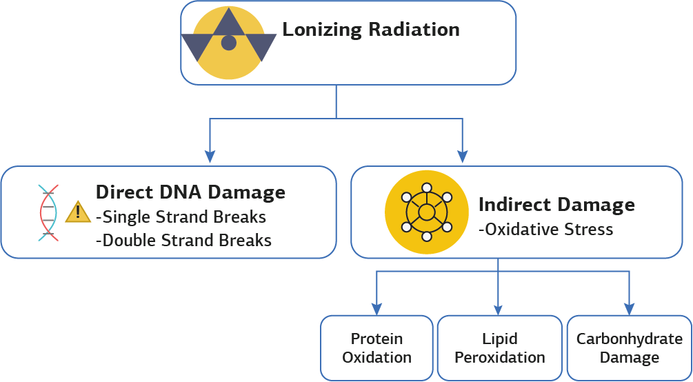
Figure 1. Effects of radiation on oxidative stress
Additional Information
Publisher’s Note
Bayrakol MP remains neutral with regard to jurisdictional and institutional claims.
Rights and Permissions

This work is licensed under a Creative Commons Attribution-NonCommercial 4.0 International License (CC BY-NC 4.0). To view a copy of the license, visit https://creativecommons.org/licenses/by-nc/4.0/
About This Article
How to Cite This Article
Huseyin Erdal, Turay Cesur. Radiation and oxidative stress: a mini-review. Ann Clin Anal Med 2025;16(Suppl 1):83-86. doi:10.4328/ACAM.22411
Publication History
- Received:
- 20.09.2024
- Accepted:
- 04.11.2024
- Published Online:
- 18.11.2024
- Printed:
- 25.03.2025