
Abstract
IntroductionMucosal appendicitis is a rare condition marked by dilation of the appendix and accumulation of mucus, often discovered incidentally during surgical interventions for appendicitis.Case PresentationThis article presents a case of appendiceal mucoceles in a 54-year-old female patient with a history of diabetes and asthma, who was diagnosed following abdominal imaging that indicated appendiceal involvement. Surgical exploration revealed an enlarged appendix, leading to a right hemicolectomy. Histopathological analysis confirmed poorly differentiated mucinous neoplasia, with clear surgical margins, suggesting a favorable prognosis for recovery.ConclusionThe case underscores the importance of early diagnosis and surgical treatment in managing appendiceal mucinous neoplasms, as timely intervention can mitigate the risks of severe complications, such as pseudomyxoma peritonei. Through this case, we emphasize the necessity for a multidisciplinary approach to enhance diagnostic and therapeutic outcomes in similar presentations.
Keywords
Introduction
Mucosal appendicitis is a rare pathological condition characterized by dilation of the appendix and accumulation of mucus. Typically, it is discovered incidentally, and its symptoms are quite nonspecific. We can divide appendiceal mucinous tumors into two main groups: benign and malignant, with the latter potentially leading to serious complications. Rokitansky first described the appendiceal mucosa in 1842, according to reports.1 Patients typically discover this pathology during surgery with a preliminary diagnosis of appendicitis, and if untreated, it can result in serious complications, such as pseudomyxoma peritonei.2 Appendiceal mucinous lesions are divided into four main subtypes: simple mucinous, hyperplastic mucinous, mucinous cystadenoma, and mucinous cystadenocarcinoma.3 This classification is critical for determining the prognosis of the disease. If mucinous cystadenocarcinoma ruptures, the mucin content can spread into the peritoneum, leading to pseudomyxoma peritonei. This situation can significantly reduce the patient’s quality of life and may lead to mortality.4 Appendiceal mucinous neoplasm is treated surgically, and depending on the type of disease, an appendectomy or more extensive resections may be required.5 Early diagnosis and surgical intervention prevent the disease from spreading and reduce the risk of complications. The post-surgical histopathological evaluation makes a definitive diagnosis. This article presents a case with appendiceal mucoceles and discusses the diagnostic and treatment process by comparing it with similar cases in the literature.
Case Presentation
A 54-year-old female patient presented to the emergency department with complaints of abdominal pain along with a known history of diabetes mellitus and asthma. The patient was using oral antidiabetic treatment and an inhaler. She has a history of three births, one of which was by cesarean section, and underwent a total thyroidectomy operation 10 years ago due to a goiter diagnosis. The emergency department’s abdominal ultrasound revealed a cyst measuring approximately 10x5 cm in the left adnexal region, prompting a referral to the obstetrics and gynecology department for further evaluation. The obstetrics and gynecology clinic conducted a detailed evaluation and determined that the cyst might not have originated from the ovary or fallopian tube, leading to the planning of an abdominal magnetic resonance (MR) examination as an advanced imaging method. The MRI result indicated a preliminary diagnosis of appendicitis with mucosal involvement (Figure 1). After completing the necessary preoperative preparations, we transferred the patient to the general surgery clinic for elective surgery. During the operation, the surgical team detected an enlarged appendix measuring approximately 11x7 cm and confirmed the diagnosis of appendiceal mucosa. Given the severity of the pathological findings, we performed a right hemicolectomy (Figure 2). Following the operation, the patient demonstrated a smooth recovery and was discharged on the 5th postoperative day in good health. The histopathological examination of the excised appendiceal tissue led to the diagnosis of poorly differentiated mucinous neoplasia in the appendix. The surgeon reported clear surgical margins and successfully discharged the patient without any findings of malignancy spread.Ethical ApprovalThis study was approved by the Ethics Committee of Necmettin Erbakan University (Date: 18.10.2024, Decision No: 2024/5255).
This case report was prepared in accordance with the CARE guideline.
Discussion
Mucosal appendicitis is a rare pathological condition characterized by dilation of the appendix and accumulation of mucus. Clinical symptoms are generally nonspecific, and most patients may be asymptomatic. Surgical intervention usually detects and diagnoses appendiceal mucosal neoplasms incidentally. However, for patients presenting with symptoms such as abdominal pain and an abdominal mass, it is an important pathology that should be considered in the differential diagnosis.1 The patient initially presented to the obstetrics and gynecology clinic with nonspecific abdominal pain, and the ultrasound results suggested a mass originating from the ovary or adnexal region. However, the use of more advanced imaging methods revealed that the cyst originated from the appendix and developed into a mucosal cyst.
Four main histological categories divide the mucoceles of the appendix: simple mucocele, hyperplastic mucocele, mucinous cystadenoma, and mucinous cystadenocarcinoma.2 This classification is critical for understanding the prognosis of the disease. Simple and hyperplastic mucosal lesions are generally benign, and the prognosis after treatment is good. However, mucinous cystadenomas and mucinous cystadenocarcinomas can turn cancerous, and when they rupture, they can cause serious problems like pseudomyxoma peritonei.3 In our case, the histopathological examination revealed poorly differentiated mucinous neoplasia, but it was reported that the surgical margins were clear. This finding suggests that the tumor has a low potential for spread and that surgical treatment is adequate.
Surgery is the primary method of diagnosing appendiceal mucinous neoplasm. However, during the preoperative period, abdominal imaging methods can contribute to the diagnosis. Ultrasound is generally the first imaging method used, but it can be difficult to differentiate appendiceal mucoceles from ovarian cysts or other adnexal lesions.4 In our case, the first ultrasound detected a cyst in the adnexal region, leading to her referral to the obstetrics and gynecology clinic. However, the advanced imaging method of magnetic resonance (MR) examination accurately identified the appendiceal mucosa. In these cases, MRI plays an important role in diagnosis because it provides high-resolution images.5 It particularly provides an advantage over other imaging methods in evaluating the size, location, and relationship of the mucosa with surrounding tissues.
Surgical treatment is the primary approach to managing appendiceal mucoceles. The surgical approach may vary depending on the size, location, and malignancy potential of the mucosal lesion. In simple mucosal cases, an appendectomy may be sufficient, while in cases with malignant potential, a right hemicolectomy or more extensive resections may be necessary.6 In our case, the large dimensions of the appendix and suspicion of malignancy led us to prefer a right hemicolectomy. This surgical approach aims to ensure the complete removal of the tumor while also minimizing the risk of malignant spread. Often, appendiceal mucosal neoplasms remain asymptomatic, with many patients receiving an incidental diagnosis during imaging studies for other purposes. However, the risk of developing pseudomyxoma peritonei is a significant complication in ruptured mucinous tumors. Pseudomyxoma peritonei occurs when mucus accumulates in the peritoneal cavity following the rupture of an appendiceal mucinous cyst, resulting in a serious clinical picture. People often confuse this condition with peritoneal carcinomatosis, and treating it can be quite challenging.7 In cases of rupture, surgical treatment often requires aggressive methods such as peritonectomy and hyperthermic chemotherapy. For this reason, the early diagnosis and surgical treatment of appendiceal mucosal lesions are of vital importance.
Histopathological examination plays a critical role in the prognosis and treatment planning of appendiceal mucinous neoplasms. Whether a mucous cyst is benign or malignant, the status of surgical margins and the spread of the cyst to surrounding tissues are determining factors for the patient’s follow-up after treatment.8 The histopathological examination in our case revealed a diagnosis of poorly differentiated mucinous neoplasia, with clear surgical margins reported. This result indicates that surgical treatment is sufficient, and the patient may not require additional treatment during follow-ups.
Limitations
This report is limited by its single-case design; therefore, the findings cannot be generalized.
Conclusion
Mucosal appendicitis is a rare pathology, but with the appropriate diagnosis and treatment methods, the prognosis is generally good. However, in cases with malignant potential, early surgical intervention and careful monitoring are necessary. In our case, the preoperative period yielded a correct diagnosis, and the appropriate surgical approach led to the patient’s complete recovery and discharge. In such cases, adopting a multidisciplinary approach can enhance the effectiveness of the diagnosis and treatment process.
Declarations
Animal and Human Rights Statement
All procedures performed in this study were in accordance with the ethical standards of the institutional and/or national research committee and with the 1964 Helsinki Declaration and its later amendments or comparable ethical standards.
Informed Consent
Written informed consent was obtained from the patient for publication of this case report and the accompanying images.
Data Availability
The datasets used and/or analyzed during the current study are not publicly available due to patient privacy reasons but are available from the corresponding author on reasonable request.
Conflict of Interest
The authors declare that there is no conflict of interest.
Funding
None.
Abbreviations
MRI: Magnetic resonance imaging
References
- Stocchi L, Wolff BG, Larson DR, Harrington JR. Surgical treatment of appendiceal mucocele. Arch Surg. 2003;138(6):585-590. doi:10.1001/archsurg.138.6.585
- Pai RK, Beck AH, Norton JA, Longacre TA. Appendiceal mucinous neoplasms: clinicopathologic study of 116 cases with analysis of factors predicting recurrence. Am J Surg Pathol. 2009;33(10):1425-1439. doi:10.1097/pas.0b013e3181af6067
- Misdraji J, Yantiss RK, Graeme-Cook FM, Balis UJ, Young RH. Appendiceal mucinous neoplasms: a clinicopathologic analysis of 107 cases. Am J Surg Pathol. 2003;27(8):1089-1103. doi:10.1097/00000478-200308000-00006
- Garg PK, Tewari M, Ahlawat S, et al. Pseudomyxoma peritonei and appendiceal mucinous neoplasms: current diagnostic and therapeutic approaches. World J Gastroenterol. 2021;27(19):2236-2250.
- Zarif J, Balaji S, Le D, et al. Appendiceal mucinous neoplasms: a review of the current literature. J Surg Oncol. 2021;123(5):1105-1115.
- Cecil TD, O’Connell J, Sugarbaker PH, Misdraji J, Carr NJ. Appendiceal mucinous neoplasms: a contemporary review. Surg Oncol Clin N Am. 2020;29(3):425-444.
- Lansom J, Alzahrani N, Liauw W, Morris DL. Cytoreductive surgery and hyperthermic intraperitoneal chemotherapy for pseudomyxoma peritonei and appendix tumours. Indian J Surg Oncol. 2016;7(2):166-176. doi:10.1007/s13193-015-0478-9
- Carr NJ, Cecil TD, Mohamed F, et al. A consensus for classification and pathologic reporting of pseudomyxoma peritonei and associated appendiceal neoplasia: the results of the Peritoneal Surface Oncology Group International modified Delphi process. Am J Surg Pathol. 2016;40(1):14-26. doi:10.1097/pas.0000000000000535
Figures
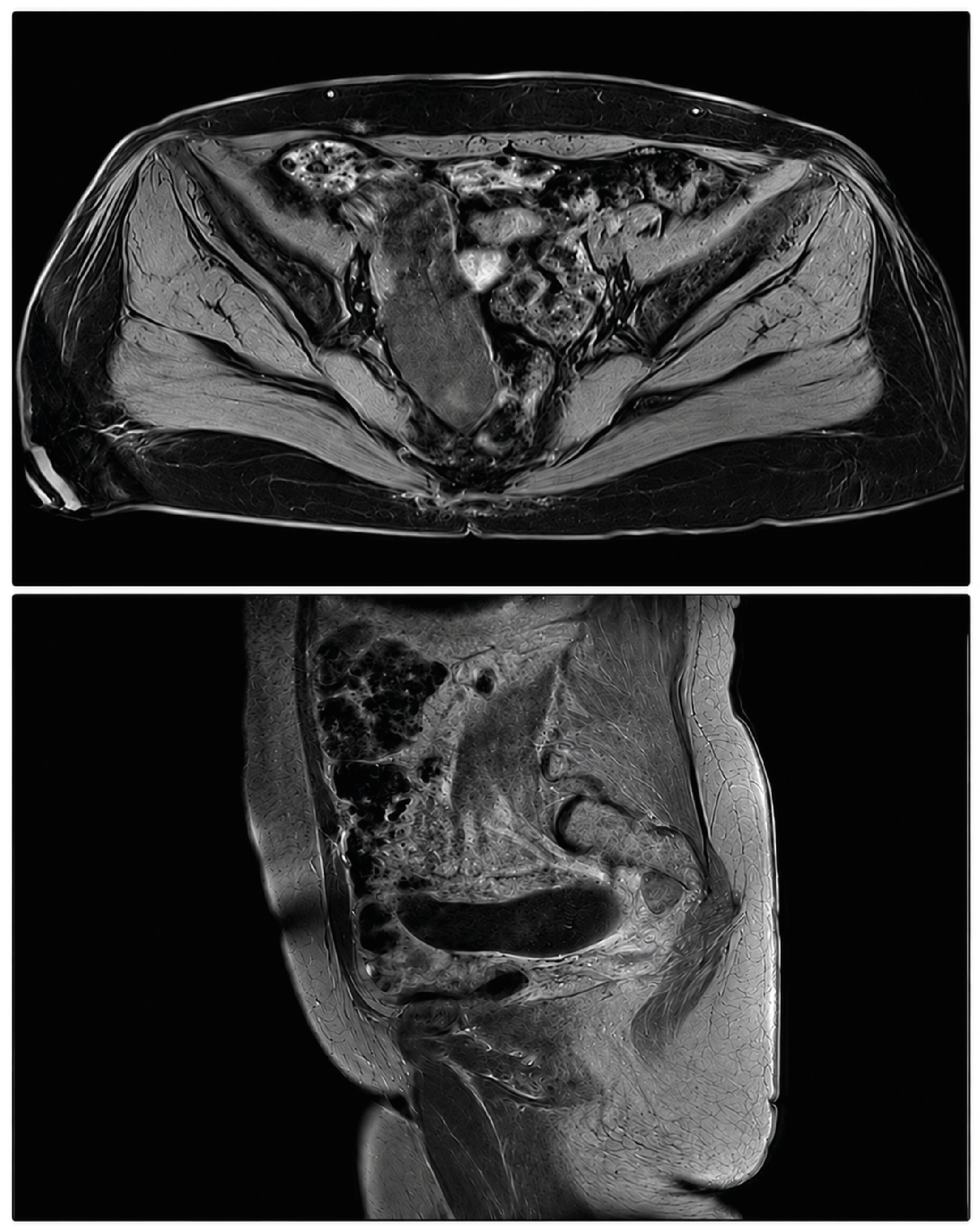
Figure 1. Appendiceal mucocele CT image
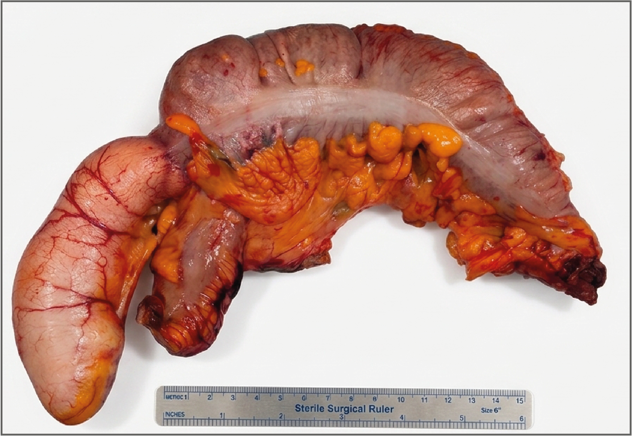
Figure 2. Appendiceal mucocele and right hemicolectomy specimen
Additional Information
Publisher’s Note
Bayrakol MP remains neutral with regard to jurisdictional and institutional claims.
Rights and Permissions

This work is licensed under a Creative Commons Attribution-NonCommercial 4.0 International License (CC BY-NC 4.0). To view a copy of the license, visit https://creativecommons.org/licenses/by-nc/4.0/
About This Article
How to Cite This Article
Arslan Hasan Kocamaz, Ömer Kişi, Abdulkadir Çelik, Celalettin Vatansev. Multidisciplinary approach to appendiceal mucoceles: a case presentation. Ann Clin Anal Med 2025;16(Suppl 2):118-120. doi:10.4328/ACAM.22424
Publication History
- Received:
- 27.09.2024
- Accepted:
- 04.11.2024
- Published Online:
- 20.11.2024
- Printed:
- 25.05.2025