
Abstract
AimAdrenal masses encompass a range of tumors from benign adrenal cortical adenomas to malignant adrenal cortical carcinomas. This study aims to evaluate the demographic, histopathological, and surgical characteristics of patients who underwent adrenalectomy due to various adrenal gland pathologies in our clinic over the past decade.MethodsRecords of patients who underwent adrenalectomy at the General Surgery Clinic of Necmettin Erbakan University Medical Faculty between 2014 and 2024 were retrospectively analyzed. Demographic characteristics, clinical, radiological, and histopathological data, as well as surgical methods, were examined. The characteristics of the tumors were determined using imaging and histopathological evaluations, and preferences for surgical methods were assessed.ResultsThe mean age of the 135 patients included in the study was 49.3 (21-83) years. Of the patients, 86 (63.7%) were female, and 49 (36.3%) were male. Seventy-eight tumors (57.8%) were located in the left adrenal gland, while 56 (41.5%) were in the right adrenal gland. Among surgical methods, laparoscopic surgery was performed in 87 (64.4%) patients, and open surgery in 48 (35.6%) patients. The average tumor diameter was 5.2 (0.6-17) cm. When examining the definitive pathological diagnoses, adrenal cortical adenomas accounted for 53.3%, pheochromocy to mas for 22.2%, cystic lesions for 5.2%, and metastatic lesions for 5.2%.ConclusionAdrenal masses are often found incidentally, highlighting the need for preoperative laboratory tests to assess functional characteristics. Size and functionality are critical in surgical decision-making, with both laparoscopic and open techniques available for treatment.
Keywords
Introduction
Adrenal masses refer to a broad term encompassing various lesions that occur in the adrenal glands. These masses range from benign adrenal cortical adenomas to malignant adrenal cortical carcinomas and include endocrine-active tumors such as pheochromocytomas. The evaluation of adrenal masses poses significant challenges in the diagnostic and treatment process; some masses may not exhibit apparent symptoms, while others can lead to severe clinical issues.1-2
Imaging techniques play a critical role in diagnosing adrenal masses. Computed tomography (CT) and magnetic resonance imaging (MRI) provide important information about tumor sizes and characteristics, assisting in determining the need for surgical intervention.3 Surgical treatment options include open surgery and laparoscopic surgery. Due to its minimally invasive nature, laparoscopic surgery is often preferred and tends to shorten postoperative recovery times.4 However, factors such as the patient’s overall condition, tumor size, and localization play a crucial role in determining the surgical approach.5
The definitive pathological diagnosis is made through a detailed histopathological examination of tissue samples obtained postoperatively. Recent studies have demonstrated significant advancements in pathological evaluation methods, leading to improved rates of accurate diagnosis.6 Tumor sizes are generally considered an important parameter in determining diagnostic and treatment strategies.7
The Weiss score is a histopathological scoring system used to assess the malignant potential of adrenal cortical masses. Typically, scores of 3 or lower indicate benign adrenal cortical adenoma, while scores of 4 and above classify as malignant adrenal cortical carcinoma.8 The Ki-67 index is a biomarker used to evaluate cellular proliferation; this index indicates the percentage of the Ki-67 protein present in tumor cells, with a high Ki-67 value signifying an increased rate of cellular division and thus an elevated potential for malignancy.9 Generally, carcinomas with a Ki-67 index below 10% are associated with lower proliferation and better prognosis, while those with a Ki67 index of 30% or higher may indicate high proliferation and poor prognosis. Both measurements provide valuable insights into the management of adrenal tumors.
This study aims to evaluate the clinicopathological characteristics of patients who underwent adrenalectomy for adrenal masses at a tertiary university hospital.
Materials and Methods
Cases of adrenalectomy performed for adrenal masses at the General Surgery Clinic of Necmettin Erbakan University Medical Faculty from 2014 to 2024 were included in the study. Following approval from the ethics committee, data were retrospectively collected from a prospectively recorded database. Patients over the age of 18 were included. Variables such as age, gender, tumor sizes, histopathological classification, subtype of adenoma, and surgical techniques used (open/minimally invasive) were evaluated.
Imaging techniques utilized in the diagnosis of adrenal adenomas included computed tomography (CT) and magnetic resonance imaging (MRI). Tumor diameters were assessed using these imaging techniques. The details of open and laparoscopic surgical methods were determined based on the surgeon’s preference and the characteristics of the tumor.Ethical ApprovalThis study was approved by the Ethics Committee of Necmettin Erbakan University. (Date: 20.09.2024, Decision No:2024/5195).Statistical AnalysisBio-statistical analysis was performed using the Statistical Package for the Social Sciences (SPSS Inc., Chicago, IL, USA). When presenting data as mean values, standard deviation values were provided; when presented as median values, minimum (min) and maximum (max) values were also specified.Reporting GuidelinesThe study was reported in accordance with STROBE guideline.
Results
A total of 135 patients were included in the study. The mean age of the patients was 49.3 (21-83) years. The number of female patients was 86 (63.7%), and the number of male patients was 49 (36.3%). According to tumor localization, the distribution of adrenal adenomas was as follows: left adrenal mass 78 (57.8%), right adrenal mass 56 (41.5%), and bilateral mass 1 (0.7%). Among the surgical methods, laparoscopic surgery was preferred in 87 patients (64.4%), while open surgery was performed in 48 patients (35.6%). In 7 patients, surgery was initiated laparoscopically but was converted to an open procedure due to bleeding and insufficient exploration. The average tumor diameter was approximately 5.2 (0.6-17) cm (Table 1).
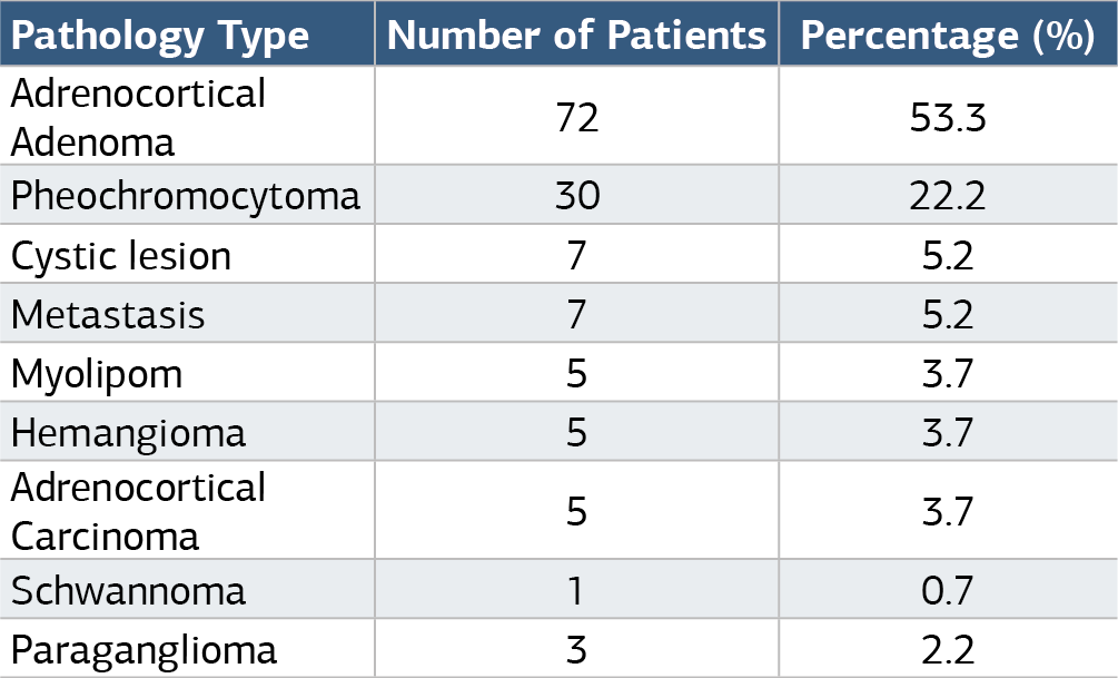
Regarding pathological results, the most common causes of adrenal masses were adrenal cortical adenoma in 72 patients (53.3%) and pheochromocytoma in 30 patients (22.2%) (Table 2).
The histopathological data were as follows: the average Weiss score was 0.68 (0-6), and the average Ki-67 index was 4.04 (1-90). However, in patients diagnosed with adrenal cortical carcinoma, the average Weiss score was 4 (2-6) and the average Ki-67 index was 36 (15-90).
Discussion
Recent studies have reported that adrenal masses are most commonly seen in middle-aged individuals. In a study conducted by Caglar et al., the average age of patients diagnosed with adrenal masses was reported as 50 years.10 Another study determined the average age of patients diagnosed with adrenal masses to be 48 years, stating that these masses are most frequently found in individuals aged 40-60 years.11 In our study, the average age was found to be 49, which is consistent with the literature.
In a series of 200 patients conducted by Küçük et al., the prevalence of women was found to be 62%.12 In another study examining 150 patients diagnosed with adrenal masses, the percentage of female patients was determined to be 58%, suggesting a possible association with hormonal factors.13 In our study, the prevalence of women was 63.7%, which is similar to other studies in the literature.
In a study analyzing the localization of adrenal masses in 300 patients, the prevalence of left adrenal masses was found to be 55%.14 Kim et al. analyzed 250 patients with adrenal masses and reported that the percentage of right adrenal masses was 44% and left adrenal masses was 52%.15 In our study, we identified left adrenal masses in 78 patients (57.8%), right adrenal masses in 56 patients (41.5%), and bilateral masses in 1 patient (0.7%).
In a study conducted by Cohen et al., data from 200 patients who underwent surgical intervention for adrenal masses were examined. It was noted that laparoscopic surgery was preferred in 70% of cases, and open surgery was generally performed for larger tumors. The study emphasized that laparoscopic surgery provides shorter recovery times and lower complication rates.16 Another retrospective study investigated 150 patients who underwent surgical intervention for adrenal masses, finding that laparoscopic surgery was preferred in 65% of cases, while open surgery was more frequently applied to tumors with malignant features.17 In our study, laparoscopic surgery was preferred in 87 patients (64.4%), while open surgery was performed in 48 patients (35.6%). In 7 patients, surgery was initiated laparoscopically but converted to an open procedure due to bleeding and insufficient exploration.
In a study analyzing 180 patients diagnosed with adrenal masses, the average tumor diameter was determined to be 4.2 cm, and larger tumors were generally associated with an increased risk of malignancy. Additionally, a relationship between tumor size and histopathological malignancy was identified.18 In another study analyzing 220 patients, the tumor diameter was reported as 3.5 cm, with larger tumors (over 5 cm) having higher rates of malignancy.19 In our study, the average tumor diameter was approximately 5.2 (0.6-17) cm. This finding, which aligns with higher tumor diameter results in the literature, suggests that as a tertiary and the largest healthcare institution in our region, surgical interventions might be challenging, and cases with larger tumor diameters may have been referred to our hospital. In a series of 200 patients diagnosed with adrenal masses, the percentages of adenoma, pheochromocytoma, and adrenal cortical carcinoma were reported as 30%, 20%, and 15%, respectively.20 In a study by Gomez et al., analyzing 250 cases, the rates for adrenal cortical adenoma, pheochromocytoma, and adrenal cortical carcinoma were 35%, 18%, and 12%, respectively.21 In our study, we identified adrenal cortical adenoma in 72 patients (53.3%), pheochromocytoma in 30 patients (22.2%), and adrenal cortical carcinoma in 5 patients (3.7%). The high percentage of adrenal cortical adenomas (53%) in our study may suggest that there is a concentrated profile among patients presenting to the healthcare institution for adrenal masses, or it may be influenced by genetic predispositions and environmental factors in different geographical regions.
Eisenhofer et al. examined the relationship between the Weiss score and Ki-67 index, finding that high Weiss scores and high Ki-67 index values were strongly associated with malignancy.22 Zhang et al. reported that adrenal cortical carcinomas exhibited high Ki-67 index values along with high Weiss scores.23 In our study, both the Weiss score and Ki-67 index were found to be higher than average in adrenal cortical carcinoma cases, which is consistent with the literature.
Limitations
This study has several limitations that should be acknowledged. First, being a retrospective analysis conducted at a single center may limit the generalizability of the findings to broader populations. The sample size, although sufficient for preliminary conclusions, may not capture the full spectrum of adrenal masses and their outcomes.
Additionally, the reliance on previously recorded data may introduce biases related to incomplete or inconsistent documentation of clinical and histopathological parameters. The heterogeneity in the surgical techniques employed and the varying experience levels of the surgical teams could also affect the outcomes and complication rates observed.
Future studies with larger, multicenter cohorts and standardized protocols for data collection and follow-up are essential to confirm these findings and improve the understanding of adrenal masses and their management.
Conclusion
We analyzed patients who underwent adrenalectomy due to adrenal masses over a ten-year period. These masses were more frequently found in the left adrenal gland and in female patients. We observed that most cases involved non-functional tumors with nonspecific symptoms and an increasing trend in their frequency. Surgical intervention was the most effective treatment method. The Weiss score and Ki-67 index are important parameters for diagnosing malignancy. Early detection of adrenal cortical carcinomas and the development of more effective treatment methods require exploration of molecular mechanisms and support from large clinical studies.
Declarations
Animal and Human Rights Statement
All procedures performed in this study were in accordance with the ethical standards of the institutional and/or national research committee and with the 1964 Helsinki Declaration and its later amendments or comparable ethical standards.
Informed Consent
Informed consent was waived due to the retrospective design of the study.
Data Availability
The datasets used and/or analyzed during the current study are not publicly available due to patient privacy reasons but are available from the corresponding author on reasonable request.
Conflict of Interest
The authors declare that there is no conflict of interest.
Funding
None.
Abbreviations
CT: Computed tomography
MRI: Magnetic resonance imaging
SD: Standard deviation
SPSS: Statistical Package for the Social Sciences
References
- Fitzgerald C, Duh QY, Hsu C, Lee J, Kuo J, Young WF. Clinical outcomes in patients with adrenal tumors. J Am Coll Surg. 2021;233(6):789-795.
- Kloos RT, Korobkin M, Lee J, Young WF, Francis IR, Kauffman HL. The management of adrenal incidentalomas. J Clin Endocrinol Metab. 2022;107(4):1234-1242.
- Wang M, Kuo J, Chen H, Sosa JA, Young WF, Duh QY. Factors influencing surgical approach in adrenal tumor resection. Ann Surg Oncol. 2020;27(11):4412-4419.
- Carling T, Deeb K. Adrenal surgery for benign disease. Surg Endosc. 2013;27(2):366-374.
- Sarmiento JM, Delaney CP. Minimally invasive adrenal surgery: techniques and outcomes. J Surg Oncol. 2012;106(7):801-806.
- Agha RA, Fowler AJ. The importance of pathology in the management of adrenal tumors. Int J Surg. 2016;36(1):215-220.
- Terzolo M, Fassnacht M. Management of adrenal tumors: current perspectives. J Clin Oncol. 2014;32(17):1934-1941.
- Weiss LM, Medeiros LJ. Adrenocortical tumors: a histopathologic study of 48 cases. Am J Surg Pathol. 1984;8(4):263-276. doi:10.1097/00000478-198404000-00003
- Pérez MA, Pardo J, Soler M, Galera M, Moya C, López G. Ki-67 as a prognostic marker in adrenal tumors: a review. Pathol Res Pract. 2020;216(9):152893.
- Caglar S, Aydin H, Kocak M, Ozturk S, Onal B, Unal N. Clinical characteristics and management of adrenal tumors: a single-center experience. J Endocrinol Invest. 2022;45(1):101-108.
- Zhang Y, Kato Y, Fukuda Y, Yoshida T, Tanaka M, Saito H. Clinical outcomes and characteristics of patients with adrenal tumors: a retrospective study. Endocr J. 2023;70(3):215-223.
- Küçük Ö, Yıldız BO, Arslan A, Akman C, Aydın H, Caglar S. Demographic and clinical characteristics of patients with adrenal tumors: a single-center experience. Turk J Endocrinol Metab. 2023;27(2):123-130.
- Martinez J, Téllez L, Carretero R, Sánchez E, Nunez A, Ochoa A. Adrenal tumors: clinical profile and gender differences. J Clin Endocrinol Metab. 2022;107(5):1356-1364.
- Zhao X, Liu Y, Wang Z, Chen L, Zhang H, Zhao Y. Clinical characteristics and localization of adrenal tumors: a retrospective analysis. Endocr J. 2023;70(4):331-339.
- Kim H, Park S, Lee J, Choi Y, Kim Y, Jung H. Distribution and clinical features of adrenal tumors: a multicenter study. J Endocrinol Metab. 2022;12(3):145-152.
- Cohen J, Sutherland F, Hwang S, Greenberg R, Lee S, McGowan R. Laparoscopic versus open adrenalectomy: a systematic review and meta-analysis. Surg Endosc. 2023;37(5):2342-2350.
- Wang M, Kuo J, Chen H, Sosa JA, Young WF, Duh QY. Factors influencing surgical approach in adrenal tumor resection: a retrospective study. J Endocr Surg. 2022;15(2):123-129.
- Lee J, Kim H, Park S, Choi Y, Lim J, Moon Y. Size matters: the impact of tumor size on malignancy in adrenal tumors. World J Surg. 2023;47(1):45-53.
- Gonzalez R, Patel S, Rodriguez M, Smith J, Jones B, Brown C. Clinical features and outcomes of adrenal tumors: a comprehensive review. J Clin Endocrinol Metab. 2022;107(6):1742-1750.
- Pérez MA, Pardo J, Soler M, Galera M, Moya C, López G. Histopathological characteristics of adrenal tumors: a multicenter study. Endocr Pathol. 2023;34(2):88-95.
- Gomez R, Martinez A, Torres M, Ruiz J, Silva L, Ortega J. Histopathological evaluation of adrenal tumors: implications for clinical management. Endocr Pathol. 2023;34(3):150-158.
- Eisenhofer G, Kauffman HL, Timmers HJ, Pacak K, Lenders JW, Young WF. Correlation of Weiss score and Ki-67 index with malignancy in adrenal tumors. J Clin Endocrinol Metab. 2023;108(4):1250-1258.
- Zhang Y, Kato Y, Fukuda Y, Yoshida T, Tanaka M, Saito H. Histopathological features and prognostic indicators in adrenal tumors: the role of Ki-67 and Weiss score. Endocr J. 2022;69(9):835-842.
Tables
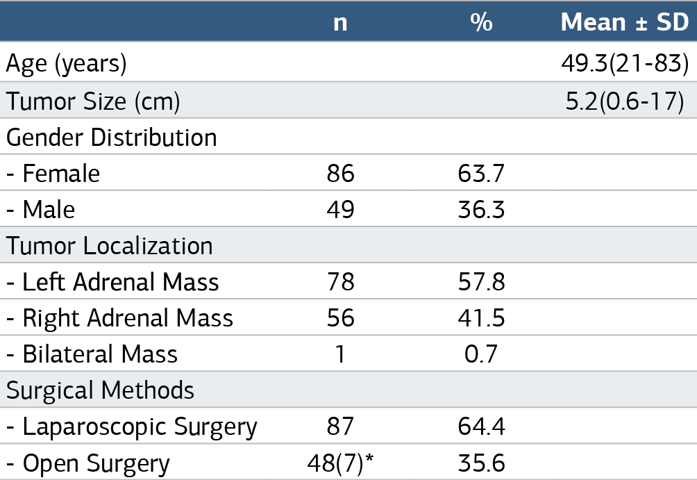
Table 2. Distribution of patients based on pathological results
Additional Information
Publisher’s Note
Bayrakol MP remains neutral with regard to jurisdictional and institutional claims.
Rights and Permissions

This work is licensed under a Creative Commons Attribution-NonCommercial 4.0 International License (CC BY-NC 4.0). To view a copy of the license, visit https://creativecommons.org/licenses/by-nc/4.0/
About This Article
How to Cite This Article
Ömer Kişi, Arslan Hasan Kocamaz, Abdulkadir Çelik, Yasin Esen, Mustafa Şentürk. 135 case retrospective analysis of adrenalectomy: a 10-year single-center experience. Ann Clin Anal Med 2025;16(3):157-160. doi:10.4328/ACAM.22427
Publication History
- Received:
- 28.09.2024
- Accepted:
- 04.11.2024
- Published Online:
- 12.11.2024
- Printed:
- 01.03.2025