
Abstract
AimThis study aims to investigate the correlation between first-trimester placental thickness and perinatal prognosis.MethodsA prospective cohort study was conducted at Siirt University Faculty of Medicine from March 2022 to March 2023 of 365 pregnant women in their first trimester (11–14 weeks of gestation). Placental volume was measured using two-dimensional (2D) ultrasound, and estimated placental volume (EPV) was calculated using Merwin’s EPV Calculator app. The patients were followed until delivery, and outcomes such as gestational age at delivery, mode of delivery, fetal weight, APGAR score, fetal gender, perinatal outcomes, preeclampsia (PE), premature rupture of membranes (PROM), gestational hypertension (GHT), gestational diabetes mellitus (GDM), intrauterine growth restriction (IUGR), preterm birth, oligohydramnios, polyhydramnios, surmaturation, presentation anomaly, intrauterine death, fetal distress, and placental abruption were evaluated.ResultsData from 365 pregnant women were analyzed. The mean maternal age was 27.2 ± 5.5 years. The distribution of placental location was 38.4% posterior, 43.0% anterior, 7.4% left sidewall, and 11.2% right sidewall. No significant differences were found in placental volume measurements based on delivery mode, fetal gender, or conditions such as PE, GDM, IUGR, PROM, preterm birth, or other perinatal pathologies. Statistical analyses showed no significant association between first-trimester placental volume and adverse perinatal outcomes (p>0.05).ConclusionNo relationship was found between first-trimester placental thickness and perinatal outcomes.
Keywords
Introduction
The relationship between first-trimester placental volume and perinatal prognosis has garnered significant interest in obstetric research. Understanding this relationship can provide early indicators of various obstetric complications, such as low birth weight, intrauterine growth restriction (IUGR), preeclampsia (PE), and gestational diabetes mellitus (GDM). Early detection of these conditions can lead to improved monitoring and intervention strategies, thereby enhancing maternal and neonatal health.
Previous studies have demonstrated a significant relationship between first-trimester placental volume and birth weight. Smaller placental volumes have been associated with small-for-gestational-age (SGA) neonates, while larger volumes have been linked to large-for-gestational-age (LGA) neonates. These findings underscore the potential of placental volume to predict neonatal weight.1
Larger first-trimester placental volumes have been found to correlate with better fetoplacental vascular function postpartum. This finding suggests that larger and/or more vascularized placentas in early pregnancy may have better adaptation mechanisms, thereby improving pregnancy outcomes.2 Additionally, studies investigating the predictive ability of first-trimester Doppler vascular flow indices have shown that lower indices are associated with pregnancies that develop PE.3
Moreover, decreased first-trimester placental volume has been linked to an increased incidence of IUGR, and the placental vascularization index has shown a positive correlation with neonatal weight.4 Placental volumetry using ultrasound in the first trimester has been reported to be useful in identifying pregnancies at risk for both low and high birth weights (SGA and macrosomia).5
In conclusion, first-trimester placental volume has been reported to have the potential to predict pregnancy complications in certain conditions. Evaluating placental volume early in pregnancy can provide valuable information about potential perinatal outcomes, paving the way for improved monitoring and intervention strategies. Despite these findings, there is a need for standardized methods to measure placental volume and to validate the predictive value of these measurements across different populations. Our study aims to investigate the relationship between first-trimester placental volume and perinatal prognosis in a well-defined cohort, thereby contributing evidence to support the clinical utility of early placental assessment in prenatal care.
Materials and Methods
For this purpose, approximately 360 pregnant women in their first trimester who were followed up at Siirt Education and Research Hospital, between March 2022 and March 2023 were included in the study. The ages, pregnancy histories, and medical histories of the pregnant women were recorded. Placental volume was measured using Merwin’s estimated placental volume (EPV) Calculator app. This application is designed to calculate the EPV from three measurements taken during the two-dimensional (2D) prenatal ultrasound, namely, the width, height, and thickness of the placenta, and is calculated using the formula V: πT/6*(4H(W-T)+W(W-4T)+4T²) (T: placental thickness, W: placental width, H: height). During the examination, all participants were in a supine position, and care was taken to include the entire placenta within the measurement frame. The study included pregnant women who applied to the prenatal clinic for routine first-trimester screening and were in the first trimester (between 11 and 14 weeks of gestation). Inclusion criteria were singleton pregnancies, maternal age between 18 and 45, and gestational age confirmed by the last menstrual period and/or early ultrasound. Exclusion criteria included known chromosomal or structural fetal abnormalities and chronic diseases, such as heart disease, chronic hypertension, diabetes, and a history of renal disease. During the examination, the gestational week, placental volume, and placental location of the patient were recorded. All patients were followed until delivery. Maternal and fetal outcomes such as gravida, parity, hemogram, biochemistry, urinalysis, gestational age at delivery, fetal weight, gender, APGAR score at 1 and 5 minutes, mode of delivery (normal spontaneous vaginal delivery (NSVD)/cesarean section (C/S)), miscarriage, neonatal death, premature rupture of membranes (PROM), preterm birth, placental abruption, fetal distress, intrauterine death, GDM, PE, postmaturity, polyhydramnios, oligohydramnios, presentation anomaly, multiple pregnancies, IUGR, postpartum hemorrhage (PPH), and placenta previa were evaluated in three pregnancy groups. Spontaneous miscarriage was defined as a miscarriage that occurred without intervention. Missed abortion was defined as the condition where the embryo/fetus loses its viability in the uterus, but uterine evacuation has not yet started. Neonatal death was defined as the death of an infant within 365 days after birth. PROM was considered as the rupture of fetal membranes before 37 weeks of gestation. Preterm birth was defined as a birth occurring between 20 0/7 and 36 6/7 weeks of gestation. Placental abruption was defined as the partial (partial abruption) or complete (complete abruption) separation of the placenta from the uterus before the birth of the baby. Fetal distress was defined as fetal oxygen deficiency, acidosis, or asphyxia and was diagnosed as fetal distress by nonstress test (NST). Intrauterine fetal death was defined as the death of a fetus with a birth weight of 500 grams or more after 20 weeks of gestation or if the gestational age was unknown. GDM was diagnosed in pregnant women with a glucose value of 140–180 mg/dl in the 1-hour 50 g glucose screening test performed between 24 and 28 weeks of gestation and if two out of four glucose reference values were exceeded in the 100 g oral glucose tolerance diagnostic test (OGTT). In high-risk groups, GDM was diagnosed directly by performing a 75 g OGTT without a screening test. PE patients included in the study were in accordance with the current recommendations of the American College of Obstetricians and Gynecologists (ACOG). Postmaturity was defined as pregnancies that extended beyond 42 weeks of gestation. Polyhydramnios was defined as amniotic fluid volume of 2000 ml or more. Oligohydramnios was defined as amniotic fluid volume of 500 ml or less. Presentation anomaly was assessed as non-cephalic presentation, such as face, brow, breech, or shoulder. IUGR was defined as an estimated fetal weight below the 10th percentile or an abdominal circumference <10% for gestational age. PPH was defined as estimated blood loss of >500 ml within the first 24 hours post-delivery. Ethical Approval The study was approved by the Ethics Committee of Siirt University (Date: 22.04.2022, Decision No: 2022/04.01). The study has been given the following ClinicalTrials.gov Identifier: NCT05429242. Statistical Analysis Statistical analyses were performed with IBM® SPSS® 26 (SPSS Inc., Chicago, IL, USA). The conformity of the variables to the normal distribution was examined using analytical methods (Kolmogorov–Smirnov/Shapiro–Wilk tests). Descriptive analyses are given as mean±standard deviation, and median, min–max are used for continuous data. Descriptive statistics were calculated from the frequency and percentage values of categorical variables obtained from sociodemographic and clinical information. In continuous data (placental volume), Student’s t-test was used for independent groups when they showed normal distribution, and the Mann–Whitney U test was used when there was non-parametric distribution to compare binary groups (prenatal pathological parameter groups). Spearman’s Rho correlation and linear regression analysis were used to predict placental volume with fetal weight and the APGAR 5 score. A p-value below 0.05 was considered statistically significant. Reporting GuidelinesThis study was reported in accordance with the STROBE guideline.Results
Data from 365 pregnant women between 11 and 14 weeks of gestation were included in the study. The overall mean age was found to be 27.2 ± 5.5 years, with a median age of 27 years, the youngest patient being 17 and the oldest 43 years old. The average gravidity was 3.2 ± 2.0, and the average parity was 1.7 ± 1.5. Of the women studied, 263 (72.1%) had vaginal deliveries, and 102 (27.9%) had C/S. Moreover, 230 (63.0%) women had no history of miscarriage, while 135 (37.0%) had experienced at least one miscarriage. The classification of placental location indicated that 140 (38.4%) were posterior, 157 (43.0%) were anterior, 27 (7.4%) were on the left sidewall, and 41 (11.2%) were on the right sidewall. The distribution of gestational weeks was as follows: 11 weeks – 33 (9.0%), 12 weeks – 108 (29.6%), 13 weeks – 107 (29.3%), and 14 weeks – 117 (32.1%). (Table 1)
When comparing placental volume measurements according to the mode of delivery, no significant differences were found between NSVD and C/S, fetal gender, PE, GDM, IUGR, PROM, preterm birth, abruption, fetal distress, intrauterine fetal demise (IUFD), postmaturity, oligohydramnios, polyhydramnios, or the presence of presentation anomalies (p>0.05) (Table 2). However, breech presentation increased placental volume by approximately 10 cm³ compared to non-breech presentation, but this difference was not statistically significant (p=0.093). The relationship between placental volume and APGAR 5 and fetal weight was examined, and no significant correlation was found (r=0.092, p=0.080; r=0.015, p=0.771, respectively).
Discussion
The placenta is a central structure in pregnancy and has very complex and powerful functions. Having the pathophysiological knowledge underlying this maternofetal structure greatly helps in understanding the development of pregnancy. If we can understand the early development of the placenta, we can understand preeclampsia, fetal growth restriction, and even the role of the placenta in maternal venous diseases.6
In our study, the relationship between placental volume and the variables APGAR 5 (Figure 1) and fetal weight (Figure 2) was examined , and no significant correlation was found (r=0.092, p=0.080; r=0.015, p=0.771, respectively) . Joshi et al. (2013) found that lower placental weights were significantly associated with lower birth weights, fetal distress, lower APGAR scores, and conditions such as PE and maternal diabetes.7 Carbone et al. (2011) observed that a combination of first-trimester placental volume, placental protein markers, and vascular indices could predict SGA outcomes but did not significantly improve predictions for PE or gestational hypertension (GHT).8
Our study also found no significant differences in placental volume measurements based on delivery method (NSVD and C/S), fetal gender, PE, GDM, IUGR, PROM, preterm birth, abruption, fetal distress, IUFD, postmaturity, oligohydramnios, polyhydramnios, and presentation anomalies (p>0.05). Stampalija T et al. investigated the relationship between placental volume in the first trimester and hypertensive pregnancy disorders (HDP) and SGA. Their study, which analyzed data from 1322 women, found that placental volume was higher in uncomplicated pregnancies and lowest in the HDP–SGA group.9 Vachon et al. found that first-trimester placental thickness increased in PE and decreased in SGA cases.10 Approximately 10% of pregnancies exhibit reduced fetal growth, often associated with pathological conditions such as IUGR. Various methods, including placental volume and vascularization assessments alongside ultrasound and Doppler studies, are used to diagnose IUGR. Modern approaches suggest integrating these methods into algorithms to better predict high-risk pregnancies.11
Soongsatitanon A et al. found that first-trimester placental volume was not an effective tool for predicting PE and/or IUGR. They observed that placental volume was particularly lower in cases of PE and/or IUGR, suggesting that further research is needed on the potential of first-trimester placental volume to predict early-onset PE.12 Chen CY et al. examined changes in placental vascular indices and volume in overweight Chinese women during the first trimester using three-dimensional power Doppler ultrasound. They found that overweight women had lower placental vascularization indices, larger placental volumes, and higher uterine artery pulsatility indices. These findings shed light on the effects of early pregnancy overweight on placental vascularization and function.13 Kim YR et al. evaluated a risk prediction model for PE using maternal age, body mass index (BMI), and EPV in the first trimester. Their study of 351 women found that 13 developed PE and that EPV was lower in women who developed PE. A model incorporating maternal age, BMI, and EPV could effectively predict PE in singleton pregnancies.14
Plasencia W et al. found that placental volume and vascularization were lower in women with PE in the first trimester. Additionally, maternal characteristics such as multiparity and low maternal weight exacerbated the adverse effects of PE on the placenta and worsened perinatal outcomes.15 Elhelaly AM et al. found no significant difference in placental volume and vascular indices between women with and without pre-gestational diabetes. However, they identified a strong relationship between HbA1C levels and vascular flow index (VFI), indicating that HbA1C could be a potential indicator for predicting placental complications in early pregnancy.16 Effendi M et al. found that first-trimester placental volume was positively associated with birth weight and placental weight but was not associated with the risk of PE. Placental volume was smaller in SGA infants and larger in larger infants.1
Studies have shown that placental diameter and thickness are highly positively correlated with fetal weight, aiding in predicting fetal weight.17-18 Significant relationships have also been found between prenatal and postnatal placental thickness and weight, APGAR scores, neonatal artery pH, and fetal growth restriction.19-20 A thick placenta detected by ultrasonography has been associated with increased mortality related to fetal anomalies and higher rates of both SGA and LGA infants, indicating increased perinatal risk.21-22
Limitations
One of the main limitations of this study is its reliance on a single-center dataset, which may limit the generalizability of the findings to broader populations. Although the sample size is sufficient for initial findings, it may not be large enough to detect all significant differences among various subgroups, necessitating larger, multicenter studies to confirm these results. Finally, the observational nature of the study presents challenges in establishing definitive relationships due to its inherent observational characteristics. The strengths of the study include its contribution to the limited literature on first-trimester placental volume studies and its comprehensive examination of perinatal diseases within a broad framework.
Conclusion
This study examined the relationship between first-trimester placental volume and various perinatal outcomes. The findings indicated no significant correlation between placental volume and perinatal diseases. Despite the lack of significant correlations in this study, the potential role of placental volume in early pregnancy as a predictor of adverse perinatal outcomes should not be overlooked. Future research should focus on larger, multicenter studies to confirm these findings and explore additional factors or more detailed measurement techniques that could enhance the predictive power of placental volume assessments.
Abbreviations
ACOG: American college of obstetricians and gynecologists
APGAR: Appearance, pulse, grimace, activity, and respiration
BMI: Body mass index
C/S: Cesarean section
EPV: Estimated placental volume
GDM: Gestational diabetes mellitus
IUGR: Intrauterine growth restriction
NSVD: Normal spontaneous vaginal delivery
PE: Preeclampsia
PROM: Premature rupture of membranes
SGA: Small for gestational age
STROBE: Strengthening the reporting of observational studies in epidemiology
References
- Effendi M, Demers S, Giguère Y, et al. Association between first-trimester placental volume and birth weight. Placenta. 2014;35(2):99-102. doi:10.1016/j.placenta.2013.11.015
- Hitzerd E, Reijnders IF, Mulders AGMGJ, et al. Larger first-trimester placental volumetric parameters are associated with lower pressure and more flow-mediated vasodilation of the fetoplacental vasculature after delivery. Front Physiol. 2020;11:6. doi:10.3389/fphys.2020.00006
- Eastwood KA, Patterson C, Hunter AJ, McCance DR, Young IS, Holmes VA. Evaluation of the predictive value of placental vascularisation indices derived from 3-dimensional power Doppler whole placental volume scanning for prediction of pre-eclampsia: a systematic review and meta-analysis. Placenta. 2017;51:89-97. doi:10.1016/j.placenta.2017.01.005
- Papastefanou I, Chrelias C, Siristatidis C, Kappou D, Eleftheriades M, Kassanos D. Placental volume at 11 to 14 gestational weeks in pregnancies complicated with fetal growth restriction and preeclampsia. Prenat Diagn. 2018;38(12):928-935. doi:10.1002/pd.5356
- Schwartz N, Quant HS, Sammel MD, Parry S. Macrosomia has its roots in early placental development. Placenta. 2014;35(9):684-690. doi:10.1016/j.placenta.2014.06.373
- Ortega MA, Fraile-Martínez O, García-Montero C, et al. The pivotal role of the placenta in normal and pathological pregnancies: a focus on preeclampsia, fetal growth restriction, and maternal chronic venous disease. Cells. 2022;11(3):568. doi:10.3390/cells11030568
- Joshi JM, Patel D, Jayun JM. A study of association between maternal placental weight and neonatal characteristics. J Evol Med Dent Sci. 2013;2(7):689-695.
- Carbone J, Goetzinger K, Liebsch J, Rampersad R, Macones G, Odibo A. Combination of first-trimester placental protein 13, 3D placental volume, vascular indices, and maternal characteristics for prediction of adverse pregnancy outcomes. Am J Obstet Gynecol. 2012;206(1). doi:10.1016/j.ajog.2011.10.364
- Stampalija T, Quadrifoglio M, Casati D, et al. First trimester placental volume is reduced in hypertensive disorders of pregnancy associated with small for gestational age fetus. J Matern Fetal Neonatal Med. 2021;34(8):1304-1311. doi:10.1080/14767058.2019.1636026
- Vachon-Marceau C, Demers S, Markey S, Okun N, Girard M, Kingdom J. First-trimester placental thickness and the risk of preeclampsia or SGA. Placenta. 2017;57:123-128. doi:10.1016/j.placenta.2017.06.016
- Albu AR, Anca AF, Horhoianu VV, Horhoianu IA. Predictive factors for intrauterine growth restriction. J Med Life. 2014;7(2):165-171.
- Soongsatitanon A, Phupong V. First trimester 3D ultrasound placental volume for predicting preeclampsia and/or intrauterine growth restriction. J Obstet Gynaecol. 2019;39(4):474-479. doi:10.1080/01443615.2018.1529152
- Chen CY, Chang HT, Chen CP, Sun FJ. First trimester placental vascular indices and volume by three-dimensional ultrasound in pre-gravid overweight women. Placenta. 2019;80:12-17. doi:10.1016/j.placenta.2019.03.014
- Kim YR, Jung I, Park G, Chang SW, Cho HY. First-trimester screening for early preeclampsia risk using maternal characteristics and estimated placental volume. J Matern Fetal Neonatal Med. 2021;34(7):1155-1160. doi:10.1080/14767058.2019.1628207
- Plasencia W, González-Dávila E, González Lorenzo A, Armas-González M, Padrón E, González-González NL. First trimester placental volume and vascular indices in pregnancies complicated by preeclampsia. Prenat Diagn. 2015;35(12):1247-1254. doi:10.1002/pd.4692
- Elhelaly AM, Elnaggar AK, Mohamed MEDM, Abdelfattah MH, Farag AH. First trimester placental volume and vascular indices in pregestational diabetic compared with nondiabetic pregnant women. J Obstet Gynaecol Res. 2020;46(8):1326-1332. doi:10.1111/jog.14271
- Ogbochukwu OJ, Dim NR, Nnamani AO, Njeze NR, Obikili EN, Dim CC. Relationship of placental diameter and thickness versus fetal growth biometrics: a cross-sectional sonographic evaluation of antenatal women in Enugu, Southeast Nigeria. Int J Med Health Dev. 2020;25(2):138-143. doi:10.4103/ijmh.ijmh_39_20
- Bedi M, Sharma H, Sandhu PS, Minhas A. Correlation of placental thickness with birth weight in singleton pregnancies. Int J Reprod Contracept Obstet Gynecol. 2021;10(10):3812-3817. doi:10.18203/2320-1770.ijrcog20213842
- Farahbod F, Zarean E, Khanjani S, et al. Relationship between placental thickness, grading, and heterogeneity in fetal growth restriction in the third trimester of pregnancy by ultrasonography and pathology tests and their relationship with estimated fetal weight and neonatal outcome. Immunopathol Persa. 2023;10(2). doi:10.34172/ipp.2023.39471
- Muraliswar RJ, Kallepally AK. Correlation of placental thickness in relation to gestational age and fetal weight by using ultrasonography. Int J Contemp Med Surg Radiol. 2019;4(3).
- Miwa I, Sase M, Torii M, Sanai H, Nakamura Y, Ueda K. A thick placenta: a predictor of adverse pregnancy outcomes. Springerplus. 2014;3:353. doi:10.1186/2193-1801-3-353
- Rawal S, Ray S, Sharma N. Correlation between ultrasonographic placental thickness and adverse fetal and neonatal outcomes. Cureus. 2024;16(3). doi:10.7759/cureus.56410
Tables
Table 1. General Descriptive Analyses
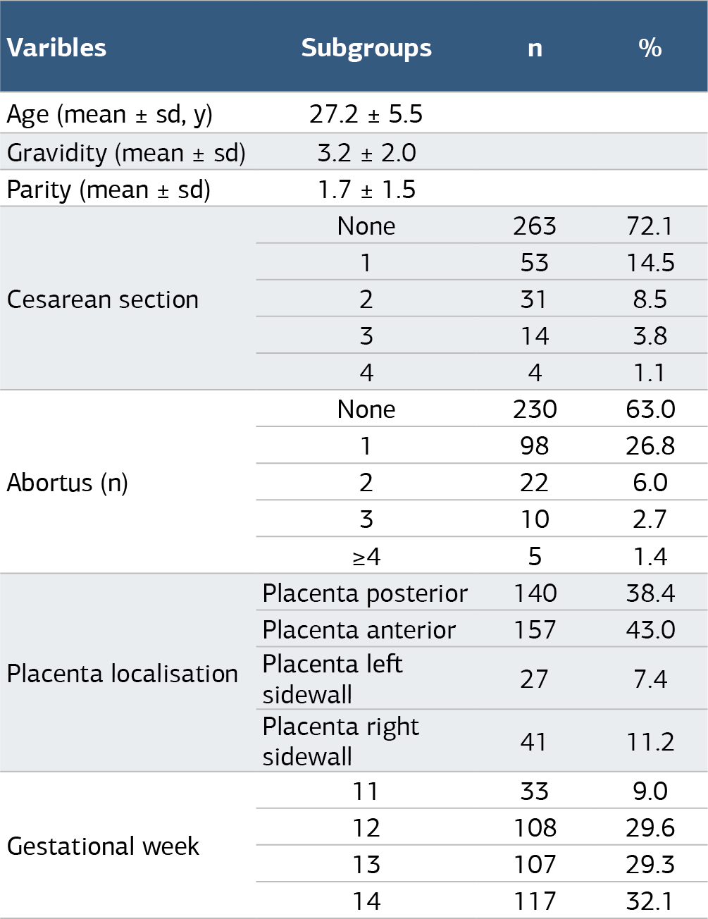
Table 2. Comparison of placental volume in variable groups
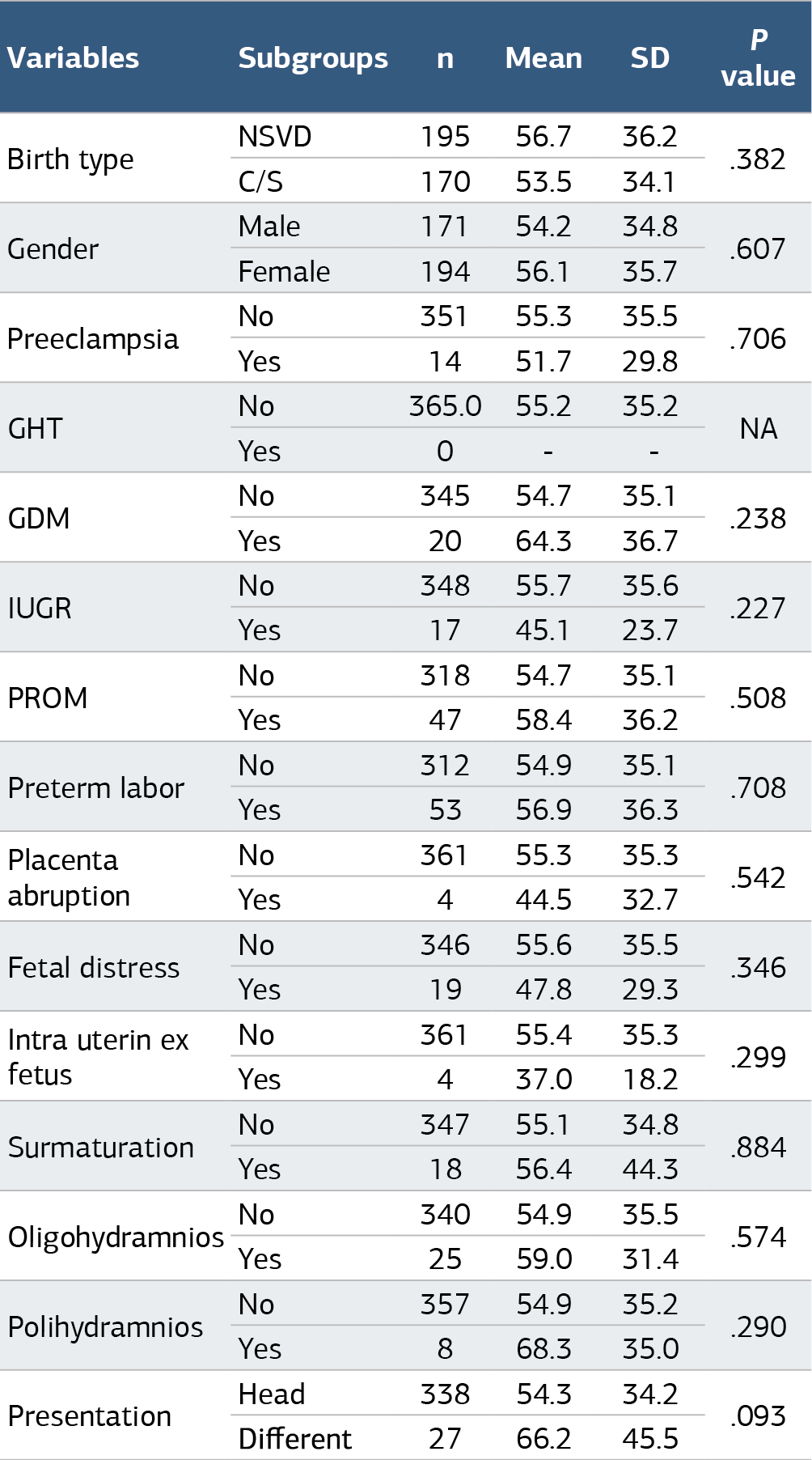
Premature rupture of membranes (PROM), Gestational hypertantion (GHT) Gestational diabetes mellitus (GDM), Intrauterine growth restriction (IUGR), Independent t test and Mann Whitney U test used. p
Figures
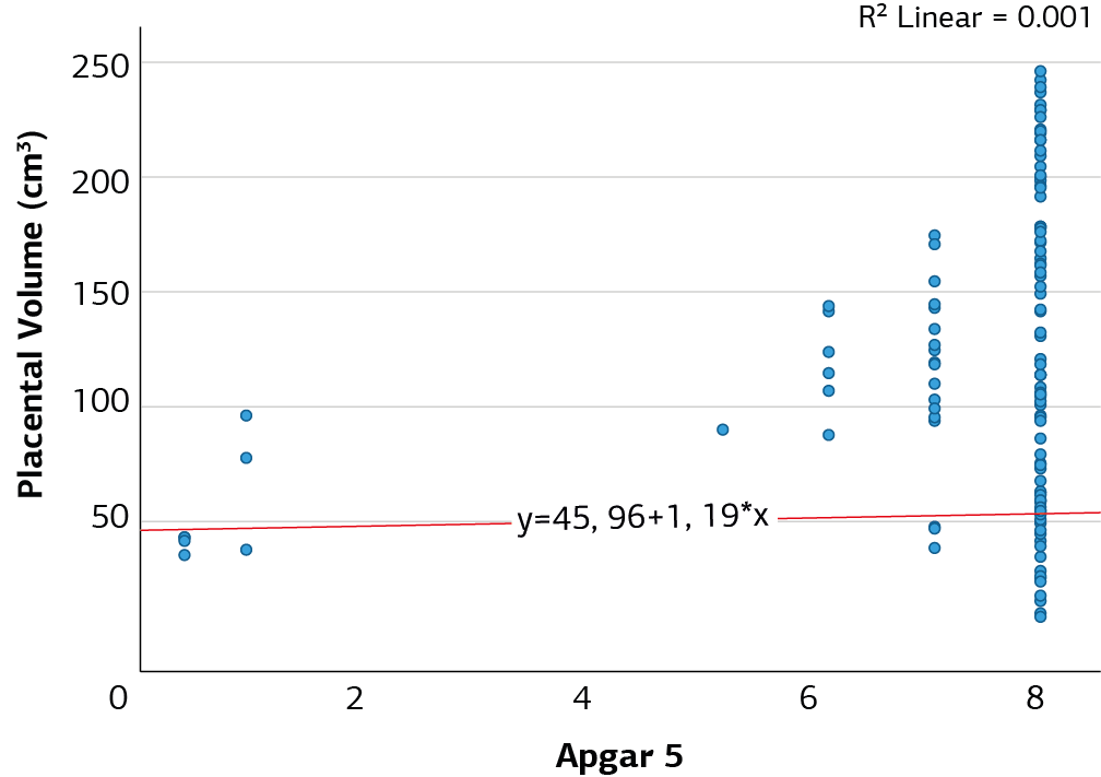
Figure 1. Relationship between APGAR parameter and placenta volume
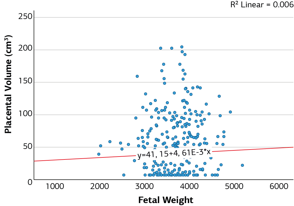
Figure 2. Relationship between fetal weight parameter and placenta volume
Additional Information
Publisher’s Note
Bayrakol MP remains neutral with regard to jurisdictional and institutional claims.
Rights and Permissions

This work is licensed under a Creative Commons Attribution-NonCommercial 4.0 International License (CC BY-NC 4.0). To view a copy of the license, visit https://creativecommons.org/licenses/by-nc/4.0/
About This Article
How to Cite This Article
Mehmet Yılmaz, Şerif Aksin, Deniz Balsak, Yasmin Aboalhasan, Fatma Zehra Kurnuç, İbrahim Batmaz. Relationship between first trimester placental thickness and perinatal prognosis: A prospective cohort study. doi:10.4328/ACAM.22441
Publication History
- Received:
- 06.10.2024
- Accepted:
- 18.11.2024
- Published Online:
- 03.12.2024
- Printed:
- 25.03.2025