
Treatment response relationship with hematologic parameters before andafter chemoradiotherapy in patients with non-small cell lung cancer
Chemoradiotherapy & hematologic markers in nsclc
Authors
Abstract
AimThis study aimed to investigate the relationship between hematological parameters and treatment response in patients with advanced non-small cell lung cancer (NSCLC) treated with curative-intent chemoradiotherapy. Specifically, we evaluated whether markers like neutrophil/lymphocyte ratio (NLR), platelet/ lymphocyte ratio (PLR), and red cell distribution width (RDW) correlate with radiologically assessed tumor size changes.
MethodsWe conducted a retrospective analysis of 49 NSCLC patients treated between 2008 and 2014 at Atatürk University School of Medicine. All patients received neoadjuvant chemotherapy followed by concurrent chemoradiotherapy. Hematological parameters, including NLR, PLR, and RDW, were collected before and after treatment. Tumor size was measured using PET-CT, MRI, or CT, and relationships between hematological markers and tumor size changes were analyzed using the Wilcoxon signed-rank test and Spearman’s correlation, with significance at p<0.05.
ResultsThe cohort included 47 males (95.91%) and two females (4.08%), with a mean age of 62.88 ± 9.91 years. Histopathologically, 79.6% had squamous cell carcinoma, and 20.4% had adenocarcinoma. Significant changes were observed in tumor size and several hematological parameters post-treatment (p<0.05). However, no significant correlations were found between tumor size reduction and NLR, PLR, or RDW, except for a weak correlation with PDW (r = 0.301, p=0.036).
ConclusionWhile NLR, PLR, and RDW reflect systemic inflammatory responses, their predictive value for tumor size reduction in NSCLC patients treated with chemoradiotherapy appears limited. Further studies with larger sample sizes are needed to confirm these findings and improve prognostic assessment.
Keywords
Introduction
In the mid-20th century, case-control-based epidemiological research established a strong association between smoking and lung cancer, which was first conclusively published in 1962.1 Despite advances in prevention efforts and the identification of risk factors, lung cancer continues to be the leading cause of cancer-related deaths worldwide in both men and women.2 According to the World Health Organization (WHO) 2004 classification, approximately 85% of all lung cancer cases are categorized as non-small cell lung cancer (NSCLC), which is further divided into histological subtypes such as squamous cell carcinoma, adenocarcinoma, and large cell carcinoma. NSCLC is often diagnosed at an advanced stage due to the lack of early symptoms, making curative surgery difficult. Consequently, treatment primarily involves chemotherapy, radiotherapy, or a combination of both (chemoradiotherapy), which offers a potentially curative option in inoperable cases.
Peripheral blood biomarkers such as the neutrophil-lymphocyte ratio (NLR) and platelet-lymphocyte ratio (PLR) have emerged as potential indicators of prognosis and treatment response in NSCLC patients.3,4 NLR and PLR are derived from routine blood tests, reflecting the systemic inflammatory response and the balance between pro-tumor and anti-tumor immune activities. Elevated NLR typically indicates an increased inflammatory response and poor prognosis, while lower PLR is often associated with better treatment outcomes. Several studies have demonstrated the prognostic significance of elevated platelet counts in NSCLC, suggesting that platelet levels may be predictive of poor outcomes.5 Similarly, changes in fibrinogen levels during chemotherapy have been shown to correlate with tumor response and survival.6 In addition to these blood-based markers, advanced imaging techniques such as perfusion CT and PET-CT have been used to monitor tumor response to treatment, offering a non-invasive method to assess tumor vascularization and metabolic activity.7
The role of the immune system in cancer progression and treatment response has also gained considerable attention. Recent studies have examined the association between immune-related blood biomarkers, such as lymphocyte count and NLR, and the effectiveness of immunotherapy in NSCLC.8,9 For example, elevated NLR and PLR have been linked with shorter overall survival and increased mortality, particularly in NSCLC patients undergoing immunotherapy.10 These findings suggest that both immune and inflammatory markers play a critical role in cancer progression and may be useful for predicting treatment outcomes.
While several studies have focused on the prognostic role of these markers, there remains a gap in the literature regarding their correlation with radiologically assessed tumor responses following chemoradiotherapy. In particular, few studies have examined the changes in NLR, PLR, and other hematological parameters both before and after treatment in relation to tumor size reduction. In this context, hematological parameters such as white blood cell (WBC) count, neutrophil and lymphocyte counts, hemoglobin (Hb), red blood cell (RBC) count, platelet count, and other derived ratios like NLR and PLR are increasingly being studied as potential predictors of treatment response and prognosis.
Our study aims to investigate the relationship between hematological parameters and treatment response in patients with advanced non-small cell lung cancer who received curative-intent chemoradiotherapy. Specifically, we examine whether hematological markers obtained before and after treatment correlate with radiologically assessed tumor responses. This study seeks to address the gap in the literature by evaluating the role of these markers in predicting tumor size changes, which could serve as a reliable indicator of treatment efficacy. By providing a clearer understanding of these correlations, we aim to contribute to the development of more personalized and cost-effective treatment strategies in NSCLC.
Understanding these relationships is crucial for the development of personalized treatment strategies in NSCLC. By identifying reliable and accessible biomarkers, we can improve patient selection for different treatment modalities, optimize therapeutic outcomes, and potentially reduce the economic burden associated with more invasive or expensive diagnostic procedures. Hematologic parameters such as NLR and PLR, along with other blood-based markers, have shown promise as potential predictors of treatment response in NSCLC, particularly in resource-limited settings where advanced imaging techniques may not always be feasible.
Our study seeks to investigate whether NLR, PLR, and other hematologic parameters obtained before and after treatment are significantly correlated with tumor size changes in NSCLC patients undergoing chemoradiotherapy. By doing so, we aim to establish these parameters as predictive markers of treatment response, which could serve as a practical tool in clinical decision-making. This study not only examines the predictive value of these hematologic markers but also explores their broader implications for personalized cancer treatment. Given the accessibility and cost-effectiveness of these biomarkers, the findings could have significant clinical importance, particularly in guiding treatment decisions and improving overall outcomes for NSCLC patients.
Materials and Methods
The study was designed as a retrospective analysis of patients diagnosed with non-small cell lung cancer (NSCLC) who were treated at Atatürk University School of Medicine, Department of Radiation Oncology, between 2008 and 2014. The initial cohort consisted of 152 patients, and following the application of strict inclusion and exclusion criteria, 49 patients were selected for the final analysis. The inclusion criteria required that patients had a confirmed diagnosis of NSCLC, had not undergone surgery, received neoadjuvant chemotherapy, and were treated with simultaneous chemoradiotherapy. Additionally, these patients had available hemogram data both before and after treatment, as well as radiologic imaging results such as PET-CT, MRI, or CT scans. Patients who underwent surgical interventions or those with incomplete hemogram or radiological follow-ups were excluded from the study.
Radiotherapy (RT) was delivered to all patients using a 3D conformal radiotherapy technique. RT was administered five days a week with a daily fraction size of 1.8-2 Gy over a treatment period of 6 to 7 weeks. The total RT dose delivered to the primary tumor was 60-66 Gy, while involved lymph nodes received 44-46 Gy, with concurrent chemotherapy administered during the RT course. The energy used for the RT was 6-18 MV, delivered by a Siemens-Primus linear accelerator (SiemensPrimus 2002, Germany). Chemotherapy regimens were tailored according to each patient’s clinical status and followed standard institutional protocols for NSCLC.
All patients were staged according to the 7th edition of the TNM staging system (2010). Histopathological evaluation classified the tumors as either squamous cell carcinoma or adenocarcinoma. In total, 39 patients (79.6%) were diagnosed with squamous cell carcinoma, and ten patients (20.4%) were diagnosed with adenocarcinoma.
Blood samples were collected from all patients both before and after chemoradiotherapy. The following hematological parameters were analyzed: white blood cell count (WBC), neutrophil count, lymphocyte count, monocyte count, basophil count, eosinophil count, red blood cell count (RBC), hemoglobin (Hb), hematocrit (Hct), platelet count (Plt), mean corpuscular volume (MCV), mean platelet volume (MPV), mean corpuscular hemoglobin (MCH), mean corpuscular hemoglobin concentration (MCHC), red cell distribution width (RDW), platelet distribution width (PDW), and plateletcrit (Pct). The neutrophil/lymphocyte ratio (NLR) and platelet/lymphocyte ratio (PLR) were also calculated for each patient based on these parameters. Tumor size was assessed using radiologic imaging, which included PET-CT, MRI, and CT scans. Radiological evaluations were performed both before and after the treatment to measure tumor response. In cases where tumor size was not reported in the radiological evaluation, axial section images from the parenchyma window were used, and the largest diameter of the tumor was manually recorded. Tumor size was expressed in square millimeters (mm²) and used for pre-and post-treatment comparisons.
Ethical ApprovalThis study was approved by the Ethics Committee of Atatürk University Faculty of Medicine (Date: 17.12.2014, Decision No: 4/32).
Statistical AnalysisThe data were statistically analyzed using SPSS v20 software. Categorical variables, such as histopathological classification and gender, were expressed as numbers and percentages. Continuous variables, including hematological parameters and tumor sizes, were expressed as means and standard deviations. The normality of the data was assessed using the Kolmogorov-Smirnov test and histogram graphing methods. For pre-and post-treatment comparisons, the Wilcoxon signed-rank test was employed due to the non-normal distribution of the data. To examine the relationships between tumor size and hematological parameters, Spearman’s rho correlation analysis was performed. A p-value of less than 0.05 was considered statistically significant for all analyses.
Reporting GuidelinesThis study was reported in accordance with the STROBE guidelines.
Results
Of the 49 patients included in the study, 47 were male (95.91%), and two were female (4.08%). The mean age of the patients was 62.88 ± 9.91 years. Upon statistical analysis, 39 patients (79.6%) were diagnosed with squamous cell carcinoma, while ten patients (20.4%) had adenocarcinoma (Figure 1). Tumor sizes and hematological parameters of all cases were statistically evaluated. There was a statistically significant difference in tumor size values before and after treatment (p=0.000). Additionally, WBC, neutrophil, lymphocyte, basophil, and eosinophil counts showed statistically significant differences (p=0.000). However, no statistically significant difference was observed in monocyte counts (p=0.066). The percentages of neutrophils, lymphocytes, basophils, and eosinophils also demonstrated statistically significant changes between pre- and post-treatment values (p=0.015).
Other hematological parameters that exhibited significant differences included RBC (p=0.000), Hb (p=0.000), Hct (p=0.000), Plt (p=0.000), MCH (p=0.000), PDW (p=0.000), RDW (p=0.000), neutrophil/lymphocyte ratio (p=0.000), platelet/ lymphocyte ratio (p=0.000), MCV (p=0.004), MCHC (p=0.002), and MPV (p=0.003).
The relationship between tumor size and hematological parameters was evaluated, but no significant correlations were found. However, there was a weak correlation with PDW (r = 0.301, p=0.036) (Table 1).
Discussion
The findings of this study contribute to the growing body of literature that examines the role of hematological parameters as prognostic markers in non-small cell lung cancer (NSCLC). Our results align with earlier studies, suggesting that certain blood-based markers, such as the neutrophil/lymphocyte ratio (NLR) and platelet/lymphocyte ratio (PLR), may provide valuable insights into treatment response and patient prognosis. Dangfan Yu et al. identified that a high preoperative platelet count is a significant prognostic marker in NSCLC patients, with poorer survival outcomes associated with elevated platelet levels.11 In our study, we also observed a significant reduction in platelet counts after chemoradiotherapy, indicating a potential link between platelet reduction and tumor response. This result reinforces the hypothesis that changes in platelet levels may reflect tumor burden and response to treatment, as also suggested by Zhao et al., who noted fibrinogen levels correlated with tumor response.6
In the context of immune and inflammatory responses, Cannon et al. demonstrated that NLR and PLR are significant predictors of survival in patients treated with stereotactic radiation for early-stage NSCLC.3 Our findings support this observation, as we found significant changes in NLR and PLR following chemoradiotherapy, indicating a strong inflammatory response that could be associated with tumor regression. Kaya et al. reported a correlation between high NLR and reduced survival in NSCLC patients, which is consistent with our findings that NLR increased after treatment.12 However, we did not observe a clear relationship between tumor size reduction and NLR, which could indicate that other factors, such as the immune status or tumor microenvironment, might play a role in modulating NLR’s predictive value.
The lack of correlation between tumor size and hematological parameters like PLR and NLR in our study aligns with the findings of Ünal et al., who also found no significant difference in chemoradiotherapy response based on these markers.4 This might suggest that while NLR and PLR are useful as general prognostic markers, they may not be reliable indicators of immediate tumor size reduction, especially in the context of chemoradiotherapy. Future studies could explore this further, possibly by combining NLR and PLR with more specific immune-related biomarkers, such as C-reactive protein,9 or through the use of advanced imaging techniques like perfusion CT to monitor real-time changes in tumor vascularity.7
One of the interesting findings of our study is the significant change in red cell distribution width (RDW) before and after treatment, which has also been highlighted by Warwick et al. as a predictor of long-term survival in NSCLC patients who underwent surgery.13 The correlation between RDW and overall patient outcomes suggests that this hematologic marker could serve as a useful tool in assessing prognosis, even in non-surgical patients undergoing chemoradiotherapy. Furthermore, the fact that RDW changes remained significant after treatment suggests that systemic inflammation and erythropoiesis may have a prolonged impact on patient survival and treatment response.13
The study by Ji et al. also supports the prognostic value of platelet counts, particularly in early-stage NSCLC.5 In our cohort, we observed significant changes in platelet counts after treatment, which could reflect a treatment-induced reduction in tumor burden. However, as we did not stratify patients based on early versus advanced stages, this relationship requires further investigation. A more detailed analysis of platelet behavior, potentially combined with advanced staging techniques like TNM, could help clarify whether platelet counts are a more valuable marker in early-stage versus advanced NSCLC.
The work of Cedrés et al. is particularly relevant to our study, as they examined NLR as a marker of prognosis in stage IV NSCLC patients.14 They found that patients with a higher NLR had significantly worse outcomes, which supports our findings that NLR increases post-treatment in response to systemic inflammation. However, our inability to correlate these changes with tumor size highlights the complexity of using NLR as a sole marker of tumor response. Instead, it may be more appropriate to use NLR in combination with other inflammatory and immune markers, such as those identified in studies of immunotherapy patients.8,10
Additionally, it is worth noting that lymphopenia, which can result from various cancer treatments, has been linked to poorer outcomes in NSCLC patients.15,16 While we did not directly assess lymphopenia in our cohort, our findings related to lymphocyte count reductions post-treatment may suggest an underlying lymphopenic response, which could influence long-term prognosis. Future research should aim to explore this link more explicitly, particularly in patients receiving chemoradiotherapy.
Limitations
A major limitation of our study is the relatively small sample size of 49 patients, which reduces the statistical power and generalizability of our findings. Additionally, the retrospective nature of the study introduces potential biases, such as selection bias and incomplete data collection, particularly in terms of radiological assessments where tumor sizes were sometimes manually measured. Advanced imaging techniques, such as perfusion CT, could provide a more objective and consistent measure of tumor response.
Conclusion
In this study, we have shown that routine hematological parameters—such as neutrophil/lymphocyte ratio (NLR), platelet/lymphocyte ratio (PLR), and red cell distribution width (RDW)—can reflect both anti-tumor immunity and systemic inflammatory responses and may have potential predictive and prognostic value in patients with non-small cell lung cancer (NSCLC). These markers, which are inexpensive and readily available from routine blood tests, provide a practical alternative to more costly examinations. However, our findings also underscore the complexity of these relationships, as no direct correlation between tumor size reduction and these hematological parameters was observed in our cohort.
Given the potential of these biomarkers, future studies should aim to evaluate them across larger and more diverse patient populations and in various cancer types. Moreover, combining these hematological parameters with other prognostic tools, such as advanced imaging techniques and immunological markers, could further improve the precision of personalized treatment strategies. In conclusion, while routine blood tests offer promising insights, more comprehensive studies are needed to fully validate their role in predicting treatment response and guiding clinical decisions in NSCLC and beyond.
Declarations
Animal and Human Rights Statement
All procedures performed in this study were in accordance with the ethical standards of the institutional and/or national research committee and with the 1964 Helsinki Declaration and its later amendments or comparable ethical standards.
Informed Consent
The requirement for informed consent was waived due to the retrospective design of the study.
Data Availability
The datasets used and/or analyzed during the current study are not publicly available due to patient privacy reasons but are available from the corresponding author on reasonable request.
Conflict of Interest
The authors declare that there is no conflict of interest.
Funding
None.
Abbreviations
CT: Computed tomography
Hb: Hemoglobin
Hct: Hematocrit
MCH: Mean corpuscular hemoglobin
MCHC: Mean corpuscular hemoglobin concentration
MCV: Mean corpuscular volume
MRI: Magnetic resonance imaging
MPV: Mean platelet volume
NLR: Neutrophil-to-lymphocyte ratio
NSCLC: Non-small cell lung cancer
PDW: Platelet distribution width
PET-CT: Positron emission tomography-computed tomography
PLR: Platelet-to-lymphocyte ratio
Plt: Platelet count
RBC: Red blood cell
RDW: Red cell distribution width
RT: Radiotherapy
WBC: White blood cell
References
-
Spiro SG, Porter JC. Lung cancer: where are we today? Current advances in staging and nonsurgical treatment. Am J Respir Crit Care Med. 2002;166(9):1166-1196. doi:10.1164/rccm.200202-070so
-
Halilçolar H, Tatar D, Ertuğrul G, Çakan A, Acıtaş MG, Kömürcüoğlu B. Epidemiology. In: Akkoçlu A, Öztürk C, eds. Multidisciplinary Approach to Lung Cancer. Bilimsel Tıp Publishing; 1999:17-22.
-
Cannon NA, Meyer J, Iyengar P, et al. Neutrophil-lymphocyte and platelet-lymphocyte ratios as prognostic factors following stereotactic radiation therapy for early-stage non-small cell lung cancer. Int J Radiat Oncol Biol Phys. 2014;90(1 suppl). doi:10.1016/j.ijrobp.2014.05.1851
-
Unal D, Eroglu C, Kurtul N, Oguz A, Tasdemir A. Are neutrophil/lymphocyte and platelet/lymphocyte rates in patients with non-small cell lung cancer associated with treatment response and prognosis? Asian Pac J Cancer Prev. 2013;14(9):5237-5242. doi:10.7314/apjcp.2013.14.9.5237
-
Ji Y, Sheng L, Du X, Qiu G, Su D. Elevated platelet count is a strong predictor of poor prognosis in stage I non-small cell lung cancer patients. Platelets. 2015;26(2):138-142. doi:10.3109/09537104.2014.888547
-
Zhao J, Zhao M, Jin B, et al. Tumor response and survival in patients with advanced non-small-cell lung cancer: the predictive value of chemotherapy-induced changes in fibrinogen. BMC Cancer. 2012;12:1-9.
-
Wang J, Wu N, Cham MD, Song Y. Tumor response in patients with advanced non-small cell lung cancer: perfusion CT evaluation of chemotherapy and radiation therapy. AJR Am J Roentgenol. 2009;193(4):1090-1096. doi:10.2214/ajr.08.1367
-
Shi Y, Liu X, Liu J, et al. Correlations between peripheral blood biomarkers and clinical outcomes in advanced non-small cell lung cancer patients who received immunotherapy-based treatments. Transl Lung Cancer Res. 2021;10(12):4477. doi:10.21037/tlcr-21-710
-
Sung M, Jang WS, Kim HR, et al. Prognostic value of baseline and early treatment response of neutrophil-lymphocyte ratio, C-reactive protein, and lactate dehydrogenase in non-small cell lung cancer patients undergoing immunotherapy. Transl Lung Cancer Res. 2023;12(7):1506. doi:10.21037/tlcr-23-7
-
Agulló-Ortuño MT, Gómez-Martín Ó, Ponce S, et al. Blood predictive biomarkers for patients with non-small-cell lung cancer associated with clinical response to nivolumab. Clin Lung Cancer. 2020;21(1):75-85. doi:10.1016/j.cllc.2019.08.006
-
Yu D, Liu B, Zhang L, Du K. Platelet count predicts prognosis in operable non-small cell lung cancer. Exp Ther Med. 2013;5(5):1351-1354. doi:10.3892/etm.2013.1003
-
Kaya V, Yildirim M, Demirpence O, Yildiz M, Yalcin AY. Prognostic significance of basic laboratory methods in non-small-cell lung cancer. Asian Pac J Cancer Prev. 2013;14(9):5473-5476. doi:10.7314/apjcp.2013.14.9.5473
-
Warwick R, Mediratta N, Shackcloth M, Shaw M, McShane J, Poullis M. Preoperative red cell distribution width in patients undergoing pulmonary resections for non-small-cell lung cancer. Eur J Cardiothorac Surg. 2014;45(1):108-113. doi:10.1093/ejcts/ezt275
-
Cedrés S, Torrejon D, Martínez A, et al. Neutrophil to lymphocyte ratio as an indicator of poor prognosis in stage IV non-small cell lung cancer. Clin Transl Oncol. 2012;14(11):864-869. doi:10.1007/s12094-012-0872-5
-
Li Y, Fan X, Pei Y, Yu Q, Lu R, Jiang G, et al. The impact of different modalities of chemoradiation therapy and chemotherapy regimens on lymphopenia in patients with locally advanced non-small cell lung cancer. Transl Lung Cancer Res. 2024;13(6):1190-1200. doi:10.21037/tlcr-24-60
-
Nakamura M, Ishikawa H, Ohnishi K, et al. Effects of lymphopenia on survival in proton therapy with chemotherapy for non-small cell lung cancer. J Radiat Res. 2023;64(2):438-447. doi:10.1093/jrr/rrac084
Figures
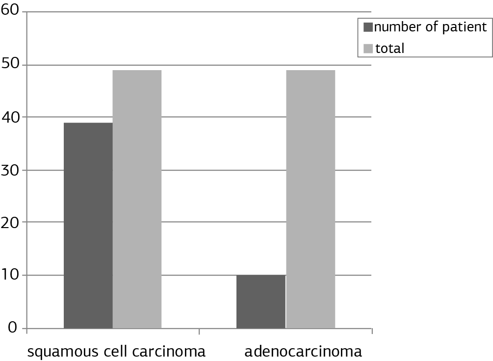
Figure 1. Histopathological distribution of patients
Tables
Table 1. Tumor size and hematologic parameters before and after chemoradiotherapy

WBC; White blood cell, RBC; Red blood cell, Hb; Hemoglobin, Hct; Hematocrit, MCV; Medium cell volume, MPV; Medium thrombocyte volume, MCH; Medium cell hemoglobin, MCHC; Medium cell hemoglobin concentration, RDW; Erythrocyte distribution width, Plt; Thrombocyte counts, Pct; Thrombocyte percentage in blood, PDW; Thrombocyte distribution width * p<0.05 significantly
Additional Information
Publisher’s Note
Bayrakol MP remains neutral with regard to jurisdictional and institutional claims.
Rights and Permissions

This work is licensed under a Creative Commons Attribution-NonCommercial 4.0 International License (CC BY-NC 4.0). To view a copy of the license, visit https://creativecommons.org/licenses/by-nc/4.0/
About This Article
How to Cite This Article
Burcu Sağlam Alan, Burak Erdemci, Hilal Kızıltunç Özmen. Treatment response relationship with hematologic parameters before andafter chemoradiotherapy in patients with non-small cell lung cancer. Ann Clin Anal Med 2025;16(5):384-388. doi:10.4328/ACAM.22477
- Received:
- November 3, 2024
- Accepted:
- December 9, 2024
- Published Online:
- December 26, 2024
- Printed:
- May 1, 2025