
Abstract
AimLymphadenopathy (LAP) can develop for many infectious, rheumatological, immunological, and hematological reasons. The most important point for patients presenting with LAP is the decision-making for further tests to identify who should undergo a biopsy and who should be followed up. This study aimed to investigate predictive markers that can be used to differentiate benign and malignant LAP.MethodsThe study included 274 patients who presented at the Hematology Polyclinic with a diagnosis of LAP. Markers were investigated to determine which affected prognosis in patients with a histopathological benign or malignant LAP diagnosis.ResultsAge, male gender, ferritin, lactate dehydrogenase (LDH), C-reactive protein (CRP), LAP located in the neck, thorax, and abdomen, the size of LAP situated in the neck, and the presence of splenomegaly, were determined at statistically significantly higher rates, and hemoglobin, hematocrit, platelet count, plateletcrit (PCT), and folate were statistically significantly lower in the group with malignant LAP. The results of multivariate analysis showed that age, gender, PCT, sedimentation value, thorax LAP, and inguinal LAP were independently significant in the differentiation of patients with benign and malignant LAP.ConclusionLAP is a frequently encountered pathology but biopsy and further tests may not be appropriate for every patient in terms of cost and workload. Therefore, predictive parameters will be of guidance to the clinician at the stage of decision-making. These parameters will avoid tiring the patient and the doctor with respect to unnecessary further tests, and the chance of early diagnosis will not be missed if it is a malignant process.
Keywords
Introduction
The lymphatic system is a complex system that functions in the filtering of toxins and other substances in the body and is involved in circulation. LAP is defined as abnormal swelling formed in the lymph nodes, which can develop for many infectious, rheumatological, immunological, and hematological reasons.1 The most important point for patients presenting with LAP is the decision-making for further tests to identify who should undergo a biopsy and who should be followed up. Many factors such as LAP localization, size compared to the site of localization, texture, whether localized or spread, mobile or fixed, and additional complaints (fever, night sweats, weight loss), can help the clinician in decision-making.2 Although many laboratory and examination findings will guide the clinician in this decision, there is still a need for additional parameters. Therefore, this study aimed to investigate the presence of markers affecting prognosis in patients with a histopathological diagnosis of benign and malignant LAP from biopsy.
Materials and Methods
The study included 274 patients who presented at the Hematology Polyclinic of Dışkapı Yıldırım Beyazıt Training and Research Hospital with a diagnosis of LAP between May 2019 and August 2022. A record was made for each patient of demographic characteristics, blood group, hemogram, anemia parameters, CRP, sedimentation rate, LAP localization and size, hepatosplenomegaly, the type of biopsy taken, and pathological result.Ethical ApprovalAll procedures performed in studies involving human participants were by the ethical standards of the institutional and/or national research committee and with the 1964 Helsinki Declaration and its later amendments or comparable ethical standards. The study was approved by the Ethics Committee of Dışkapı Yıldırım Beyazıt Training and Research Hospital (Date: 07.03.2022, Decision No: 132/11).Statistical AnalysisData obtained in the study were analyzed statistically using SPSS vn. 28.0 software. Descriptive statistics were stated as mean ± standard deviation (SD), median, minimum and maximum values, or number (n) and percentage (%). The distribution of the variables was examined with the Kolmogorov Smirnov test. In the analysis of independent quantitative data, the Independent Samples t-test and the Mann-Whitney U-test were used. The Chi-square test was applied in the analysis of independent qualitative data. The effect level of parameters was investigated with univariate analysis and multivariate logistic regression analysis. A value of p<0.05 was accepted as the level of statistical significance.Reporting GuidelinesThe study was reported in accordance with STROBE guideline.
Results
A total of 274 patients were analyzed, comprising 127 (46.4%) males and 147 (53.6%) females with a mean age of 50.2±16.5 years. The demographic characteristics and descriptive parameters of the patients are shown in Table 1.
Neck LAP was seen in 75.2% of the patients; the average size was 23.9± 10.6 and the most common locations were left cervical and right cervical (31.1%, and 27.7% respectively).
Supraclaviculer LAP was seen in 16.8 % of the patients; the average size was 22.9±10.1 and the most common locations were left supraclaviculer (67.4%). Thorax LAP was seen in 26.3 % of the patients; the average size was 21.2 ± 13.6 and the most common locations were hilar (47.2%). Axillary LAP was seen in 62.3 % of the patients; the average size was 26.7 ± 12.8 and the most common locations were left axillary (54%). Abdomen LAP was seen in 20.4 % of the patients; the average size was 34.8 ± 25.3 and the most common locations were paraortic (53.6%). Inguinal LAP was seen in 57.7 % of the patients; the average size was 25.3 ± 15.0 and the most common locations were right ınguinal (57.6 %). Splenomegaly was detected in 15% of the patients and hepatomegaly in 25.5%.
The patients who presented with LAP were analyzed in two groups 130 (47.4%) patients with a histopathological diagnosis of benign LAP and 144 (52.6%) with malignant LAP. In the group with malignant LAP, age, male gender, ferritin, LDH, CRP, LAP located in the neck, thorax, and abdomen, the size of LAP situated in the neck, and the presence of splenomegaly, were determined at statistically significantly higher rates (p<0.05), and hemoglobin, hematocrit, platelet count, PCT, and folate were statistically significantly lower (p<0.05) (Table 2).
The comparisons of the groups in respect of LAP localization and size. The rates of LAP localization in the thorax (36.1%,15.4%) and abdomen (29.9%, 10.0%) the size of LAP with neck localization (26.4 ±12.1, 21.8±8.6), and the presence of splenomegaly (20.1%, 9.2%) were determined to be statistically significantly higher in the malignant group. LAP with a neck (84.6%, 66.7%) axillary (74.6%, 52.1%), and inguinal (71.5%, 45.1%) localisation were determined to be statistically significantly high in the benign group (p<0.05).
In the univariate analysis model, age, gender, hemoglobin, hematocrit, platelets, PCT, ferritin, LDH, sedimentation, CRP, LAP located in the neck, the largest diameter of LAP in the neck, thorax LAP, axillary LAP, abdomen LAP, inguinal LAP, and the presence of splenomegaly, were observed to have a statistically significant effect on the differentiation of patients with benign and malignant LAP (p<0.05). The results of multivariate analysis showed that age, gender, PCT, sedimentation value, thorax LAP, and inguinal LAP were independently significant in the differentiation of patients with benign and malignant LAP (p<0.05) (Table 3).
Discussion
LAP develops due to an increase in inflammatory or neoplastic cells in the lymph node. Especially if it does not regress with antibiotic treatment or if there is suspicion of systemic disease, a biopsy must be taken. Although fine needle aspiration biopsy (FNAB) is helpful in benign-malignant differentiation, it is usually not sufficient for a definitive diagnosis.3 When there is persistent LAP with a pre-diagnosis of lymphoma, excisional biopsy at the right time is the gold standard method. An approach that prevents unnecessary operations and does not delay the diagnosis of malignancy is the most appropriate.4 The most important point at this stage is the availability of predictive markers that will guide the clinician.
Many studies in the literature have shown a relationship between age and malignant LAP.4-5 With increasing age, longer exposure to carcinogens, the formation of defects in tumor-suppressing genes, and a decrease in immunity increase the risk of malignancy. In the current study, the mean age of the group diagnosed with malignant LAP was seen to be 55.0±16.8 years, which was determined to be statistically significantly higher than the mean age of the group with benign LAP. In a study by Chau et al., male gender was reported to be associated with malignancy,6 and there are data supporting this in other studies in the literature.7 Consistent with these findings in the literature, male gender was determined to be statistically significantly correlated with malignancy in the current study.
For hemoglobin and hematocrit levels to be low, there needs to be infiltration that is chronic or has affected the bone marrow. These conditions are directly associated with malignancy. After discounting other causes of anemia, low hemoglobin seen together with LAP should alert the clinician in respect of malignancy. Supporting this theory, a relationship was determined between low hemoglobin/hematocrit and malignancy in the current study. In a study of pediatric patients, Sen et al. similarly determined a relationship between anemia and malignancy.8-9
LDH is an enzyme used in the cascade obtaining energy in the body. It is found in almost all tissues of the body and is determined to be high in the blood when there is tissue destruction for various reasons.10 It is a parameter used in the prediction of malignant processes when especially high elevations are evaluated together with other supporting factors.5,11-12 The results of the current study showed that the LDH level was found to be a statistically significant parameter in the prediction of malignant LAP.
There are various studies in literature related to the association with malignancies of thrombocytes and the thrombocyte parameters of PCT, mean platelet volume(MPV), and platelet distribution width( PDW), which can be easily evaluated in the full blood count. In a 2015 study, Özakşit et al. found no statistically significant difference in PCT between groups with benign and malignant adnexal masses,13 while Oncel et al. reported that PCT values were lower in lung cancer patients, consistent with the results of the current study.14
LAP, especially generalized LAP accompanying thrombocytopenia, can reflect a malignant disease or a severe infectious process.15 Similar to the current study, Al Kadah et al. found a relationship between thrombocytopenia and malignant lymph nodes.16 PCT, which expresses the thrombocyte percentage, is expected to be low consistent with thrombocytopenia.
CRP is synthesized from hepatocytes in many rheumatological, oncological, hematological inflammatory, or infectious processes. It has been emphasized in many studies that there is an increase in CRP to accelerate the response to an inflammatory process in the environment, and this has been associated with a poor prognosis in malignant processes.17 In the current study, a statistically significant relationship was determined between CRP and malignant lymph nodes. A positive correlation between ferritin and the development of tumor cells and inflammation has been shown in many studies in the literature.18-19 The iron content and ferritin expression have been determined to be high in malignant cells. Supporting these data, it was seen that elevated ferritin can be used as a predictive parameter in the evaluation of LAP in the current study.
Folic acid deficiency is a cause of hypoproductive macrocytic anemia. In a study of 937 cancer patients, folic acid deficiency was determined at the rate of 7%, and in the hematological malignancy subgroup, this rate was found to be 9.5%.18 Similar data were obtained in the current study and folic acid deficiency was found to be associated with mortality. In cancer patients, the reason for folic acid deficiency may be reduced oral intake, or folic acid deficiency as a cause of increased DNA mutation may cause malignancy.
Other than laboratory parameters in the evaluation of LAP, lymph node localization, and size are among the factors affecting prognosis In literature, a relationship has been found between malignancy and supraclavicular localization and involvement of more than two lymph node regions.6 In the current study, LAPs with neck, thorax, and abdomen localization, and the size of LAP localized in the neck were determined to be statistically significantly high in the malignant LAP group.20 Celenk et al. found a relationship between left-side localized LAP and malignancy.7 However, there are studies in the literature that have shown no difference between left and right-side localization in respect of the association with malignancy,4 or that have reported results showing a relationship between bilateral localization and malignancy.21 In the current study, no statistically significant data were obtained on this point.
Splenomegaly develops in conditions such as diseases that result in hyperplasia (eg., infections, connective tissue disorders), infiltrative or deposition diseases, hemolytic anemia, or portal hypertension. In support of this, the current study data showed that if there is also splenomegaly when evaluating patients with LAP, that there could be malignancy should be kept in mind.9
Limitations
There are some limitations to our study. Since the study was conducted retrospectively, new-generation potential markers could not be evaluated. If the number of patients was larger, the statistics could have been stronger.
Conclusion
LAP can be frequently encountered as a reason for presentation at many departments of polyclinics such as Internal Medicine, Hematology, Ear, Nose, and Throat, and General Surgery. In a pathology encountered this often, biopsy and further testing of every patient may not be appropriate for reasons of both cost and workload. Therefore, predictive parameters will be of guidance to the clinician at the stage of decision-making. These parameters will avoid tiring the patient and the doctor with respect to unnecessary further tests, and the chance of early diagnosis will not be missed if it is a malignant process. This study aimed to investigate parameters that could show a predictive effect. In the results of the multivariate analysis, age, gender, PCT, sedimentation value, thorax LAP, and inguinal LAP were observed to be independently significant in the differentiation of patients with benign and malignant LAP (p<0.05). Rather than the use of these parameters singly, evaluation of them together would seem to be more beneficial with respect to specificity.
Declarations
Animal and Human Rights Statement
No animal experiments were conducted. All human procedures complied with ethical standards.
Informed Consent
Written informed consent was obtained from all participants.
Data Availability
The data supporting the findings of this study are available from the corresponding author upon reasonable request.
Conflict of Interest
The authors declare that there is no conflict of interest.
Funding
None.
Acknowledgements
None.
Abbreviations
CRP: C-reactive protein
FNAB: Fine needle aspiration biopsy
LAP: Lymphadenopathy
LDH: Lactate dehydrogenase
MPV: Mean platelet volume
PCT: Plateletcrit
PDW: Platelet distribution width
References
- Ali M, Elhatw A, Hegazy M, et al. The evaluation of lymphadenopathy in a resource-limited setting. Cureus. 2022;14(10). doi:10.7759/cureus.30623
- Moor JW, Murray P, Inwood J, Gouldesbrough D, Bem C. Diagnostic biopsy of lymph nodes of the neck, axilla and groin: rhyme, reason or chance? Ann R Coll Surg Engl. 2008;90(3):221-225. doi:10.1308/003588408x242105
- Chong Y, Park G, Cha HJ, et al. A stepwise approach to fine needle aspiration cytology of lymph nodes. J Pathol Transl Med. 2023;57(4):196-207. doi:10.4132/jptm.2023.06.12
- Metin U, Mengi E, Kara CO, Bir F. Risk analysis in persistent cervical lymphadenopathies: malignant or benign? North Clin Istanb. 2021;8(4):354-358. doi:10.14744/nci.2020.29895
- Jung YH, Woo IS, Han CW. Clinical characteristics and outcomes in diffuse large B-cell lymphoma patients aged 70 years and older: a single-center experience with a literature review. Korean J Intern Med. 2015;30(5):684-693. doi:10.3904/kjim.2015.30.5.684
- Chau I, Kelleher M, Cunningham DE, et al. Rapid access multidisciplinary lymph node diagnostic clinic: analysis of 550 patients. Br J Cancer. 2003;88(3):354-361. doi:10.1038/sj.bjc.6600738
- Celenk F, Gulsen S, Baysal E, Aytac I, Kul S, Kanlikama M. Predictive factors for malignancy in patients with persistent cervical lymphadenopathy. Eur Arch Otorhinolaryngol. 2016;273(1):251-256. doi:10.1007/s00405-015-3717-3
- Han SV, Park M, Kwon YM, et al. Mild anemia and risk for all-cause, cardiovascular and cancer deaths in apparently healthy elderly Koreans. Korean J Fam Med. 2019;40(3):151-158. doi:10.4082/kjfm.17.0089
- Şen HS, Ocak S, Yılmazbaş P. Children with cervical lymphadenopathy: reactive or not? Turk J Pediatr. 2021;63(3):363-371. doi:10.24953/turkjped.2021.03.003
- Korkmaz MF, Türe E, Dorum BA, Kılıç ZB. The epidemiological and clinical characteristics of 81 children with COVID-19 in a pandemic hospital in Turkey: an observational cohort study. J Korean Med Sci. 2020;35(25). doi:10.3346/jkms.2020.35.e236
- Kamiya N, Ishikawa Y, Takeshima T, et al. Usefulness of lactate dehydrogenase in differentiating abnormal cervical lymphadenopathy. J Gen Fam Med. 2021;22(2):75-80. doi:10.1002/jgf2.386
- Kim JJ, Kim HM, Kim H, et al. Circulating tumor DNA reflects histologic and clinical characteristics of various lymphoma subtypes. Cancer Res Treat. 2023;56(1):314-323.
- Ozaksit G, Tokmak A, Kalkan H, Yesilyurt H. Value of the platelet to lymphocyte ratio in the diagnosis of ovarian neoplasms in adolescents. Asian Pac J Cancer Prev. 2015;16(5):2037-2041. doi:10.7314/apjcp.2015.16.5.2037
- Oncel M, Kiyici A, Oncel M, Sunam GS, Sahin E, Adam B. Evaluation of platelet indices in lung cancer patients. Asian Pac J Cancer Prev. 2015;16(17):7599-7602. doi:10.7314/apjcp.2015.16.17.7599
- Popatia R. Berman’s Pediatric Decision Making. JAMA. 2012;307(6):617-618. doi:10.1001/jama.2012.104
- Al Kadah B, Popov HH, Schick B, Knöbber D. Cervical lymphadenopathy: study of 251 patients. Eur Arch Otorhinolaryngol. 2015;272(3):745-752. doi:10.1007/s00405-014-3315-9
- Meyer MM, Brandenburg L, Hudel H, et al. Who is afraid of CRP? Elevated preoperative CRP levels might attenuate the increase in inflammatory parameters in response to lung cancer surgery. J Clin Med. 2020;9(10):3340. doi:10.3390/jcm9103340
- Zhang D, Wen X, Wu W, Guo Y, Cui W. Elevated homocysteine level and folate deficiency associated with increased overall risk of carcinogenesis: meta-analysis of 83 case-control studies involving 35 758 individuals. PLoS One. 2015;10(5). doi:10.1371/journal.pone.0123423
- Rychtarcikova Z, Lettlova S, Tomkova V, et al. Tumor-initiating cells of breast and prostate origin show alterations in the expression of genes related to iron metabolism. Oncotarget. 2017;8(4):6376-6398. doi:10.18632/oncotarget.14093
- Lee JE, Jeong WG, Nam BD, et al. Impact of mediastinal lymphadenopathy on the severity of COVID-19 pneumonia: a nationwide multicenter cohort study. J Korean Med Sci. 2022;37(22). doi:10.3346/jkms.2022.37.e78
- Loh Z, Hawkes EA, Chionh F, Azad A, Chong G. Use of ultrasonography facilitates noninvasive evaluation of lymphadenopathy in a lymph node diagnostic clinic. Clin Lymphoma Myeloma Leuk. 2021;21(2). doi:10.1016/j.clml.2020.09.012
Tables
Table 1. The demographic and basic descriptive statistics of the patients
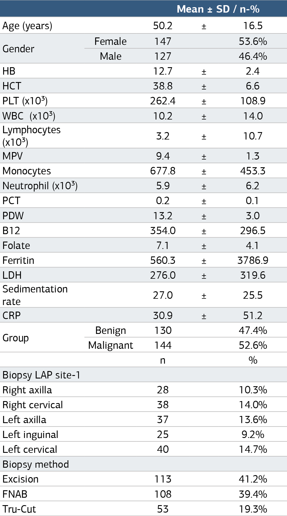
HB:hemoglobin, HTC: hematocrit, PLT:Platelet, WBC: White Blood Cell, MPV: Mean Platelet Volume PCT: Plateletcrit, PDW: Platelet Distribution Width, LDH: Lactate Dehydrogenase, CRP: C- Reactive Protein, FNAB: Fine Needle Aspiration Biopsy
Table 2. Comparisons of the parameters of the benign and malignant lap groups
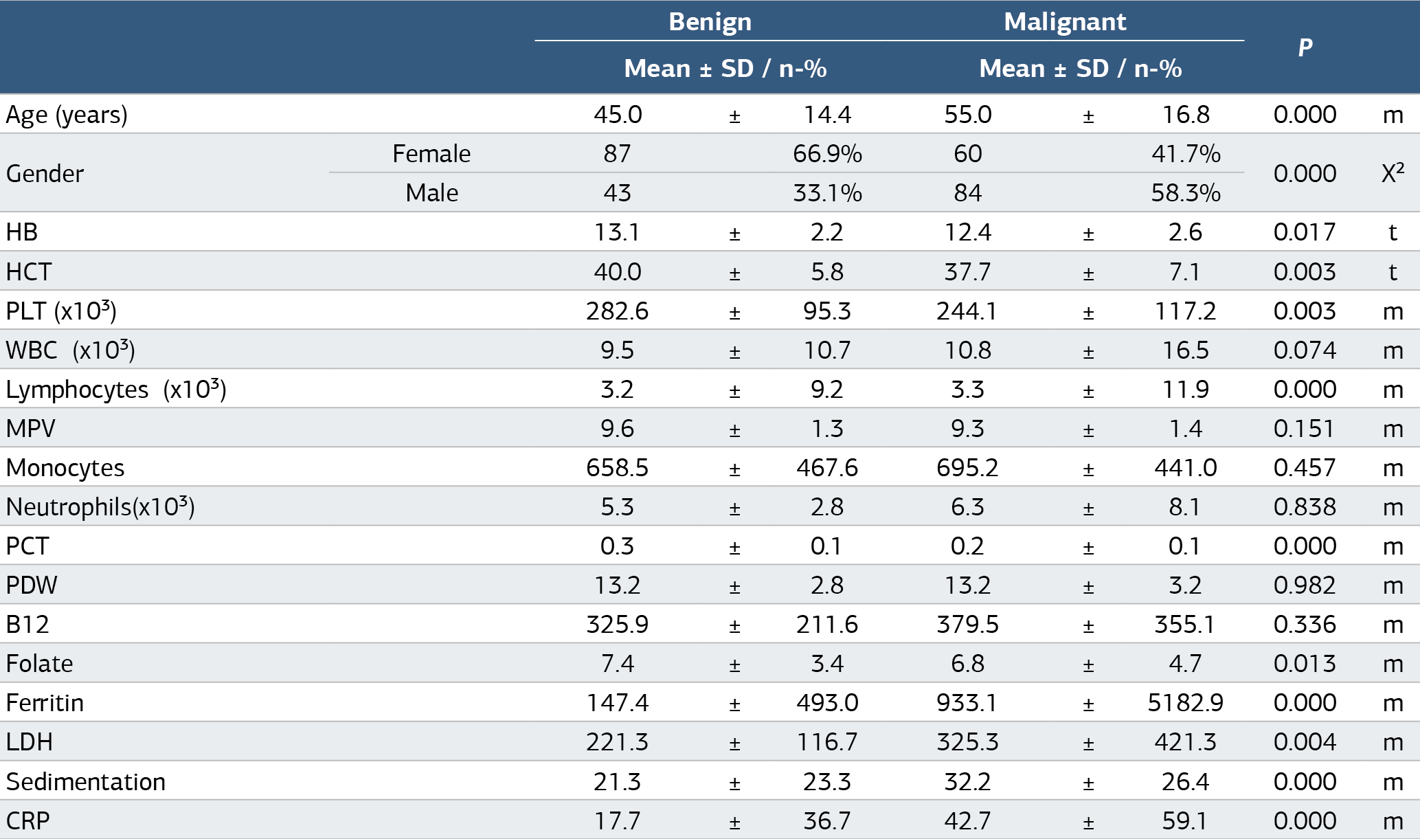
t Independent Samples t-test / m Mann-Whitney U-test / X² Chi-square test HB:hemoglobin, HTC: hematocrit, PLT:Platelet, WBC: White Blood Cell, MPV: Mean Platelet Volume PCT: Plateletcrit, PDW: Platelet Distribution Width, LDH: Lactate Dehydrogenase, CRP: CReactive Protein
Table 3. Logistic regression analysis of the parameters
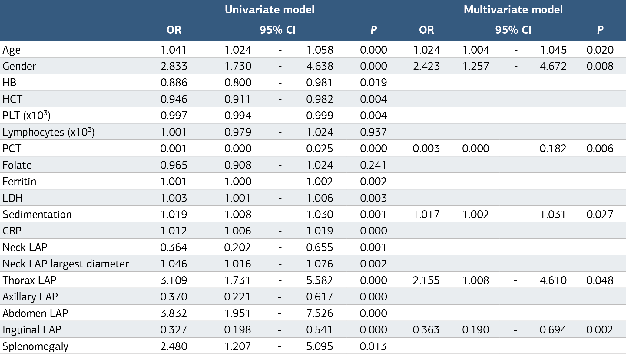
HB: hemoglobin, HTC: hematocrit, PLT:Platelet, WBC: White Blood Cell, MPV: Mean Platelet Volume PCT: Plateletcrit, LDH: Lactate Dehydrogenase, CRP: C- Reactive Protein
Additional Information
Publisher’s Note
Bayrakol MP remains neutral with regard to jurisdictional and institutional claims.
Rights and Permissions

This work is licensed under a Creative Commons Attribution-NonCommercial 4.0 International License (CC BY-NC 4.0). To view a copy of the license, visit https://creativecommons.org/licenses/by-nc/4.0/
About This Article
How to Cite This Article
Fatma Yılmaz, Abdülkerim Yıldız, Merih Reis Aras, Hatice Kozan Kalkışım, Ahmet Demircan, Hacer Berna Afacan Öztürk, Murat Albayrak. An investigation of the predictive markers that can be used in the differentiation of benign and malignant lymphadenopathy. Ann Clin Anal Med 2025;16(3):177-181. doi:10.4328/ACAM.22536
Publication History
- Received:
- 25.12.2024
- Accepted:
- 04.02.2025
- Published Online:
- 11.02.2025
- Printed:
- 01.03.2025