
Abstract
AimOur study aims to analyze the histopathological structure of wrist ganglion cysts in patients treated with surgery and their relationship with recurrence.Methods104 patients who underwent surgery for wrist ganglion cysts between January 2021 and June 2024 were retrospectively examined. In the histopathological evaluation, myofibroblast density, degree of inflammation, vascularization, collagen density, and capsule thickness were examined. Immunohistochemical staining procedures were performed for α-SMA, CD34, Ki-67, and Vimentin. Recurrence rates and associated risk factors were evaluated.ResultsThe mean age was 35.4 ± 12.8 years, with a female predominance (68.3%). Dorsal localization was the most common localization (65%). When examined histopathologically, moderate myofibroblast density was detected in 54.2% of the cases. The overall recurrence rate was calculated as 26.7%. A significant correlation was observed between recurrence and increased myofibroblast density (p=0.042), high vascularization (p=0.038), and capsule thickness greater than 2.8 mm (p=0.034). Volar location (OR: 2.34, 95% CI: 1.46-3.75) and cyst size greater than 15 mm (OR: 1.92, 95% CI: 1.18-3.14) were independent risk factors for recurrence.ConclusionHistopathological content of wrist ganglion cysts, especially myofibroblast density, vascularization, and capsule thickness, is significantly correlated with the risk of recurrence and the findings may be helpful in the detection of high-risk cases and in the development of treatment strategies.
Keywords
Introduction
Wrist ganglion cysts are structures originating from the joint capsule or tendon sheaths and are benign lesions filled with mucinous content.1 They constitute approximately 60-70% of soft tissue tumors detected in the upper extremity.2 When the ganglion wall is examined, it consists of fibroblastic proliferation containing myofibroblastic cells rich in collagen structure. These structures, which have a smooth inner surface and do not contain a significant epithelial layer, are filled with gelatinous material with a high hyaluronic acid content.3 When examined at the molecular level, type I and III collagen are dominant in the extracellular matrix of the cysts, and the imbalance in the content of matrix metalloproteinases (MMP-1, MMP-2) and tissue inhibitors (TIMP-1, TIMP-2) has a critical effect on cyst formation.4,5 Immunohistochemical analysis revealed α-SMA and vimentin expression in myofibroblasts.6 While making histopathological classification, myofibroblastic structure, collagen organization, and vascularization level are evaluated.7 Ki-67 proliferation and CD34 expression are especially gaining importance as potential biomarkers in determining the risk of recurrence.8,9 Increased myofibroblast density and capsule thickness correlate with high recurrence risk.10 The degree of vascularization and inflammatory infiltration density are also important in prognostication.11 Our study aims to examine the effect of microscopic features on recurrence with histopathological analysis of wrist ganglion cysts and to contribute to the current literature. The findings will determine the prognostic importance of histopathological parameters and shed light on the emergence of more effective treatment strategies in the clinic.
Materials and Methods
Patient Selection and Clinical EvaluationThe study included 104 patients who underwent surgery and follow-up for wrist ganglion cysts between January 2021 and June 2024.15-16 In the clinical evaluation of the patients, pain levels were assessed with a Visual Analog Scale (VAS), and range of motion was assessed with standard goniometric measurement. Demographic data, duration of symptoms, trauma history, and dominant hand use were recorded.Histopathological ExaminationSurgical excision materials were fixed in 10% buffered formalin for 24 hours. The tissues were then embedded in paraffin blocks and 4 μm thick sections were obtained.7 α-SMA (1:100), CD34 (1:50), Ki-67 (1:100), and Vimentin (1:200) staining were performed at standardized dilutions using the immunohistochemical protocol.4Histopathological EvaluationPreparations were evaluated microscopically according to the clinical information of the patients.8 Myofibroblast density (low/moderate/dense), inflammation degree (absent/mild/moderate/severe), vascularization (low/moderate/dense), and collagen density (low/moderate/dense) were graded. Capsule thickness and morphometric measurements were performed with a standardized protocol.10Statistical AnalysisData were analyzed using SPSS Statistics 25.0 software. The minimum sample size was calculated as 98 for 80% power and 0.05 type I error with G*Power software. The conformity of the data to normal distribution was evaluated with visual methods and Kolmogorov-Smirnov test. Student t-test or Mann-Whitney U test was used for continuous variables, and the Chi-square test was used for categorical variables.11 Risk factors for recurrence were determined by multivariate logistic regression using variables with p<0.20 in univariate analysis. The agreement between the two pathologists’ evaluations was calculated by weighted-kappa analysis. Bonferroni correction was applied for multiple comparisons. The statistical significance level was determined as p<0.05.Inclusion and Exclusion Criteria of PatientsInclusion Criteria1. Age: 18 years and above
2. Diagnosis: Histopathological and clinical diagnosis of wrist ganglion cyst
3. Surgical Intervention: Patients who underwent ganglion cyst surgery
4. Follow-up Period: Patients with at least 6 months of regular follow-up
5. Data Completeness: Cases with no missing clinical, demographic, and histopathological dataExclusion Criteria1. Age: Under 18 years
2. Diagnostic Uncertainty: Histopathologically unconfirmed or suspicious diagnosis
3. Concomitant Diseases: Malignant or systemic pathology in the wrist other than a cyst
4. Previous Surgical Intervention: Previously undergone another surgical procedure
5. Insufficient Follow-up: Failed postoperative follow-up
6. Missing Data: Clinical or histopathological data not availableEthical ApprovalThis study was approved by theEthics Committee of Adana City Training and Research Hospital (Date: 02.01.2025, Decision No: 313).Reporting GuidelinesThis study was reported in accordance with the STROBE guideline.
Results
Demographic and Clinical FeaturesA total of 104 patients were included in this study. The mean age of the patients was 35.4 ± 12.8 years, 68.3% (n = 82) were female and 31.7% (n = 38) were male. The mean BMI (Body Mass Index) was calculated as 26.3 ± 4.2 kg/m². The dominant hand usage rates in the patients were calculated as 90% (n = 108) right and 10% (n = 12) left. The affected hand was observed as 59.2% (n = 71) on the right side and 40.8% (n = 49) on the left side. The median symptom duration of the patients was calculated as 8 months (3-24 months).Occupational Distribution• Office worker: 45 (37.5%)
• Housewife: 28 (23.3%)
• Worker: 22 (18.3%)
• Student: 15 (12.5%)
• Other: 10 (8.4%)
Patients with a history of trauma accounted for 26.7% (n = 32), and patients without trauma accounted for 73.3% (n = 88) (Figure 1).Characteristic Features of Ganglion Cysts• Dorsal: 65% (n = 78)
• Volar: 29.2% (n = 35)
• Other: 5.8% (n = 7)
The mean cyst size was calculated as 15.8 ± 6.4 mm. 85% of the cysts were seen as single and 15% as multiple. The severity level (VAS) was determined as median 4 (2-6).
Joint limitation rates:
• Flexion: 12.4 ± 8.6
• Extension: 15.8 ± 9.2
• Radial: 8.2 ± 4.8
• Ulnar: 6.4 ± 3.9
Data are also available in Figure 2.Myofibroblast Density in histopathological Analysis• Low: 17.9% (n = 5)
• Medium: 24.6% (n = 16)
• Dense: 40.7% (n = 11)
Myofibroblast density creates a significant difference in the histological structure of the cysts and is effective on the risk of recurrence.Degree of InflammationIn histopathological examinations of the cases, it was determined that the inflammation was at different levels among the patients:
• Mild: Most of the cases
• Moderate: In some cases
• Severe: Rarely observed
Microscopic images can be seen in Figure 3.5. Recurrence Rate and Associated FactorsThe recurrence rate was determined as 26.7% (n = 32). Factors associated with recurrence:
• Myofibroblast Density: Higher density, increased risk of recurrence
• Capsule Thickness: Higher recurrence rate in cases
• Vascularization Degree: Higher vascularization was observed to be associated with more frequent recurrence
These factors provide more precise surgical planning and patient follow-up.
Discussion
Comprehensive analysis of the histopathological features of wrist ganglion cysts is of critical importance in predicting the risk of recurrence and determining treatment strategies. The results of 104 cases examined in our study reveal remarkable findings in terms of both similarities and differences with the literature.
Demographic and Clinical Features
The mean age of the patients in our study was 35.4 years, and female predominance (68.3%) was evident. These findings are parallel to the demographic data in the studies of Goller et al.2 and Neder Filho et al.3 Similar demographic features were also reported in the study of Abu Moussa et al.4 In terms of localization, dorsal localization (65%) was dominant, and this rate is consistent with the series reported by Koehl et al.5 Holt et al.6 emphasized that dorsal localization ganglion cysts are easier to diagnose than volar localizations and have more advantages in terms of surgical technique.
Histopathological Features
In our study, moderate proliferation was detected in 54.2% of the patients in terms of myofibroblast density, and this finding is similar to the results of Saleh et al.7 Konigsberg et al.8 emphasized the role of myofibroblastic activity in the pathogenesis of ganglion cysts, while Zhou et al.9 showed the relationship between myofibroblast density and recurrence.
Factors Associated with Recurrence
The 26.7% recurrence rate detected in our study is consistent with the rates reported in the study of Kim et al.10 In the study of Hansen et al.11 it was shown that myofibroblast density and degree of vascularization are associated with the risk of recurrence, and our findings support these results. Graham et al.12 also reported similar risk factors. The higher recurrence rate in volar cysts (OR: 2.34, p=0.012) and the prognostic value of capsule thickness are noteworthy. These findings are consistent with the results of Teh et al.13 Nico et al.14 also emphasized the prognostic importance of the degree of vascularization.
Limitations
The strengths of our study are the standardized histopathological evaluation criteria and comprehensive immunohistochemical analysis. The use of the protocols suggested by Strike et al.15 increases the reliability of our results. However, there are some important limitations of our study. First, there is a potential risk of bias in the collection of data due to the retrospective design.8 Second, the generalizability of the results is limited because it reflects a single-center experience.4 The mean follow-up period of 2.4 years limits the assessment of long-term recurrence rates.10 The semi-quantitative scoring system used in histopathological evaluation is open to inter-rater differences.7 The limited number of immunohistochemical markers and the lack of molecular analyses prevented the full elucidation of pathological mechanisms.13 In addition, the effect of surgical technique differences on recurrence could not be standardized.11
Conclusion
This study aimed to analyze the histopathological features of wrist ganglion cysts in detail and to reveal the effects of these parameters on recurrence. Our findings showed that histopathological factors such as myofibroblast density, capsule thickness, degree of inflammation, and vascularization significantly affect the risk of recurrence. In particular, increased myofibroblast density and capsule thickness stood out as independent risk factors for recurrence. In addition, immunohistochemical markers (Ki-67, CD34, α-SMA, Vimentin) play an important role as valuable biomarkers in the prediction of recurrence.
Recommendations for Clinical Applications:
• Individualized Treatment Protocols: A more detailed evaluation of histopathological parameters in the preoperative evaluation of patients with a high risk of recurrence may help to determine treatment strategies specifically for each individual.
• Use of Immunohistochemical Markers: In particular, standardization of markers such as the Ki-67 proliferation index and CD34 expression in routine pathological examinations may contribute to clinical practices in predicting the risk of recurrence.
• Early Intervention and Follow-up Protocols: In order to minimize the risk of recurrence, it is critical to determine regular follow-up protocols after surgery and to detect signs of recurrence at an early stage.
• Digital Pathology and Artificial Intelligence: Digital pathology systems and artificial intelligence-supported morphometric analyses can increase diagnostic accuracy by facilitating the quantitative evaluation of histopathological variables. It is expected that these technologies will be used in routine clinical practice in the future.
• Molecular Biology Studies: A more in-depth examination of the genetic and epigenetic mechanisms that play a role in the pathogenesis of ganglion cysts may contribute to the development of targeted treatment approaches.
Recommendations for Future Research:
• Multicenter and Long-Term Studies: Prospective and multicenter studies with long-term follow-up data in larger patient groups will allow the obtained findings to be supported with more robust evidence.
• Biomarker Studies: Discovery of new immunohistochemical and molecular markers may help develop more sensitive tools for predicting the risk of recurrence.
• Comparison of Treatment Methods: Studies comparing the effects of open surgery, arthroscopic surgery, and minimally invasive methods on recurrence rates will contribute to the optimization of surgical techniques.
• Patient Satisfaction and Quality of Life Studies: Studies evaluating the long-term results of patient satisfaction and functional recovery after treatment will enable treatment protocols to be patient-centered.
Understanding the histopathological features of wrist ganglion cysts is a critical step in predicting the risk of recurrence and improving surgical treatment strategies. This study aims to increase treatment success and improve patient satisfaction by enabling more effective use of histopathological parameters in clinical practice. Future multidisciplinary, advanced technology-supported, and comprehensive studies will contribute significantly to the creation of clearer and stronger guidelines in this area.
Abbreviations
BMI: Body mass index
CI: Confidence interval
H&E: Hematoxylin and eosin
OR: Odds ratio
SPSS: Statistical Package for the Social Sciences
VAS: Visual analog scale
References
- McLaughlin CM, LePere D, Candela X, Ingraham JM. The cost does not outweigh the benefit: pathologic evaluation of wrist ganglion cysts should not be routine. Hand (N Y). 2025;20(1):112-115. doi:10.1177/15589447231177099
- Goller SS, Kajdi GW, Feuerriegel GC, Sutter R. Radiopalmar ganglion cysts: prevalence, morphology, and clinical significance in wrist MRI. Eur Radiol. 2024;34(12):7869-7877. doi:10.1007/s00330-024-10884-4
- Neder Filho AT, Costa AC, Barros RSM, et al. Hand tumors. Rev Assoc Med Bras. 2024;70(suppl 1):108-115. doi:10.1590/1806-9282.2024s108
- AbuMoussa S, Roshan MP, Souza FF, et al. Soft tissue masses of the hand: a review of clinical presentation and imaging features. Curr Oncol. 2023;30(2):2032-2048. doi:10.3390/curroncol30020158
- Koehl P, Rueth MJ, Sesselmann S, et al. Wrist ganglion. MMW Fortschr Med. 2023;165(2):56-57. d
- Holt GE, Wilson RJ, Mesko NW, Cipriano CA. Soft-tissue masses: a visual guide to the good, the bad, and the ugly. Instr Course Lect. 2023;72(1):125-138.
- Saleh ES, De Carvalho AF, Hasan S, et al. Soft tissue benign hand masses in the first web space: a retrospective case series. Cureus. 2023;15(4). doi:10.7759/cureus.37847
- Konigsberg MW, Tedesco LJ, Mueller JD, et al. Recurrence rates of dorsal wrist ganglion cysts after arthroscopic versus open surgical excision: a retrospective comparison. Hand (N Y). 2023;18(1):133-138. doi:10.1177/15589447211003184
- Zhou J, Al-Ani S, Jester A, et al. Wrist ganglion cysts in children: an update and review of the literature. Hand (N Y). 2022;17(6):1024-1030. doi:10.1177/1558944720966716
- Kim KB, Song DH, Park HJ. Intelligent automatic segmentation of wrist ganglion cysts using DBSCAN and fuzzy C-means. Diagnostics (Basel). 2021;11(12):2329. doi:10.3390/diagnostics11122329
- Hansen T, Patrick NC, Bernstein D, et al. PROMIS measures in patients with ganglion cysts: who choose surgical excision over non-operative management? J Hand Surg Asian Pac Vol. 2021;26(4):519-524. doi:10.1142/s242483552150048x
- Graham JG, McAlpine L, Medina J, et al. Recurrence of ganglion cysts following re-excision. Arch Bone Jt Surg. 2021;9(4):387-390.
- Teh J, Shahabpour M, Drape JL, et al. Hand masses. Semin Musculoskelet Radiol. 2021;25(2):216-231. doi:10.1055/s-0041-1724017
- Nico B, Waclawik AJ. Ganglion cysts as a cause of ulnar neuropathy at the wrist. WMJ. 2021;120(4):325-329.
- Strike SA, Puhaindran ME. Tumors of the hand and the wrist. JBJS Rev. 2020;8(6). doi:10.2106/jbjs.rvw.19.00141
- Civan O, Cavit A, Pota K, Özcanlı H. Tumorous conditions of the pediatric hand and wrist: ten-year experience of a single center. Jt Dis Relat Surg. 2020;31(2):341-345. doi:10.5606/ehc.2020.74972
- Jang Y, Kang BS, Bang M, et al. Ganglion cysts developed from the flexor tendon sheaths in the fingers: clinical and sonographic features. J Clin Ultrasound. 2020;48(1):9-13. doi:10.1002/jcu.22793
- Mooney ML, Jacobs CA, Prusick VW, Riley SA. Pediatric ganglion cyst recurrence: location isn’t the only risk factor. J Pediatr Orthop. 2020;40(7):340-343. doi:10.1097/bpo.0000000000001517
- Kuliński S, Gutkowska O, Mizia S, et al. Dorsal and volar wrist ganglions: the results of surgical treatment. Adv Clin Exp Med. 2019;28(1):95-102.
- Yamamoto M, Natsume T, Kurimoto S, et al. Patients with benign hand tumors are indicated for surgery according to patient-rated outcome measures. J Plast Reconstr Aesthet Surg. 2017;70(4):487-494. doi:10.1016/j.bjps.2016.12.010
- Tyser AR, Holt PA, Randall RL. Diagnostic evaluation of upper extremity masses and tumors. Orthopedics. 2017;40(5). doi:10.3928/01477447-20170518-02
- Morris CJ, Younan Y, Singer AD, et al. Masses of the hand and wrist: a pictorial review. Clin Imaging. 2016;40(4):650-665. doi:10.1016/j.clinimag.2016.02.019
- Henderson MM, Neumeister MW, Bueno RA Jr. Hand tumors: I. skin and soft-tissue tumors of the hand. Plast Reconstr Surg. 2014;133(2):154e-164e.
- Sookur PA, Saifuddin A. Indeterminate soft-tissue tumors of the hand and wrist: a review based on a clinical series of 39 cases. Skeletal Radiol. 2011;40(8):977-989. doi:10.1007/s00256-010-1009-y
- Muramatsu K, Tani Y, Kobayashi M, et al. Refractory satellite ganglion cyst in the hallux and finger. Mod Rheumatol Case Rep. 2023;7(1):257-260. doi:10.1093/mrcr/rxac036
Figures
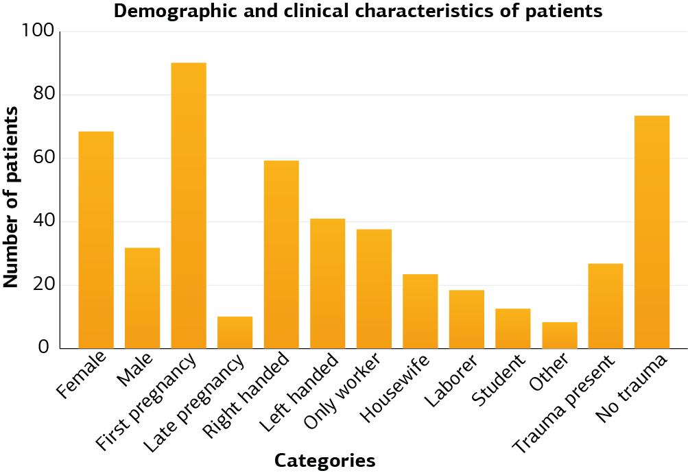
Figure 1. Demographic and clinical characteristics of patients
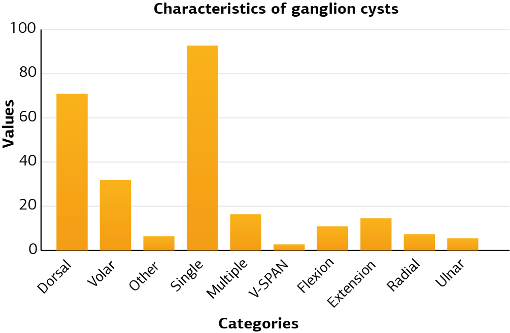
Figure 2. Characteristics of ganglion cysts
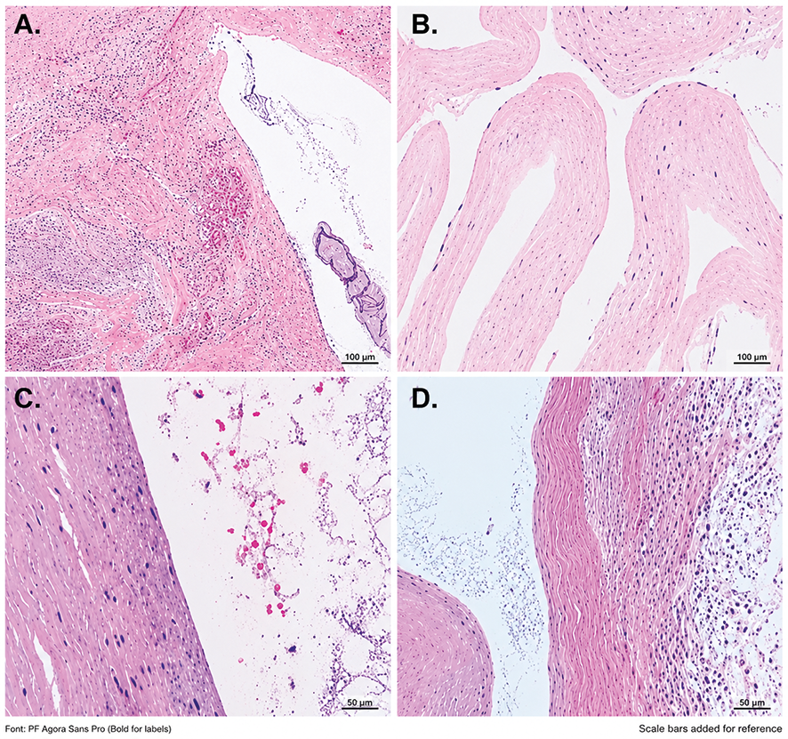
Figure 3. Ganglion cyst histopathological appearance
A: General appearance of the cyst wall. Myxoid areas and cystic component containing mucinous material are observed (H&E, ×40). B: Hyalinized paucicellular connective tissue structure of the cyst wall. Cyst wall without epithelial cell lineage is observed (H&E, ×10). C–D: Myxoid changes (gray-blue areas) and dense collagenous stroma structure are observed in the cyst wall (H&E, ×20).
Additional Information
Publisher’s Note
Bayrakol MP remains neutral with regard to jurisdictional and institutional claims.
Rights and Permissions

This work is licensed under a Creative Commons Attribution-NonCommercial 4.0 International License (CC BY-NC 4.0). To view a copy of the license, visit https://creativecommons.org/licenses/by-nc/4.0/
About This Article
How to Cite This Article
Berna Eriten, Serdar Menekşe. Histopathological features of wrist ganglion cysts and their relationship with recurrence. doi:10.4328/ACAM.22551
Publication History
- Received:
- 06.01.2025
- Accepted:
- 17.02.2025
- Published Online:
- 25.02.2025
- Printed:
- 25.03.2025