
Abstract
AimThis study aimed to investigate the protective effect of alpha lipoic acid (ALA) against the toxicity caused by 6-mercaptopurine (6-MP), which is used in the treatment of childhood cancers, in the testicular tissue of pre-pubertal and pubertal rats.MethodsIn this study, thirty-six Wistar Albino male rats, approximately 28-day old, were used and divided into 6 groups (n=6). According to the groups, 6-MP (10 mg/kg/day) and ALA (100 mg/kg/day) were administered to the rats orally for 14 days by dissolving them in physiological saline. The rats in Group 1 (Control), Group 2 (6-MP), and Group 3 (6-MP+ALA) were decapitated after 14 days of gavage. Group 4 (Control), Group 5 (6-MP), and Group 6 (6-MP+ALA) were decapitated after waiting for 14 more days to reach puberty after 14 days of gavage. Histopathological changes in the testicular tissue were evaluated by hematoxylin and eosin staining. Johnsen’s scores were also compared between groups. In the immunofluorescence analysis, the apoptotic index of the cells was determined by staining with cleaved caspase-3.ResultsAccording to our results, 6-MP administered to pre-pubertal and pubertal rats decreased seminiferous tubule diameter and seminiferous epithelial thickness, but did not affect seminiferous epithelial thickness/diameter ratio. In the testicular tissue of ALA-treated groups, this damage caused by 6-MP was found to be reversed. In addition, 6-MP decreased the Johnsen’s score, but the Johnsen’s score recovered significantly in the ALA-treated groups. The apoptotic index increased in the pre-pubertal group treated with 6-MP and decreased in the group treated with ALA.Conclusion6-MP may cause damage to the testis of pre-pubertal rats, and ALA may protect against this damage.
Keywords
Introduction
The pre-pubertal testes are not immune to chemotherapy, and prolonged exposure to chemotherapy can cause chromatin aberrations and DNA damage in sperm and germ cells.1 Cancer treatment with chemotherapy reduces sperm count and can even lead to azoospermia, which can last for several years or become permanent.2 There is still little information on the effects of many chemotherapy drugs on the prepubertal testis, and most of the available data are mainly from adult studies.3 The 6-mercaptopurine (6-MP) is a safe and cost-effective drug that has been used in humans for the treatment of several types of cancer as well as autoimmune diseases.4 6-MP, an antimetabolite, competes with purine derivatives for the enzyme hypoxanthine-guanine phosphoribosyl transferase and is incorporated into DNA during synthesis. This phenomenon has been shown to cause cytotoxicity that kills cancer cells while damaging healthy cells. In some childhood cancer survivors, 6-MP has been reported to cause caspase 3 activation and loss of Leydig cell function, depending on the dose of exposure.5 Early spermatocytes are more susceptible to 6-MP exposure compared to late and post-meiotic cells.6 Alpha lipoic acid (ALA) is an important molecule in physiological systems containing thiol groups and has antioxidant activity. ALA, a cofactor of the mitochondrial multienzyme dehydrogenase complex, exhibits antioxidant effects such as regeneration of vitamin E, ascorbic acid and glutathione when applied exogenously. ALA exhibits free radical scavenging effects in metal chelation and inhibition of lipid peroxidation.7
Chemotherapy-related toxicity is a major concern for younger cancer patients, who have higher survival rates. A common long-term adverse effect of cancer treatment in children is infertility. Testicular dysfunction is common during adolescence after chemotherapy, and there is currently no known treatment to prevent this damage.8-9-10 Antimetabolites such as 6-MP are used to treat a variety of cancers, but their treatmentrelated adverse effects on male reproductive function are also overlooked. The aim of the current study was to address the increasing concern about long-term side effects, particularly infertility, among cancer survivors. Therefore, this study aimed to investigate the protective effect of ALA against the toxicity caused by 6-MP, which is used in the treatment of childhood cancers, in the testicular tissue of pre-pubertal and pubertal rats.
Materials and Methods
Animal CareIn this study, thirty-six Wistar Albino male rats, approximately 28 day old (pre-pubertal male rats), were used and divided into 6 groups (n=6). During the experiments, the animals were kept in an environment with a temperature of 21 ± 1ºC and a 12-hour light/dark period and were fed with normal tap water and standard rat chow ad libitum. The 6-MP (Biochemical CAS number: 6112-76-1, Sigma-Aldrich, USA) and ALA (Biochemical CAS No: 1077-28-7, Abcam, UK) were administered orally (gavage) to animals by dissolving them in physiological saline for 14 days.
According to the literature, puberty in male rats begins around 40-45 days and ends at 70-77 days of age. Between 56-70 days of age is considered late puberty/adolescence.11 Therefore, the study started with 28-day old rats. Group-1, Group-2 and Group-3 were decapitated after 14 days of gavage, that is, when the rats were 42 days old. Group-3, Group-4 and Group-5 were decapitated after waiting for them to reach puberty, that is, after 14 days of gavage, after waiting for another 14 days, when the rats were 56 days old.Experimental Groups and Experimental DesignGroup 1 (Control): Rats in this group were not administered 6-MP or ALA. Only the same amount of solvent, saline solution, was administered orally (gavage). Rats in this group were decapitated 14 days after the start of the experiment. Group 2 (6-MP): Rats in this group were administered orally (gavage) 6-MP (10 mg/kg/day).12 for 14 days.13 Rats were decapitated after 14 days of gavage. Group 3 (6-MP+ALA): Rats in this group were administered orally (gavage) 6-MP (10 mg/ kg/day).12 and ALA (100 mg/kg/day).7 for 14 days.13 Rats were decapitated after 14 days of gavage. Group 4 (Control): Rats in this group were not administered 6-MP or ALA. Only the same amount of saline solution with solvent was given orally (gavage). The rats in this group were allowed to reach puberty and were decapitated 28 days after the start of the experiment. Group 5 (6-MP): The rats in this group were administered orally (gavage) 6-MP (10 mg/kg/day).12 for 14 days.13 After 14 days of 6-MP administration, the rats were allowed to reach puberty for 14 days and were decapitated at the end of the 28th day. Group 6 (6-MP+ALA): The rats in this group were administered orally (gavage) 6-MP (10 mg/kg/day).12 and ALA (100 mg/kg/day).7 for 14 days.13 After 14 days of 6-MP and ALA administration, the rats were allowed to reach puberty for 14 days and were decapitated at the end of the 28th day.Histopathological AnalysisTesticular tissues were kept in 10% formaldehyde solution at room temperature for 24 hours and then used in histopathological and histomorphometry analyses. After 24 hours of fixation, testicular tissues were divided into two equal parts and subjected to manual tissue processing, and the testicular tissues that completed processing were embedded in paraffin. From the paraffin-embedded testis blocks, 4 µm thick sections were cut on poly-L-lysine slides using a microtome. The sections were stained with hematoxylin and eosin (HE) for histomorphology measurements. Seminiferous tubule diameter, epithelial thickness measurements and Johnsen’s scoring were performed on HE-stained testicular tissue sections. For diameter measurements, the shortest axis of 50 randomly selected seminiferous tubules were determined and measured at two different depths. In 50 seminiferous tubules with smooth contours in each section, seminiferous epithelial thickness was measured in two different areas from the basal lamina to the lumen, including Sertoli and germ cells. The diameter of the seminiferous tubules, the thickness of the seminiferous epithelium and the ratio of the thickness of the seminiferous epithelium to the diameter of the seminiferous tubules were calculated separately for each group. In addition, Johnsen’s scoring was performed on HE-stained sections. For this purpose, 20 seminiferous tubule sections from randomly selected transfers were analyzed in each section.14 A light microscope (Nikon Eclipse E200), DS-Fi-1 camera (Nikon Instruments Inc., Melville, NY) and NIS-Elements BR 2.30 image analysis system (Nikon Corp., Tokyo, Japan) were used for analysis.Immunofluorescence AnalysisSections of 4 µm thickness were cut from the paraffinembedded tissue blocks on positively charged microscope slides. For deparaffinization, sections were passed through xylene (5 min x 3), 100% alcohol (3 min), 96% alcohol (3 min), 80% alcohol (3 min), 70% alcohol (3 min), distilled water and then PBS. Sections were incubated with 5% normal donkey serum cat no: GTX30972 (GeneTex, USA) for 15 minutes. After incubation, the tissue sections were spotted on the slide using a hydrophobic pen and rabbit clonal primary antibody cleaved caspase-3 (1:200) catalogue no: E-AB-30004 (Elabscience, USA) was added and incubated overnight at +4°C. After washing with PBS for 10 minutes x 3, the sections were incubated with anti-rabbit Alexa Fluor 594 (1:200) cat no: ab150076 (Abcam, UK) secondary antibody for 1 hour at room temperature in the dark. After washing with PBS buffer for 10 min x 3 times, the sections were cover slipped with DAPI (diamidin-2-phenylindole dihydrochloride) cat no: GTX30920 (GeneTex, USA) containing coverslip. The sections were digitally photographed with a fluorescent microscope (Nikon Eclipse E600, Nikon Corp., Tokyo, Japan).Ethical ApprovalThis study was conducted with approval from Ethics Committee of the Karabük University Experimental Animal Ethics Committee (Date: 03.09.2024, Decision No: 2024/09/15).Statistical AnalysisStatistical analyses were conducted on IBM SPSS Statistics 24.0 for Windows software. Normal distribution was determined with the Kolmogorov-Smirnov test. One-way analysis of variance (One-way ANOVA) was employed to compare the variables between the groups. Multiple comparisons were conducted with the appropriate post-hoc tests (Tukey or Tamhane’s test). The findings were expressed as means ± standard deviations (SD), and p<0.05 was accepted as statistical significance.Reporting GuidelinesThe study was reported in accordance with STROBE guideline.
Results
Histopathological Results The seminiferous tubule diameter of group 2 treated with 6-MP decreased compared to group 1 and group 3 (p<0.001). However, the seminiferous tubule diameter of group 3 treated with prophylactic ALA increased compared to group 2 and was statistically similar to group 1 (p<0.001). Similarly, the seminiferous tubule diameter of group 5 receiving 6-MP decreased compared to group 4 and group 6, but in group 6 receiving prophylactic ALA, the seminiferous tubule diameter increased compared to group 5 and was statistically similar to group 4 (p<0.001). The seminiferous epithelial thickness of group 2 receiving 6-MP decreased compared to group 1 and group 3 (p<0.001). However, the seminiferous epithelial thickness of group 3 receiving ALA as a prophylactic agent increased compared to group 2 and was statistically similar to group 1 (p<0.001). Similarly, the seminiferous epithelial thickness of group 5 treated with 6-MP decreased compared to group 4 and group 6 (p<0.001). The seminiferous epithelial thickness of group 6, which received ALA as a prophylactic agent, increased compared to group 5 and was statistically similar to group 4 (p<0.001). The groups were compared in terms of thickness/ diameter and there was no statistical difference between group 1, group 2 and group 3, and between group 4, group 5 and group 6 (p>0.05) (Table 1 and Figure 1). Johnsen’s Score The Johnsen score of group 2 given 6-MP decreased compared to group 1 and group 3 (p<0.001). However, the Johnsen’s score of group 3, which received ALA as a prophylactic, increased compared to group 2 and was statistically similar to group 1 (p<0.001). Similarly, Johnsen’s score of group 5 given 6-MP decreased compared to group 4 and group 6, but in group 6 given ALA as prophylactic, Johnsen’s score increased compared to group 5 and was statistically similar to group 4 (p<0.001) (Figure 2). Apoptotic Index Caspase 3 is often activated to catalyze the cleavage of certain downstream molecules. This ultimately leads to DNA fragmentation and programmed cell death (apoptosis).15 In our study, immunofluorescence staining of cleaved caspase-3 was evaluated as an apoptotic index. The level of cleaved caspase-3 was increased in group 2 treated with 6-MP compared to group 1 and group 3 (p<0.002). However, the level of cleaved caspase-3, which is considered an apoptotic index, was increased in group 3 receiving ALA as a protective agent compared to group 2 and was statistically similar to group 1 (p<0.001). Comparing groups 4, 5 and 6 for cleaved caspase-3 levels, which is considered an apoptotic index, there was no statistical difference between groups (p>0.05) (Table 2 and Figure 3).Discussion
Because 6-MP interferes with purine metabolism, particularly in rapidly growing and dividing cells, it raises concerns about spermatogenesis and pregnancy outcome. Although studies in rodents have reported that exposure to high doses of 6-MP significantly inhibits male reproduction, experimental data on the effect of low doses on fertility and reproductive outcome are still lacking.16 Therefore, our study was designed to evaluate the protective effects of ALA against 6-MP toxicity on testicular tissue of pre-pubertal male rats.
In mice treated with 6-MP (2, 5 and 8 mg/kg), sperm morphology and sperm production in the seminiferous tubules were not affected. However, pregnancy rates in female mice mated with these mice were inversely correlated with increasing doses of 6-MP, indicating latent sperm damage.16 Chronic low-dose 6-MP treatment was shown to have no effect on testicular weight, seminiferous tubule stage quantification, number of mature spermatids and serum testosterone levels in rats.17 Panghal et al. investigated the toxicity of 6-MP administered for one week and intermittently for three weeks in the testicular tissue of juvenile rats. In their results, they reported that oneweek and intermittent 3-week exposure cycles to 6-MP did not change the testis-to-body weight ratio but decreased the epididymis-to-body weight ratio in a dose-dependent manner, but this change was insignificant. It has also been reported that 6-MP causes structural abnormalities in the basement membrane, reduction in the spermatogonia and spermatid population, degeneration of the seminiferous tubules and enlargement of the interstitial space. It has also been shown to reduce seminiferous tubule diameter and Johnsen’s score. In addition, 6-MP has been shown to decrease sperm count, sperm motility and the number of sperm with normal head morphology.12 In childhood cancer survivors, 6-MP has been reported to cause Leydig cell failure. Furthermore, in a mouse model, 6-MP has been shown to induce caspase 3 activation and cause Leydig cell loss.5
Recent studies have shown that ALA plays a protective role in the testis against damage caused by anticancer drugs.18 various toxic agents.19-20-21-22-23 ionizing radiation.24 or ischemiareperfusion.25 In our study, the protective effect of ALA against the damage caused by 6-MP administered to pre-pubertal rats before or after puberty was investigated. According to our results, 6-MP administered to pre-pubertal rats decreased the seminiferous tubule diameter and seminiferous epithelial thickness of pre-pubertal and pubertal rats but did not alter the ratio of seminiferous epithelial thickness to diameter . In the testicular tissue of ALA-treated groups, this damage caused by 6-MP was found to be reversed. In addition, 6-MP decreased the Johnsen’s score, but the Johnsen’s score almost recovered in the ALA-treated groups. In our study, cleaved caspase-3 levels were evaluated to determine the apoptotic index, and it was found that the apoptotic index increased in the 6-MP treated groups and decreased in the ALA treated groups. In conclusion, 6-MP may have induced oxidative stress/DNA damage by inducing the formation of oxidant substances in reproductive cells, and cells may have been induced to undergo apoptosis to recover from this stress. ALA may also have a protective effect against 6-MP-induced damage in the testes of rats.
Limitations
A limitation of our study is the absence of biochemical analyses.
Conclusion
There is a paucity of studies in the literature investigating the effects of 6-MP on reproductive cells/functions following its use before puberty. Therefore, our study is an important contribution to the literature. The results of our study suggest that 6-MP may cause damage to the testes of pre-pubertal rats and that ALA may have a protective effect against this 6-MPinduced damage.
Abbreviations
6-MP: 6-Mercaptopurine
ALA: Alpha-lipoic acid
HE: Hematoxylin and eosin
PBS: Phosphate-buffered saline
References
- Beaud H, Albert O, Robaire B, Rousseau MC, Chan PTK, Delbes G. Sperm DNA integrity in adult survivors of paediatric leukemia and lymphoma: a pilot study on the impact of age and type of treatment. PLoS One. 2019;14(12). doi:10.1371/journal.pone.0226262
- Meistrich ML. Effects of chemotherapy and radiotherapy on spermatogenesis in humans. Fertil Steril. 2013;100(5):1180-1186. doi:10.1016/j.fertnstert.2013.08.010
- Delessard M, Saulnier J, Rives A, Dumont L, Rondanino C, Rives N. Exposure to chemotherapy during childhood or adulthood and consequences on spermatogenesis and male fertility. Int J Mol Sci. 2020;21(4):1454. doi:10.3390/ijms21041454
- Sahasranaman S, Howard D, Roy S. Clinical pharmacology and pharmacogenetics of thiopurines. Eur J Clin Pharmacol. 2008;64(8):753-767. doi:10.1007/s00228-008-0478-6
- Morgan JA, Lynch J, Panetta JC, et al. Apoptosome activation, an important molecular instigator in 6-mercaptopurine-induced Leydig cell death. Sci Rep. 2015;5:16488. doi:10.1038/srep16488
- Generoso WM, Preston RJ, Brewen JG. 6-Mercaptopurine, an inducer of cytogenetic and dominant-lethal effects in premeiotic and early meiotic germ cells of male mice. Mutat Res. 1975;28(3):437-447. doi:10.1016/0027-5107(75)90237-7
- Tanbek K, Ozerol E, Yilmaz U, Yilmaz N, Gul M, Colak C. Alpha-lipoic acid decreases neuronal damage on brain tissue of STZ-induced diabetic rats. Physiol Behav. 2022;248:113727. doi:10.1016/j.physbeh.2022.113727
- Matthews ML, Hurst BS, Marshburn PB, Usadi RS, Papadakis MA, Sarantou T. Cancer, fertility preservation, and future pregnancy: a comprehensive review. Obstet Gynecol Int. 2012;2012:953937. doi:10.1155/2012/953937
- Pampanini V, Hassan J, Oliver E, Stukenborg JB, Damdimopoulou P, Jahnukainen K. Fertility preservation for prepubertal patients at risk of infertility: present status and future perspectives. Horm Res Paediatr. 2020;93(11-12):599-608. doi:10.1159/000516087
- Dohle GR. Male infertility in cancer patients: review of the literature. Int J Urol. 2010;17(4):327-331. doi:10.1111/j.1442-2042.2010.02484.x
- Campion SN, Carvallo FR, Chapin RE, et al. Comparative assessment of the timing of sexual maturation in male Wistar Han and Sprague-Dawley rats. Reprod Toxicol. 2013;38:16-24. doi:10.1016/j.reprotox.2013.02.003
- Panghal A, Jena G. Single versus intermittent cycle exposure effect of 6-mercaptopurine in juvenile Sprague-Dawley rat: a germ cell-specific mechanistic study. Naunyn Schmiedebergs Arch Pharmacol. 2024;397(5):3155-3168. doi:10.1007/s00210-023-02797-8
- Jin S, Xu J, Zou Y, et al. Microbiome changes involves in mercaptopurine mediated anti-inflammatory response in acute lymphoblastic leukemia mice. Int Immunopharmacol. 2023;123:110782. doi:10.1016/j.intimp.2023.110782
- Yilmaz N, Hudaykuliyeva J, Gul S. Phoenixin-14 may ameliorate testicular damage caused by torsion-detorsion by reducing oxidative stress and inflammation in prepubertal rats. Tissue Cell. 2024;88:102405. doi:10.1016/j.tice.2024.102405
- Slee EA, Harte MT, Kluck RM, et al. Ordering the cytochrome c-initiated caspase cascade: hierarchical activation of caspases-2, -3, -6, -7, -8, and -10 in a caspase-9-dependent manner. J Cell Biol. 1999;144(2):281-292. doi:10.1083/jcb.144.2.281
- Ligumsky M, Badaan S, Lewis H, Meirow D. Effects of 6-mercaptopurine treatment on sperm production and reproductive performance: a study in male mice. Scand J Gastroenterol. 2005;40(4):444-449. doi:10.1080/00365520510011597
- Karl PI, Katz R, Daum F, Fisher SE. 6-Mercaptopurine and spermatogenesis in the young rat. Dig Dis Sci. 1991;36(11):1569-1573. doi:10.1007/bf01296399
- Pinar N, Cakirca G, Ozgur T, Kaplan M. The protective effects of alpha-lipoic acid on methotrexate-induced testis injury in rats. Biomed Pharmacother. 2018;97:1486-1492.
- Prathima P, Venkaiah K, Pavani R, et al. Alpha-lipoic acid inhibits oxidative stress in testis and attenuates testicular toxicity in rats exposed to carbimazole during embryonic period. Toxicol Rep. 2017;4:373-381. doi:10.1016/j.toxrep.2017.06.009
- Goudarzi M, Haghi Karamallah M, Malayeri A, et al. Protective effect of alpha-lipoic acid on di-(2-ethylhexyl) phthalate-induced testicular toxicity in mice. Environ Sci Pollut Res Int. 2020;27(12):13670-13678. doi:10.1007/s11356-020-07817-1
- Prathima P, Pavani R, Sukeerthi S, Sainath SB. Alpha-lipoic acid inhibits testicular and epididymal oxidative damage and improves fertility efficacy in arsenic-intoxicated rats. J Biochem Mol Toxicol. 2018;32(2). doi:10.1002/jbt.22016
- Lebda M, Gad S, Gaafar H. Effects of lipoic acid on acrylamide-induced testicular damage. Mater Sociomed. 2014;26(3):208-212. doi:10.5455/msm.2014.26.208-212
- Gules O, Eren U. Protective role of alpha-lipoic acid against polychlorobiphenyl (Aroclor 1254)-induced testicular toxicity in rats. Rom J Morphol Embryol. 2016;57(2):451-459.
- Manda K, Ueno M, Moritake T, Anzai K. Alpha-lipoic acid attenuates x-irradiation-induced oxidative stress in mice. Cell Biol Toxicol. 2007;23(2):129-137. doi:10.1007/s10565-006-0137-6
- Ozbal S, Ergur BU, Erbil G, Tekmen I, Bagriyanik A, Cavdar Z. The effects of alpha-lipoic acid against testicular ischemia-reperfusion injury in rats. ScientificWorldJournal. 2012;2012:489248. doi:10.1100/2012/489248
Tables
Table 1. Histopathological results

Group 1 (Control), Group 2 (6-MP), Group 3 (6-MP+ALA), Group 4 (Control), Group 5 (6-MP), and Group 6 (6-MP+ALA). a Significant decrease acccording to Group 1. b Significant increase acccording to Group 2. c Significant decrease acccording to Group 4. d Significant increase acccording to Group 5
Table 2. Apoptotic index

Group 1 (Control), Group 2 (6-MP), Group 3 (6-MP+ALA), Group 4 (Control), Group 5 (6-MP), and Group 6 (6-MP+ALA). a Significant increase acccording to Group 1. b Significant decrease acccording to Group 2
Figures
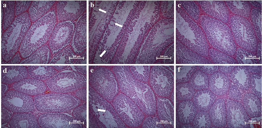
Figure 1. Histopathological results a. Group 1 (Control), b. Group 2 (6-MP), c. Group 3 (6-MP+ALA), d. Group 4 (Control), e. Group 5 (6-MP), f. Group 6 (6-MP+ALA). Scale bar=200 μm. Arrows show the degenerated seminiferous tubule epithelium in groups 2 and 5 Magnification ratio is 20X
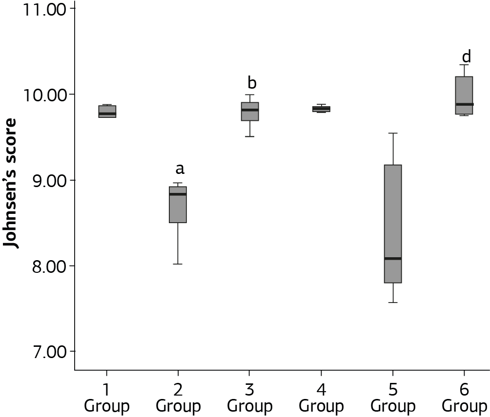
Figure 2. Johnsen’s score Group 1 (Control), Group 2 (6-MP), Group 3 (6-MP+ALA), Group 4 (Control), Group 5 (6-MP), and Group 6 (6-MP+ALA). a Significant decrease acccording to Group 1. b Significant increase acccording to Group 2. c Significant decrease acccording to Group 4. d Significant increase acccording to Group 5
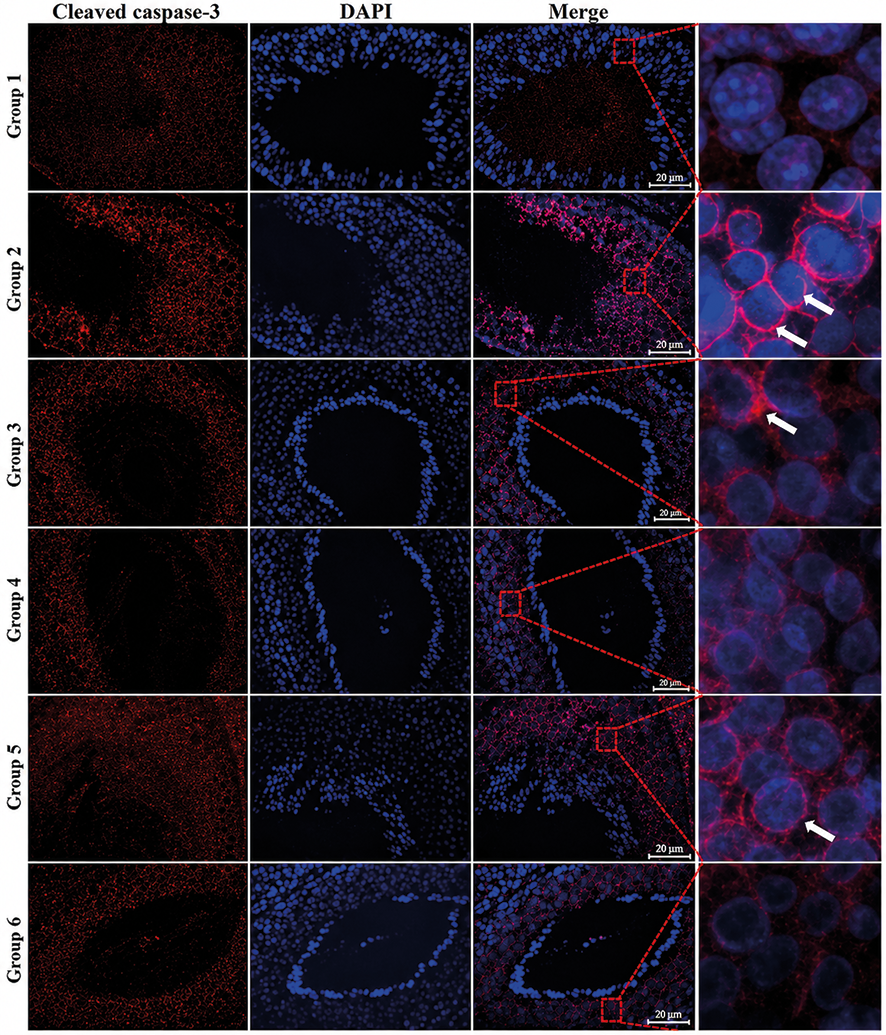
Figure 3. Apoptotic index Immunofluorescence staining of cleaved caspase-3. Cleaved caspase-3 (red) with the nuclei counterstained by DAPI (blue). Scale bar=200 μm. Magnification ratio is 40X
Additional Information
Publisher’s Note
Bayrakol MP remains neutral with regard to jurisdictional and institutional claims.
Rights and Permissions

This work is licensed under a Creative Commons Attribution-NonCommercial 4.0 International License (CC BY-NC 4.0). To view a copy of the license, visit https://creativecommons.org/licenses/by-nc/4.0/
About This Article
How to Cite This Article
Umit Yilmaz, Semir Gul. The protective effect of alpha lipoic acid against 6-mercaptopurine-inducedtesticular damage in pre-pubertal and pubertal rats. doi:10.4328/ACAM.22570
Publication History
- Received:
- 18.01.2025
- Accepted:
- 19.02.2025
- Published Online:
- 25.02.2025
- Printed:
- 01.03.2025