
Abstract
IntroductionMetaplastic breast cancer (MBC) is a rare subtype of invasive breast cancer characterized by mixed epithelial and mesenchymal differentiation. It has a poorer prognosis than triple-negative breast cancer, often responds poorly to conventional treatments, and early diagnosis is essential for treatment planning and prognosis.Case PresentationWe present a patient who developed recurrent metaplastic breast carcinoma on the left anterior chest wall seven months after left total mastectomy for MBC. The clinical and radiological findings demonstrated rapid local recurrence.ConclusionLiterature on MBC remains limited, and patients are underrepresented in clinical studies. This case highlights the aggressive behavior of MBC and the importance of early diagnosis and close follow-up.
Keywords
Introduction
Metaplastic breast carcinoma (MBC) is a rare and heterogeneous group of primary breast malignancies, constituting less than 1% of all invasive breast carcinomas.1 This carcinoma is distinguished by the presence of non-epithelial cellular components alongside epithelial carcinoma. Recently, the WHO breast tumors working group introduced a descriptive classification for MBC, encompassing low-grade adenosquamous carcinoma, fibromatosis-like metaplastic carcinoma, spindle cell carcinoma, metaplastic carcinoma with mesenchymal differentiation, and mixed metaplastic carcinoma.2
MBC is frequently identified as a subtype of triple-negative breast cancer (TNBC).3 Unfortunately, it is associated with a poorer prognosis compared to non-metaplastic TNBC. The recurrence risk is twice as high, and both disease-free survival (DFS) and overall survival (OS) rates are lower than those observed in non-metaplastic triple-negative breast cancer (TNBC) and other invasive carcinomas.2,4 These findings highlight the intricate histological and molecular characteristics of MBCs.5 Typically, MBCs are high-grade tumors that present as large masses. While most cases arise de novo, there have been reports of occurrences originating from pre-existing lesions, including complex sclerosing lesions, papillomas, and breast adenomas.3
Comparative studies evaluating the clinicopathological and prognostic outcomes of MBC and invasive ductal carcinomas indicate a worse prognosis for MBC, with a heightened risk of disease recurrence and poorer overall survival.6 Factors such as the stage at diagnosis, estrogen receptor (ER) status, and adjuvant therapy significantly influence recurrence rates. MBC carries a high risk of recurrence following initial treatment, with limited evidence supporting the efficacy of selective estrogen receptor modulators or aromatase inhibitors post-treatment.6 Due to the substantial risk of local recurrence, reported to range between 35-62% within the first 2-5 years, modified radical mastectomy (MRM) or mastectomy is preferred over breast conservation surgery (BCS).6
The aim of this case report was to present the clinical and radiological features of rapidly recurrent metaplastic breast carcinoma following total mastectomy.
Case Presentation
In 2017, a 67-year-old female patient was admitted to our hospital with a mass in her left breast. After the biopsy, histopathological examination diagnosed TNBC (ER (-), PR (-), C-erbB2 (-), and Ki-67: 10-15%). A decision was made to operate after neoadjuvant chemotherapy (NAC). However, the patient declined surgery because the mass regressed completely after NAC. She presented to us again in 2023 because the mass had enlarged again, and the patient underwent a left total mastectomy procedure. As a result of the pathology examination of the patient’s mastectomy material, it was evaluated as grade 2 metaplastic carcinoma. (ER (-), PR (-), C-erbB2 (-), p53 (-), p63 (-), CK5/6 (+), Ki-67: 5-6%). Less than 1 year after surgery (approximately 7 months) in 2024, the patient presented to our clinic with the complaint of increasing hardness in the left anterior chest wall. During the physical examination, while the right breast examination was normal, a hard, irregular lump-shaped mass lesion of approximately 5 cm, fixed to the anterior chest wall, was detected at the junction of the left anterior chest wall and axilla.
Ultrasonography examination revealed a hypoechoic mass lesion with a heterogeneous internal structure and regular lobulated contours on the left anterior chest wall that did not fit into the probe area and measured approximately 60x50 mm at its widest portion (Figure 1). In the lateral aspect of this lesion, 2-3 more lesions of similar nature, the largest of which was approximately 55x35 mm in size, were observed. In the left axilla, 2-3 lymphadenopathies with a pathological appearance and an internal structure similar to the primary mass, the largest of which was approximately 35 mm in diameter, were observed. The biopsy result of the mass was evaluated as grade 3 metaplastic carcinoma. (ER 11-20%, PR 11-20%, HER2 (-), p53 wild type, GATA-3 focal (+), CK5/6 (+), Ki-67 20%).
This report presents a single descriptive case without including identifiable personal information.
Axillary lymph node evaluation with PET-CT (July 2024) revealed multiple lymph nodes in levels I, II, and III. At level I, multiple lymph nodes with hypermetabolic activity, some with a conglomerated appearance, measuring approximately 5.5 x 5 cm, were observed (Figure 2). Chemotherapy could not be administered due to the patient’s comorbid conditions (hypertension, Diabetes mellitus, chronic renal failure). The patient and her relatives did not accept the recommended radiotherapy.Ethical ApprovalEthical approval was not required.
This case report was prepared in accordance with the CARE reporting guideline.
Discussion
Although the global epidemiology of MBC remains relatively unclear, increasing case reports continue to provide valuable insights. These tumors tend to present at a more advanced stage compared to other breast cancer types, and most MBC subtypes are linked to poorer survival outcomes.5 Large, newly developed, or rapidly growing lesions with complex echogenicity observed on ultrasound necessitate further evaluation through image-guided biopsy for a definitive diagnosis.6
MBC is a rare and highly heterogeneous malignancy, exhibiting significant histological variability and marked aggressiveness. It demonstrates a diminished response to conventional chemotherapy and has a worse overall prognosis compared to TNBC and other invasive breast carcinoma subtypes. Managing MBC is particularly challenging due to its aggressive nature, high recurrence rates, and resistance to standard treatment approaches. Unfortunately, only a limited number of clinical trials specifically focus on this distinct cancer subtype.6 Further research is essential to better understand its origin, imaging characteristics, and potential therapeutic strategies to improve patient outcomes.
Limitations
This report describes a single patient; therefore, the findings cannot be generalized.
Conclusion
Our knowledge of MBC remains very limited, and MBC patients are underrepresented in clinical studies. Further international studies and larger datasets are needed to guide patient treatment and improve survival rates. Although the diagnosis of MBC can be challenging, early detection is critical to patient management.
Declarations
Animal and Human Rights Statement
This case report describes a single patient and was conducted in accordance with the ethical principles of the Declaration of Helsinki and its later amendments or comparable ethical standards.
Informed Consent
Written informed consent was obtained from the patient for publication of this case report and the accompanying images.
Data Availability
The datasets used and/or analyzed during the current study are not publicly available due to patient privacy reasons, but are available from the corresponding author on reasonable request.
Conflict of Interest
The authors declare that there is no conflict of interest.
Funding
None.
Author Contributions (CRediT Taxonomy)
Conceptualization: M.B., E.A.
Investigation: M.B., E.A., C.K.
Data Curation: M.B., C.K.
Visualization: M.B., C.K.
Writing – Original Draft Preparation: M.B.
Writing – Review & Editing: E.A., C.K., D.E.T.Ş.
Supervision: D.E.T.Ş.
AI Usage Disclosure
The authors declare that no AI-assisted technologies were used.
Abbreviations
BCS: Breast-conserving surgery
DFS: Disease-free survival
ER: Estrogen receptor
MBC: Metaplastic breast cancer
MRM: Modified radical mastectomy
NAC: Neoadjuvant chemotherapy
OS: Overall survival
PET-CT: Positron emission tomography-computed tomography
PR: Progesterone receptor
TNBC: Triple-negative breast cancer
References
- Boler DE, Abulut M, Guler G. Metaplastic breast carcinoma: clinicopathologic analysis of 30 cases. J Oncol Sci. 2016;2(2):38-42.
- Reddy TP, Rosato RR, Li X, Mani SA. A comprehensive overview of metaplastic breast cancer: clinical features and molecular aberrations. Breast Cancer Res. 2020;22(1):121. doi:10.1186/s13058-020-01353-z
- Altaf FJ, Mokhtar GA, Emam E, Ezzat AA, Eltorki E. Metaplastic carcinoma of the breast: an immunohistochemical study. Diagn Pathol. 2014;9:139. doi:10.1186/1746-1596-9-139
- Yin L, Duan JJ, Bian XW, Yu SC. Triple-negative breast cancer molecular subtyping and treatment progress. Breast Cancer Res. 2020;22(1):61. doi:10.1186/s13058-020-01296-5
- Thomas A, Douglas E, Reis-Filho JS, Gurcan MN, Wen HY. Metaplastic breast cancer: current understanding and future directions. Clin Breast Cancer. 2023;23(8):775-783. doi:10.1016/j.clbc.2023.04.004
- Al-Hilli Z, Choong G, Keeney MG, Visscher DW, Ingle JN, Goetz MP, et al. Metaplastic breast cancer has a poor response to neoadjuvant systemic therapy. Breast Cancer Res Treat. 2019;176(3):709-716. doi:10.1007/s10549-019-05264-2
Figures
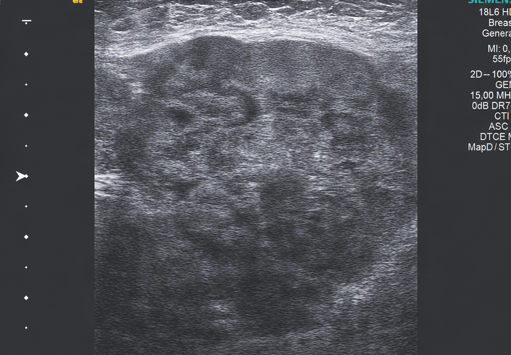
Figure 1. Ultrasonographic image of the recurrent mass that developed on the patient’s anterior chest wall 7 months after surgery. A hypoechoic mass lesion with a heterogeneous internal structure and regular lobulated contours measuring approximately 60x50 mm at its widest portion, not fitting into the probe area on the left anterior chest wall in the ultrasonography image. The biopsy result was evaluated as grade 3 metaplastic carcinoma. (ER 11-20%, PR 11-20%, HER (-), p53 and “wild type”, GATA-3 focal (+), CK5/6 (+), Ki-67 20%)
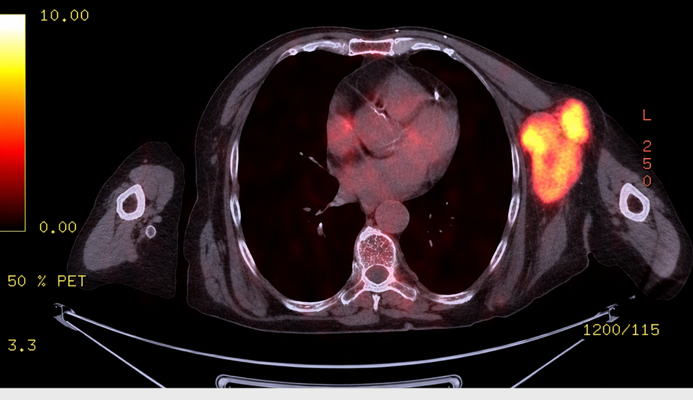
Figure 2. PET-CT (July 2024) image of the patient. Multiple hypermetabolic lymph nodes with a conglomerated appearance were seen in the left axilla
Additional Information
Publisher’s Note
Bayrakol MP remains neutral with regard to jurisdictional and institutional claims.
Rights and Permissions

This work is licensed under a Creative Commons Attribution-NonCommercial 4.0 International License (CC BY-NC 4.0). To view a copy of the license, visit https://creativecommons.org/licenses/by-nc/4.0/
About This Article
How to Cite This Article
Mete Birışık, Emre Aksu, Cuma Keklik, Deniz Esin Tekcan Şanlı. Rapidly recurrent metaplastic breast carcinoma after mastectomy: A Case Report. Ann Clin Anal Med 2026;17(8):871-873. doi:10.4328/ACAM.22679
Publication History
- Received:
- 28.03.2025
- Accepted:
- 03.07.2025
- Published Online:
- 11.07.2025
- Printed:
- 01.08.2026