
Abstract
Aim We aimed to investigate the origin and branching variations of the ophthalmic artery (OA) by digital subtraction angiography (DSA) and flat-panel detector computed tomography angiography (FDCTA).Methods 150 patients with FDCTA images were selected for retrospective analysis of the following OA features: the angle between OA and ICA at origin, the distance of the OA origin to the tuberculum sella, the distance of the central retinal artery origin to the orbital apex, distance between OA origin and entry point to the optic channel, the diameters of the OA at its origin and the osseous optic channel entry and exit.Results There was a significant but weak correlation between the distance from the tuberculum sella and the mean thickness of the OA’s origin on axial and sagittal planes (P < .05). A significant but weak positive correlation between the optical channel entry distance after the OA origin and OA thickness at origin on axial and sagittal planes and optical channel insertion and exit, and the width of the osseous optic canal exit and entry (r = 0.204, 0.225, 0.215, 0.315, 0.257 and P < .05, respectively) were found. A strong correlation between the width of the osseous optic canal at the exit and entry sites was found (r = 0.655, P < .001). Conclusion FDCTA can provide details about the anatomy of the OA. We gained data about the variety due to age and gender. We mentioned the advantages and disadvantages that may arise in clinical practice related to OA’s variant anatomy.
Keywords
Introduction
The anatomy of the orbital vascular territory has a complex structure and may show a wide range of variations. Ophthalmic artery (OA), a branch of the internal carotid artery (ICA), is the main source of orbital blood supply.1,2 The ophthalmic segment of the ICA (C6) includes the section between the distal dural ring and the origin of the posterior communicating artery.3 Two major arteries originate from the ophthalmic segment: OA and the superior hypophyseal artery.4 OA usually arises from the anteromedial aspect of the ICA and courses through the optic canal along with the optic nerve (in an inferolateral position to the optic nerve) towards the orbit.5,6
The C-arm flat-panel detector computed tomography angiography (FDCTA) is an advanced imaging modality that is used as an adjunct to digital subtraction angiography (DSA) and provides computed tomography-like images with a high spatial resolution.7-11 The anatomy can be delineated with a higher resolution with this imaging method.12
OA is an important route in the management of a variety of orbital pathologies (especially of neoplastic and vascular origin). Although surgical methods have been frequently used in the treatment of such pathologies, endovascular methods have gained wider use in recent years. For proper access during endovascular treatment and to avoid possible complications, a thorough understanding of OA’s origin, branching variations, course, and anastomotic connections is essential.
The purpose of this study was to investigate the origin and branching variations of OA by DSA and FDCTA.
Materials and Methods
IndividualsOur institutional review board approved the study protocol. Informed consent was obtained from each patient. We included a total of 160 patients who had undergone FDCTA and/or DSA examinations between 2010-2016 for diagnostic purposes or endovascular treatment for ICA aneurysms, retinoblastoma, pituitary adenoma, meningioma, chordoma, vasculitis, cavernoma, arteriovenous malformation, capillary telangiectasis-developmental venous anomaly, stenosis, and carotid-cavernous fistula. 150 patients with FDCTA images were retrospectively analyzed.Data AcquisitionThe FDCTA images were obtained with a flat-panel biplane angiography system (Allura Xper 20/20; Philips Healthcare) subsequent to a DSA study. High-resolution FDCTA images were obtained from the fluoroscopic 2D images. An automatic power injector (Angiomat, Liebel Flarsheim) was used for contrast injections. Raw data was transferred to the XtraVision workstation, where reconstructions were performed.Imaging AnalysisA neuroradiologist and a radiologist reviewed reconstructed FDCTA images retrospectively and independently. 150 patients with FDCTA images were selected for retrospective analysis of the following OA features: the angle between OA and ICA at origin, the distance of the OA origin to the tuberculum sella, the distance of the central retinal artery origin to the orbital apex, distance between OA origin and entry point to the optic channel, the diameters of the OA at its origin (axial and longitudinal) and at the osseous optic channel entry and exit. While evaluating these 6 parameters with FDCTA; the distance of the OA origin to the tuberculum sella was measured in the sagittal reconstruction plan, the distance of the central retinal artery origin to the orbital apex, and distance between OA origin and entry point to the optic channel were measured in the axial reconstruction plan, and the diameters of the OA at its origin (axial and longitudinal) were measured in the axial and sagittal reconstruction plan.
The age and gender of all patients were recorded. The diameters of the OA at its origin were measured as two values in the axial and longitudinal planes, and in statistical analysis, the mean value of these diameters was taken. 150 patients with FDCTA images were evaluated as a group, and descriptive statistical analysis was performed.Ethical ApprovalThe study protocol was approved by the Clinical Research Ethics Committee of Cerrahpasa University, School of Medicine (Date: 2016-09-07, No: 326832).Statistical AnalysisStatistical analysis of the data was performed using IBM SPSS version 20 (SPSS Inc.). The Shapiro-Wilk test was used to determine the normal distribution of the data. The data with normal distribution was presented as mean and standard deviation, and non-parametric data as median, minimum, and maximum values. Independent Student-t test for parametric data and Mann-Whitney U test for non-parametric data. Pearson’s correlation analysis was used for parametric data, and Spearman’s correlation analysis for non-parametric data. The statistical significance threshold (P value) was set at 0.05.Reporting GuidelinesThis retrospective observational study was reported in accordance with the STROBE guidelines.
Results
A total of 150 patients (99 women, 51 men) were evaluated by FDCTA, with a mean age of 48 years. Of the 150 ophthalmic arteries, 76 of the ophthalmic arteries were on the right side and 74 were on the left. 80 ophthalmic arteries originated from a superomedial aspect, while 55 anteromedially, 13 anteriorly, and 2 inferomedially. The distance of the OA origin to the tuberculum sella, the distance of the central retinal artery origin to the orbital apex, distance between the OA origin and entry point to the optic channel had a normal distribution, other parameters were not normally distributed. Demographic data and morphological features of the ophthalmic artery are shown in Table 1.
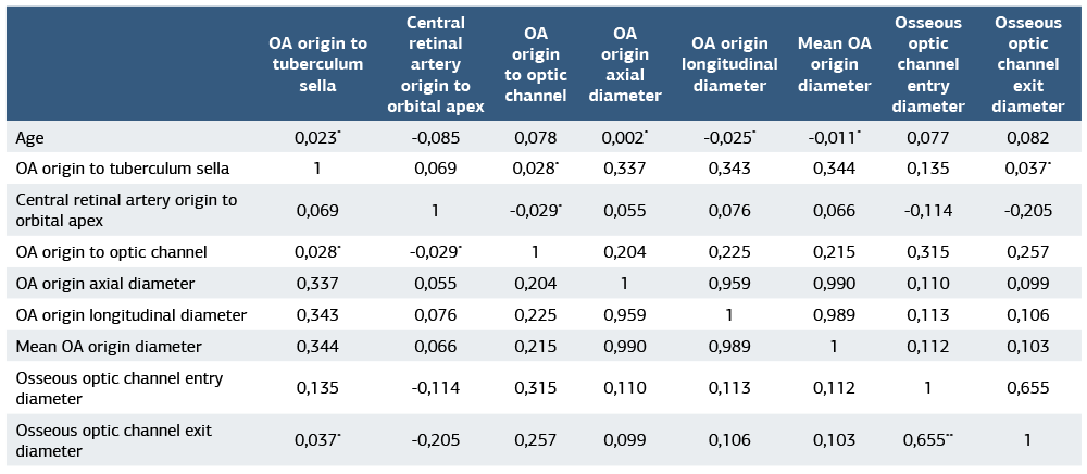
There was no significant difference between the distance to the tuberculum sella, the distance of the central retinal artery origin to the orbital apex, and the osseous optical channel diameter at its entry and exit based on gender. However, the size of the OA on axial and sagittal planes was significantly higher in men than in women (P < .05) (Table 2). There was no significant correlation between age and distance to the tuberculum sella, distance from the central retinal artery to the orbital apex, optical channel insertion distance between OA origin and entry point to the optic channel, size of the OA at its origin on axial and sagittal planes and the osseous optic channel diameter at its entry and exit. There was a significant but weak correlation between the distance from the tuberculum sella and the mean thickness of the OA’s origin on axial and sagittal planes (r = 0.337, 0.343, 0.344 and P < .05, respectively). There was a significant but weak negative correlation between the distance of the central retinal artery to orbital apex and the width of the osseous optic canal exit (r = -0.205, P = .012). A significant but weak positive correlation between the optical channel entry distance after the OA origin and OA thickness at origin on axial and sagittal planes and optical channel insertion and exit, and the width of the osseous optic canal exit and entry (r = 0.204, 0.225, 0.215, 0.315, 0.257 and P < .05, respectively) were found. A strong correlation between the width of the osseous optic canal at the exit and entry sites was found (r = 0.655, P < .001). The values for correlations are shown in Table 3. Of the 10 patients whose DSA images were evaluated only, 2 had PDOA variation (Figure 1), 1 had both PDOA and PVOA variation, 2 had meningo-ophthalmic collaterals (Figure 2), 2 had meningo- lacrimal variant, 1 had lacrimal variant (Figure 3), and 2 had OA-meningeal branch anastomosis.
Discussion
Ophthalmic artery branches do not form a fixed pattern but show significant individual differences. The OA may arise from the anteromedial (53,6%) or superomedial part (31,5%) of the ICA; rare cases of superolateral origin have been reported.13-15 According to Hayreh and Dass.16 in 83.6% of the cases the OA arises from subdural space and follows an intradural course, according to Hokama et al.,17 in 95% of the cases it follows an intradural course, and in 5% of the cases it follows a totally or partially extradural course. Most of the variations of the OA are based on theories like migration, partial and complete regression during embryogenesis, and persistence of primitive vascular structures or persistence of anastomotic connections.18
Knowledge of the detailed anatomy of the OA is essential to understand the pathophysiology of diseases, to determine the diagnostic approach, and treatment modalities. In the treatment of aneurysms of the intracranial or intracanalicular segment of OA, the course of OA should be known.19,20 Endovascular embolization or surgical treatment may be preferred in treating OA aneurysms in the intracranial segment. The surgical approach to these aneurysms at this level can have some difficulties due to its proximity to the optic nerve, the optic channel, and the anterior clinoid process. Knowing the anatomy of OA minimizes these difficulties. The treatment of retinoblastoma is another example highlighting the importance of the OA anatomy. In the treatment of retinoblastoma, super selective intra- arterial chemotherapy with selective embolization of the OA is gaining wider use. Compared to the side effects of systemic chemotherapy, the side effects of endovascular treatment, such as inflammation, hematoma, avascular retinopathy, and cataracts, are relatively safe.21,22 Knowing the origin of the OA for the catheterization of OA or central retinal artery origin pattern from the OA in the super selective catheterization of central retinal artery for retinoblastoma chemoembolization decreases the complication rate.23
The clinical significance of the anastomotic connections between the ophthalmic artery and the external carotid artery branches lies in the possible risk of blindness that may develop during endovascular embolization of a hypervascular tumor or arteriovenous malformation fed by external carotid artery branches. If these connections are not recognized, the embolic agent injected through the external carotid system can diffuse into the ophthalmic artery and central retinal artery. From a different perspective, the administration of the embolic agent from the external carotid arterial system for the endovascular treatment of epistaxis may cause retrograde occlusion of the internal carotid artery through these anastomotic connections between the branches of the external carotid artery and the branches of OA.24,25These anastomoses can be used to advantage during certain endovascular interventional procedures. For example, in cases where it is not possible to access the ophthalmic artery by standard techniques, the ophthalmic artery can be accessed using these anastomotic connections via the internal maxillary artery-middle meningeal artery.24,25
The anatomy and variations of the ophthalmic artery can be profound. Knowing these anatomical features and possible variations increases the probability of success and prevents complications during endovascular treatment involving this region and the surrounding structures, as well as surgeries involving orbita and surrounding structures like optical apex surgery, skull base surgery, pituitary gland surgery, and decompression treatments. The aim of this study was to obtain detailed information about the normal anatomy and variations of the OA.
According to this study, based on 150 ophthalmic arteries that were evaluated by FDCTA, there was no significant difference between the distance to the tuberculum sella, the distance of the central retinal artery origin to the orbital apex, and the osseous optical channel diameter at its entry and exit based on gender. However, the size of the OA on axial and sagittal planes was significantly higher in men than in women. There was no significant correlation between age and distance to the tuberculum sella, distance from the central retinal artery to the orbital apex, optical channel insertion distance between OA origin and entry point to the optic channel, size of the OA at its origin on axial and sagittal planes and the osseous optic channel diameter at its entry and exit. There was a significant but weak correlation between the distance from the tuberculum sella and the mean thickness of the OA’s origin on axial and sagittal planes. There was a significant but weak negative correlation between the distance of the central retinal artery to the orbital apex and the width of the osseous optic canal exit. A significant but weak positive correlation between the optical channel entry distance after the OA origin and OA thickness at origin on axial and sagittal planes, and optical channel insertion and exit, and the width of the osseous optic canal exit and entry was found. A strong correlation between the width of the osseous optic canal at the exit and entry sites was found. Of the 10 patients whose DSA images were evaluated only, 2 had PDOA variation, 1 had both PDOA and PVOA variation, 2 had meningo-ophthalmic collaterals, 2 had meningo-lacrimal variant, 1 had lacrimal variant, and 2 had OA-meningeal branch anastomosis.
Limitations
The limitations of this study were the retrospective nature of the study, the relatively low number of patients, the absence of a control group, and the heterogeneity of the patient and disease groups. However, it is not appropriate to establish a control group because it is unethical to perform procedures such as DSA and/or FDCTA on healthy people without any indications.
Conclusion
In many orbital pathologies (especially those of neoplastic and vascular origin), the OA plays an important role in management options. Although surgical methods are frequently used in the treatment of such pathologies, endovascular methods have become very popular in recent years. It is of great importance to know the origin and branching variations of the OA, the course, and anastomotic connections in detail in order to optimize the success of the treatment and to avoid possible complications. In our study, we found that FDCTA can provide details about the anatomy of the OA. Also, we gained data about the variety due to age and gender. Furthermore, we tried to mention the advantages and disadvantages that may arise in clinical practice related to OA’s variant anatomy.
Declarations
Ethics Declarations
This study was conducted in accordance with the ethical principles of the Declaration of Helsinki and its later amendments. The study protocol received approval from the Clinical Research Ethics Committee of Cerrahpasa University, School of Medicine (Date: 2016-09-07, No: 326832). The authors declare that there is no conflict of interest and no external funding was received for this study.
Animal and Human Rights Statement
All procedures performed in this study were in accordance with the ethical standards of the institutional and/or national research committee and with the 1964 Helsinki Declaration and its later amendments or comparable ethical standards.
Informed Consent
Written informed consent was waived due to the retrospective design of the study.
Data Availability
The data supporting the findings of this article are available from the corresponding author upon reasonable request, due to privacy and ethical restrictions. The corresponding author has committed to share the de-identified data with qualified researchers after confirmation of the necessary ethical or institutional approvals. Requests for data access should be directed to bmp.eqco@gmail.com
Conflict of Interest
The authors declare that there is no conflict of interest.
Funding
None.
Author Contributions (CRediT Taxonomy)
Conceptualization: E.E., S.G.K., O.K.
Methodology: E.E., S.G.K.
Formal Analysis: E.E., D.E.T.S.
Investigation: E.E., S.G.K., D.E.T.S., Y.K.
Data Curation: E.E., D.E.T.S.
Writing – Original Draft Preparation: E.E.
Writing – Review & Editing: S.G.K., N.K., C.I., O.K.
Supervision: N.K., C.I., O.K.
Abbreviations
AJNR: American journal of neuroradiology
CT: Computed tomography
DSA: Digital subtraction angiography
FDCTA: Flat-Panel detector computed tomography angiography
ICA: Internal carotid artery
OA: Ophthalmic artery
SPSS: Statistical package for the social sciences
References
- Gibo H, Lenkey C, Rhoton AL. Microsurgical anatomy of the supraclinoid portion of the internal carotid artery. J Neurosurg. 1981;55(4):560–574. doi:10.3171/jns.1981.55.4.0560
- Bouthillier A, van Loveren HR, Keller JT. Segments of the internal carotid artery: a new classification. Neurosurgery. 1996;38(3):425–433. doi:10.1097/00006123-199603000-00001
- Osborn A. The cerebral veins. In: Diagnostic cerebral angiography. 2nd ed. Philadelphia: Lippincott Williams & Wilkins; 1998.p.31–55.
- Ökten Aİ, Güzel A. Beynin arteryel anatomisi [Arterial anatomy of the brain]. Türk Nöroşir Derg. 2012;22(3):171–188.
- Aburn NS, Sergott RC. Orbital colour Doppler imaging. Eye. 1993;7(5):639–647. doi:10.1038/eye.1993.147
- Lieb WE, Cohen SM, Merton DA, Shields JA, Mitchell DG, Goldberg BB. Colour Doppler imaging of the eye and orbit - technique and normal vascular anatomy. Arch Ophthalmol. 1991;109(4):527–531. doi:10.1001/archopht.1991.01080040095036
- Kalender WA, Kyriakou Y. Flat-detector computed tomography (FD-CT). Eur Radiol. 2007;17(11):2767–2779. doi:10.1007/s00330-007-0651-9
- Linsenmaier U, Rock C, Euler E, et al. Three-dimensional CT with a modified C-arm image intensifier: feasibility. Radiology. 2002;224(1):286–292. doi:10.1148/radiol.2241010646
- Fahrig R, Fox AJ, Lownie S, Holdsworth DW. Use of a C-arm system to generate true three-dimensional computed rotational angiograms: preliminary in vitro and in vivo results. AJNR Am J Neuroradiol. 1997;18(8):1507–1514.
- Akpek S, Brunner T, Benndorf G, Strother C. Three-dimensional imaging and cone beam volume CT in C-arm angiography with flat panel detector. Diagn Interv Radiol. 2005;11(1):10–13.
- Cowen AR, Davies AG, Sivananthan MU. The design and imaging characteristics of dynamic, solid-state, flat-panel x-ray image detectors for digital fluoroscopy and fluorography. Clin Radiol. 2008;63(10):1073–1085. doi:10.1016/j.crad.2008.06.002
- Gupta R, Grasruck M, Suess C, et al. Ultra-high resolution flat-panel volume CT: fundamental principles, design architecture, and system characterization. Eur Radiol. 2006;16(6):1191–1205. doi:10.1007/s00330-006-0156-y
- Hayreh SS. The ophthalmic artery. Section I. Normal gross anatomy. In: Newton TH, Potts G, editors. Radiology of the skull and brain. Vol 2. St. Louis: Mosby; 1974.p.1333–1350.
- Özgen T, İnci S. Temel nöroşirürji cilt I. [Fundamental neurosurgery volume I.] Ankara: Türk Nöroşir Derg; 1995:464-471.
- N’da HA, Peltier J, Zunon-Kipre Y, et al. An unusual case of superolateral origin of ophthalmic artery; an anastomotic case report. Surg Radiol Anat. 2014;36(1):95–97. doi:10.1007/s00276-013-1132-2
- Hayreh SS, Dass R. The ophthalmic artery: I. Origin and intra-cranial and intra-canalicular course. Br J Ophthalmol. 1962;46(2):65–98. doi:10.1136/bjo.46.2.65
- Hokama M, Hongo K, Gibo H, Kobayashi S. Microsurgical anatomy of the ophthalmic artery and the distal dural ring for the juxta-dural ring aneurysms via the pterional approach. Neurol Res. 2001;23(4):331–335. doi:10.1179/016164101101198703
- Louw L. Different ophthalmic artery origins: embryology and clinical significance. Clin Anat. 2015;28(5):576–583. doi:10.1002/ca.22470
- Nishio S, Matsushima T, Fukui M, Sawada K, Kitamura K. Microsurgical anatomy around the origin of the ophthalmic artery with reference to contralateral pterional surgical approach to the carotid-ophthalmic aneurysm. Acta Neurochir (Wien). 1985;76(3-4):82–89. doi:10.1007/bf01418465
- Yanaka K, Matsumaru Y, Kamezaki T, Nose T. Ruptured aneurysm of the ophthalmic artery trunk demonstrated by three-dimensional rotational angiography: case report. Neurosurgery. 2002;51(4):1066–1070. doi:10.1097/00006123-200210000-00038
- Gobin YP, Dunkel IJ, Marr BP, Brodie SE, Abramson DH. Intra-arterial chemotherapy for the management of retinoblastoma: four-year experience. Arch Ophthalmol. 2011;129(6):732–737. doi:10.1001/archophthalmol.2011.5
- Abramson DH, Dunkel IJ, Brodie SE, Kim JW, Gobin YP. A phase I/II study of direct intraarterial (ophthalmic artery) chemotherapy with melphalan for intraocular retinoblastoma initial results. Ophthalmology. 2008;115(8):1398– 1404. doi:10.1016/j.ophtha.2007.12.014
- Perrini P, Cardia A, Fraser K, Lanzino G. A microsurgical study of the anatomy and course of the ophthalmic artery and its possibly dangerous anastomoses. J Neurosurg. 2007;106(1):142–150. doi:10.3171/jns.2007.106.1.142
- Akdemir Aktaş H, Ergun KM, Tatar İ, Arat A, Hayran KM. Evaluation of the anastomoses between the ophthalmic artery and the middle meningeal artery by superselective angiography. Surg Radiol Anat. 2020;42(11):1355–1361. doi:10.1007/s00276-020-02546-z
- Geibprasert S1, Pongpech S, Armstrong D, Krings T. Dangerous extracranial- intracranial anastomoses and supply to the cranial nerves: vessels the neurointerventionalist needs to know. AJNR Am J Neuroradiol. 2009;30(8):1459– 1468. doi:10.3174/ajnr.a1500
Tables
Table 1. Demographic characteristics and morphological features of the ophthalmic artery
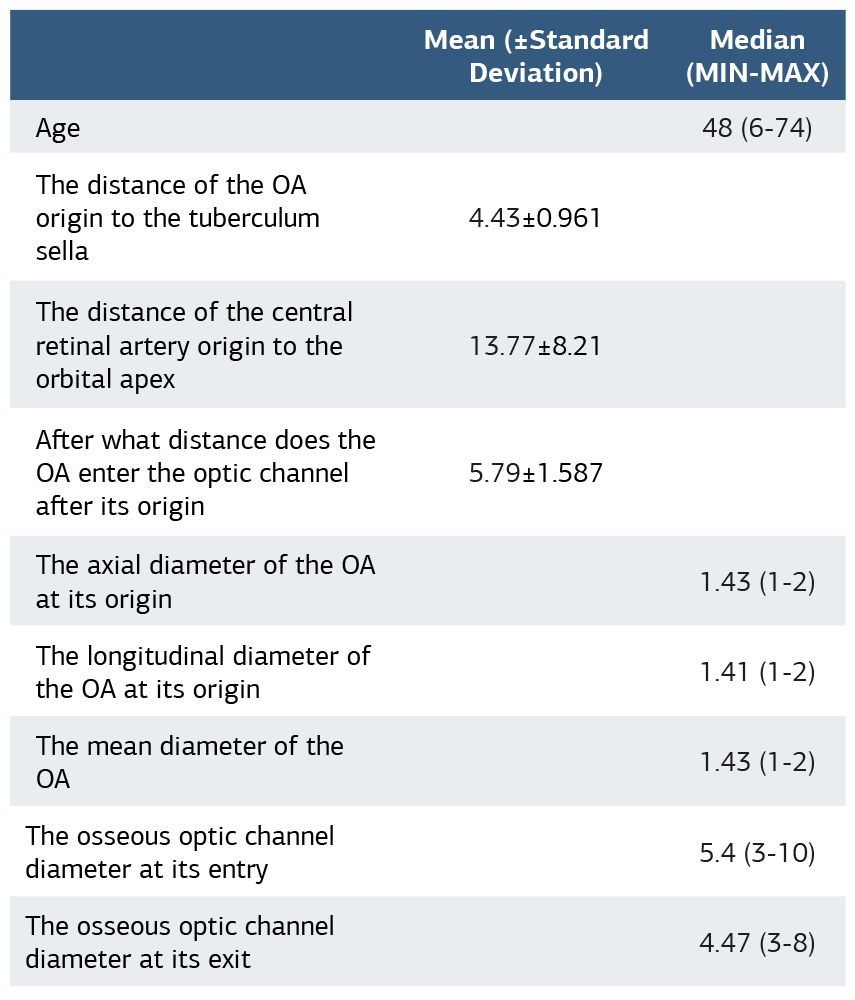
Table 2. Mean-median values of the morphological features of the OA based on gender
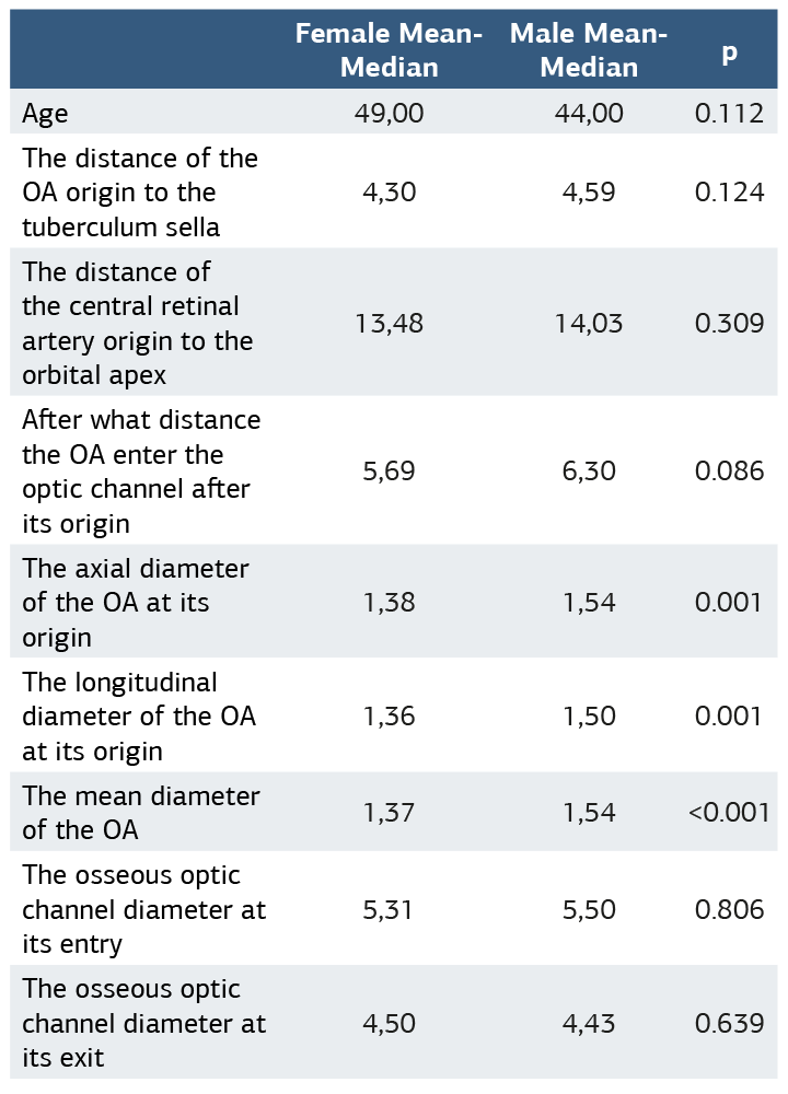
Table 3. The values of correlation
Figures
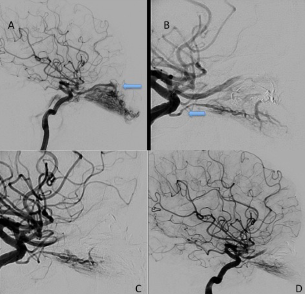
Figure 1. An example of PDOA. In the left globe, a hypervascular mass is observed and the mass is fed by (A) the left ophthalmic artery and (B) the left dorsal ophthalmic artery. (C) After embolization of OA, feeding from the dorsal ophthalmic artery is continued, and (D) retinal crescent staining is preserved
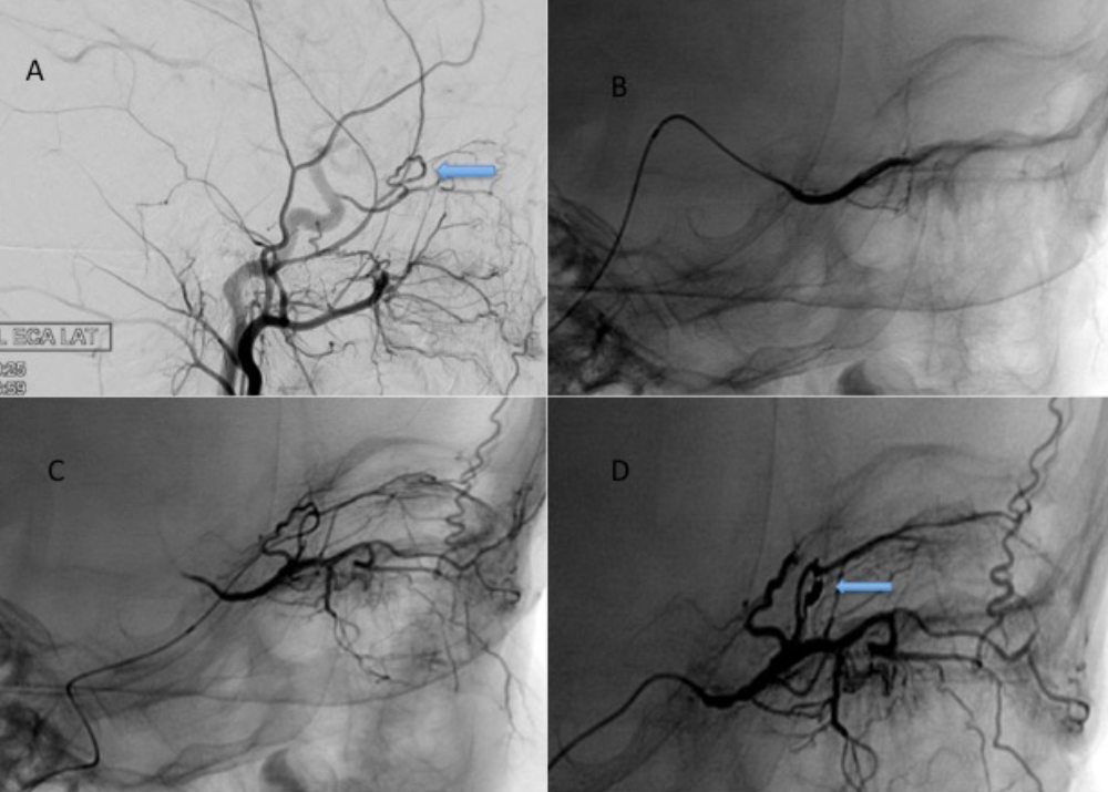
Figure 2. An example of meningo-ophthalmic collateral. After the contrast medium injection of the left external carotid artery, (A) the meningo-ophthalmic collateral (arrow sign) between the middle meningeal artery and the ophthalmic artery is observed. OA shows an inverse filling due to the meningo- ophthalmic collateral. (D) After the coil embolization of the meningo-ophthalmic collateral (arrow sign), it is seen that the OA is normally filled anterogradely after the superselective catheterization of the OA

Figure 3. DSA images of a 50-year-old woman who underwent preoperative tumor embolization due to left parafalcine meningioma. In ECA injections (A) and (B), posteroanterior images show anastomosis between the sphenoidal branch of the middle meningeal artery and the lacrimal artery (arrow sign)
Additional Information
Publisher’s Note
Bayrakol MP remains neutral with regard to jurisdictional and institutional claims.
Rights and Permissions

This work is licensed under a Creative Commons Attribution-NonCommercial 4.0 International License (CC BY-NC 4.0). To view a copy of the license, visit https://creativecommons.org/licenses/by-nc/4.0/
About This Article
How to Cite This Article
Emel Esmerer, Sedat Giray Kandemirli, Deniz Esin Tekcan Sanli, Yasemin Kayadibi, Naci Kocer, Civan Islak, Osman Kizilkilic. Evaluation of the origin and branching patterns of the ophthalmic artery using DSA and FDCTA. Ann Clin Anal Med 2026;17(4):318-323. doi:10.4328/ACAM.22723
Publication History
- Received:
- 24.04.2025
- Accepted:
- 14.07.2025
- Published Online:
- 30.07.2025
- Printed:
- 01.04.2026