
Clinical characteristics of four children with azygos lobe: A Case Report
Children with an azygos lobe
Authors
Abstract
Introduction Normally, the azygos vein is localized in the posterior part of the upper lobe of the right lung. Still, during the process of embryogenesis, the azygos vein passes through the upper lobe of the lung. As a result of the parietal and visceral pleura being pulled together during this process, the azygos lobe is formed. The clinical features of the azygos lobe in children have not been summarized so far.
Case Presentation This paper describes the presence of an azygos lobe in 4 pediatric patients presenting with cough and/or wheezing. Additionally, no pathological conditions such as bronchiectasis, pneumothorax, or neoplasm associated with the azygos lobe were observed in the evaluated patient series.
Conclusion The azygos lobe does not require specific treatment unless it leads to clinically significant disease. However, to prevent misdiagnosis in radiological evaluations, clinicians should consider this anatomical variation.
Keywords
Introduction
The azygos lobe is a congenital variation of the lung, and its prevalence in the general population has been reported to be approximately 0.2–1.2%.1 Normally, the azygos vein is located posteriorly in the upper lobe of the right lung; however, during the embryogenesis process, the azygos vein arch is displaced into the right lung and alters the normal relationship between the medial aspect of the right lung, located next to the azygos fissure, and the right side of the mediastinum.2 The azygos lobe has been reported to resemble various pathological conditions, which can lead to misdiagnosis and incorrect treatment in adults, creating difficulties during thoracic surgeries. For example, an azygos lobe may be mistaken for a bulla, abscess, tumor, or localized pneumothorax.2,3
While the clinical characteristics of the azygos lobe in adults are well defined, the clinical implications of this variation in children are more ambiguous, and systematic analyses are limited. This study presents the clinical features of four pediatric patients diagnosed with an azygos lobe at our clinic over the past five years.
Case Presentation
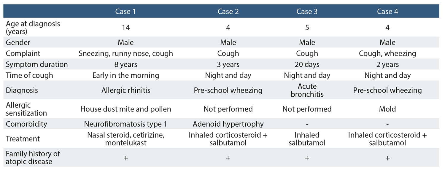
The demographic and clinical characteristics of the four patients with an azygos lobe are summarized in Table 1. Additionally, the postero-anterior chest X-rays are shown in Figure 1.
Ethical ApprovalWritten informed consent was obtained from the parents/legal guardians. Ethical committee approval was not required for case reports.
Reporting GuidelinesThis case is reported in accordance with the CARE guidelines.
Discussion
In the early phases of fetal development, the right posterior cardinal vein, which eventually becomes the azygos vein, is positioned next to the apex of the right lung. Typically, this vein shifts above the lung and settles in the area between the right upper lobe and the main stem bronchi. However, in individuals with an azygos lobe, the vein extends into the right upper lobe, carrying with it four layers of pleura. As a result, a section of the right upper lobe becomes enclosed between this new fissure and the mediastinum, forming what is known as the azygos lobe.4
The azygos lobe is usually discovered incidentally during a chest X-ray or thoracic CT scan, as seen in our patients. The azygos lobe tends to be asymptomatic and does not present with symptoms in most cases. However, in some instances, it may lead to misinterpretations and be mistaken for lung infections or other pulmonary pathologies.5
A review of the current literature reveals that the clinical features of the azygos lobe are mostly described in adult case reports. Some studies suggest that the azygos lobe is not predisposed to diseases. However, Ndiaye et al. have noted that if the fissure is deep enough to compress the bronchus draining the azygos lobe, it could lead to atelectasis or bronchiectasis.6
Limitations
This study has several limitations. First, it is a single-center, retrospective case series involving only four pediatric patients, which limits the generalizability of the findings. Second, the relatively short follow-up period and limited clinical data restrict the ability to assess long-term outcomes and potential complications associated with the azygos lobe in children. Further large-scale, prospective studies are needed to better understand the clinical significance of the azygos lobe in the pediatric population.
Conclusion
The clinical features of the azygos lobe in children have not been well documented until now. In this study, it was incidentally identified in four pediatric patients with respiratory symptoms such as cough and wheezing, without any associated pathological conditions like bronchiectasis, pneumothorax, or neoplasm. Although the azygos lobe is a rare anatomical variant and typically asymptomatic, recognizing it is important to avoid radiological misinterpretation and unnecessary interventions. Greater awareness among clinicians, especially radiologists and surgeons, is essential, and further pediatric-focused research may help clarify its clinical relevance.
Declarations
Ethics Declarations
Written informed consent forms were obtained from the parents/legal guardians. Ethical committee approval was not required for case reports.
Animal and Human Rights Statement
All procedures performed in this study were in accordance with the ethical standards of the institutional and/or national research committee and with the 1964 Helsinki Declaration and its later amendments or comparable ethical standards.
Informed Consent
Written informed consent was obtained from the patients for publication of this case report. The patients’ identities have been protected.
Data Availability
The datasets used and/or analyzed during the current study are not publicly available due to patient privacy reasons but are available from the corresponding author on reasonable request.
Conflict of Interest
The authors declare that there is no conflict of interest.
Funding
None.
Author Contributions (CRediT Taxonomy)
Conceptualization: Y.K., D.A.
Investigation: A.A., M.M.Ö., N.G.
Data Curation: Y.K., M.M.Ö., N.G.
Writing – Original Draft Preparation: Y.K., D.A.
Writing – Review & Editing: Y.K., A.A.
Supervision: M.M.Ö., N.G., A.A., D.A.
Scientific Responsibility Statement
The authors declare that they are responsible for the article’s scientific content, including study design, data collection, analysis and interpretation, writing, and some of the main line, or all of the preparation and scientific review of the contents, and approval of the final version of the article.
References
-
González R, Farias J, Seguel E, Alarcón E. Azygous lobe. J Am Coll Surg. 2009;209(5):673. doi:10.1016/j.jamcollsurg.2009.04.020
-
Felson B. The azygos lobe: its variation in health and disease. Semin Roentgenol. 1989;24(1):56-66. doi:10.1016/0037-198x(89)90054-0
-
Darlong LM, Ram D, Sharma A, et al. The azygous lobe of the lung: in the case of lung cancer. Indian J Surg Oncol. 2017;8(2):195-197. doi: 10.1007/s13193-016- 0617-y
-
Kolbenstvedt A, Kolmannskog F, Aakhus T. The appearance of an anomalous azygos vein on computed tomography of the chest. Radiology. 1979;130(2):386. doi:10.1148/130.2.386
-
Akhtar J, Lal A, Martin K, Popkin J. Azygos lobe: a rare cause of right paratracheal opacity. Respir Med Case Rep. 2018;23:136-137. doi:10.1016/j.rmcr.2018.02.001
-
Ndiaye A, Ndiaye NB, Ndiaye A, Diop M, Ndoye JM, Dia A. The azygos lobe: an unusual anatomical observation with pathological and surgical implications. Anat Sci Int. 2012;87(3):174-178. doi:10.1007/s12565-011-0119-5
Figures
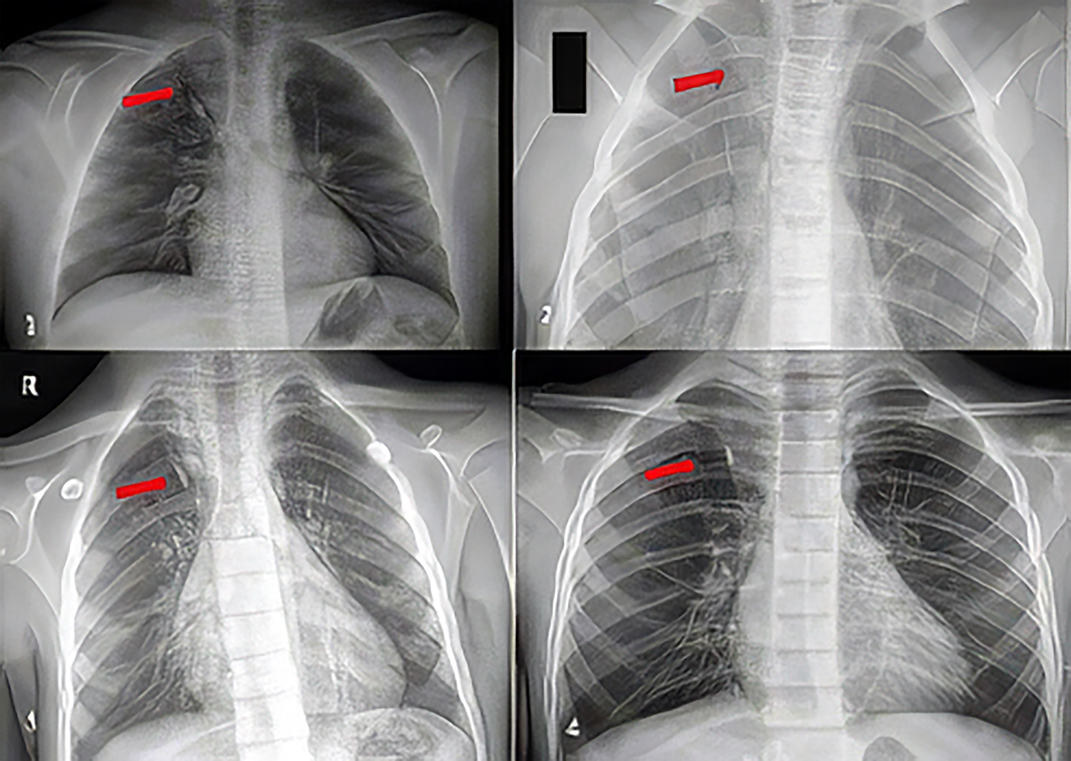
Figure 1. Chest X-rays of the patients (arrows indicate the azygos lobe)
Tables
Table 1. Demographic and clinical characteristics of the patients
Additional Information
Publisher’s Note
Bayrakol MP remains neutral with regard to jurisdictional and institutional claims.
Conference Presentation
This study was presented as a poster presentation at the 19th Pediatric Allergy, Immunology and Asthma Congress, Antalya, Türkiye, 2025
Rights and Permissions

This work is licensed under a Creative Commons Attribution-NonCommercial 4.0 International License (CC BY-NC 4.0). To view a copy of the license, visit https://creativecommons.org/licenses/by-nc/4.0/
About This Article
How to Cite This Article
Yaşar Kandur, Mesut Melih Özercan, Nesimi Günal, Ayşegül Alpcan, Dilek Azkur. Clinical characteristics. Ann Clin Anal Med 2026;17(Suppl 1):S74-76
- Received:
- June 19, 2025
- Accepted:
- July 21, 2025
- Published Online:
- September 19, 2025
- Printed:
- February 20, 2026