
Abstract
AimThis study evaluated the effects of the devastating Turkish earthquake in 2023 on the prevalence of keratoconus and corneal topographic parameters, with a focus on identifying potential associations between earthquake-induced environmental and psychological stressors and changes in keratoconus presentations.MethodsThis retrospective analysis was conducted at a single center by comparing clinical data and corneal topography measured before and after the earthquake. Parameters evaluated included K1, K2, Kmax, corneal thickness, and asymmetry indices (the keratoconus index (KI), index of height of decentration (IHD), central keratoconus index (CKI), and Kmax-K2). Statistical analyses were performed to assess alterations in the prevalence of keratoconus and topographic indices.ResultsAlthough the overall increase in the prevalence of keratoconus from 14.0% (n = 28) pre-earthquake to 21.0% (n = 42) post-earthquake was not statistically significant (P > .05), significant changes were observed in corneal asymmetry indices. Specifically, the KI increased at a value of P = .03, the IHD at P = .02, and the CKI at P = .01. The difference between Kmax and K2 was also significant (P = .04).ConclusionThe findings indicate a trend toward a higher prevalence of keratoconus coupled with significant alterations in corneal asymmetry measurements following the Turkish earthquake of 2023. These results support the hypothesis that earthquake-related environmental and psychological factors may affect the progression of keratoconus, underscoring the need for further multicenter, long-term studies to clarify these associations and underlying mechanisms.
Keywords
Introduction
Keratoconus is an ophthalmic disease characterized by progressive thinning, increased curvature, and conical deformation of the cornea, affecting visual acuity. Numerous mechanisms are involved in the pathogenesis, including genetic factors, biomechanical disorders, and oxidative stress. While genetic predisposition occupies an important place in the emergence of the disease, decreased microstructural stability and the biomechanical properties of the cornea also contribute to its progression.1,2 Studies have revealed that oxidative stress plays a determinant role in the pathogenesis of keratoconus. Higher than normal free radical production and an insufficient antioxidant defense system in corneal tissue cause the tissue to be exposed to oxidative damage. This can contribute to the progression of the disease by leading to microstructural deterioration in corneal cells and stroma.2,3 Additionally, oxidative stress triggering the release of proinflammatory cytokines can lead to increased local inflammation and further deterioration of the corneal structure.4
Acute and chronic stress conditions caused by major natural disasters such as earthquakes lead to increased systemic oxidative stress and proinflammatory responses. Researchers have suggested that, under such conditions, increased stress hormones (e.g., cortisol) and inflammatory mediators may accelerate the progression of chronic diseases such as keratoconus by weakening the microstructural integrity of corneal tissue.5 Additionally, the indirect effects of psychological stress include eye rubbing or traumatic behaviors resulting from increased discomfort. These in turn can have additional destructive effects on the corneal structure due to mechanical trauma.2,5 In the wake of major natural disasters such as earthquakes, routine eye examinations and corneal topography imaging that monitor the progression of keratoconus may be delayed due to intense pressure on patients and healthcare infrastructure. Failure to diagnose early-stage keratoconus or disruption of regular monitoring programs may lead to the disease only being detected at more advanced stages and to limitations on treatment options.
In the early stages of keratoconus, standard keratometry measurements, and especially corneal topography data, objectively reflect the micro-level asymmetry and irregularities of the disease. Corneal topography parameters are particularly important tools in determining the process of corneal conical deformation and thinning, as well as monitoring the progression of the disease.6 In particular, changes in topography observed during examinations in periods of trauma or high stress, such as after an earthquake, can be critically important in objectively evaluating whether the disease is progressing. This study compared the changes in corneal topography parameters of patients with suspected keratoconus before and after the February 6, 2023, Turkish earthquake. It also set out to reveal the role of psychological stress factors on the clinical course of the disease by evaluating the potential effects of the earthquake on keratoconus pathogenesis and progression.
Materials and Methods
This retrospective and comparative cohort study was conducted in Kahramanmaraş, one of the regions in Türkiye most severely affected by the 2023 earthquake. Patients who presented for ophthalmological examinations in the pre- (June–August 2022) and post-earthquake (June–August 2023) periods were evaluated in the scope of the research. One hundred patients (200 eyes) in the pre-earthquake group and one hundred patients in the post-earthquake group, all with uncorrectable changes in visual acuity, corneal surface irregularities, and increased astigmatism detected during clinical examinations, were included in the study. Individuals in whom keratoconus was suspected as a result of abnormalities detected in standard keratometric measurements (K1 and K2 values) and who therefore underwent advanced corneal topographic examinations constituted the study population. All topographic measurements in the pre- and post-earthquake periods were performed by the same technician using a Pentacam HR (Oculus, Wetzlar, Germany) device. All patients were evaluated by the same ophthalmology specialist (M.B.). Patients with a history of corneal surgery, corneal trauma, corneal infection, contact lens use, or systemic diseases such as connective tissue disorders were excluded from the study. Demographic data such as age and gender, and clinical data including visual acuity, refraction values, intraocular pressure, biomicroscopic examination findings, and fundus examination findings were recorded. Best-corrected visual acuity (BCVA) was quantified as a decimal value based on the ratio of the test distance to the standard distance for the letter read on the Snellen chart. Patients who underwent topography due to suspicion of keratoconus were diagnosed according to the Amsler-Krumeich diagnostic classification. These were divided into four stages using eccentric corneal steepening, degree of myopia/astigmatism, mean central K value, and corneal thickness parameters.
Various corneal topography parameters were measured. Anterior surface parameters included Q front, representing the anterior surface asphericity; K1 front, indicating the anterior surface flat keratometry; K2 front, denoting the anterior surface steep keratometry; and Kmax front, the anterior surface maximum keratometry. Additionally, the difference between the maximum and steep keratometry is expressed as Kmax-K2, while Astig front defines the anterior surface corneal astigmatism. The overall anterior curvature is further characterized by Km front, the mean keratometry of the anterior surface. For the posterior surface, the parameters included Q back for posterior surface asphericity; K1 back and K2 back, representing the posterior surface flat and steep keratometry, respectively; Kmax back as the posterior surface maximum keratometry; Astig back, indicating the posterior surface corneal astigmatism; and finally, Km back, which quantifies the mean posterior surface keratometry. Corneal thickness is evaluated through three parameters: Thinnest L, the thinnest corneal point; Pachy apex, the corneal apex thickness; and T-A, the difference in thickness between the thinnest point and the apex. Topographic indices further include the index of surface variance (ISV), index of vertical asymmetry (IVA), keratoconus index (KI), index of height asymmetry (IHA), index of height decentration (IHD), R min (the minimum corneal curvature radius), and the central keratoconus index (CKI).Ethical ApprovalThis study was approved by the Ethics Committee of Harran University (Date: 30.12.2024, Decision No: 24.21.30).Statistical AnalysisDescriptive statistics were expressed as mean, standard deviation, median, minimum, maximum, frequency, and ratio values. The distribution of variables was assessed using the Kolmogorov-Smirnov and Shapiro-Wilk tests. The Mann-Whitney U test was applied for the analysis of non-normally distributed quantitative independent data. The chi-square test was used for the analysis of qualitative independent data. SPSS version 28.0 was used for all analyses.Reporting GuidelinesThis study was reported according to the STROBE guidelines.
Results
Four hundred eyes of 200 patients (pre- and post-earthquake) were evaluated in this study. The patients’ mean age was 25.6 ± 11.5 years, BCVA was 0.87 ± 0.12, K1 front was 43.2 ± 2.4, K2 front was 45.4 ± 2.8, and Kmax front was 46.8 ± 3.7 diopters. In addition, 82.5% of the eyes were classified as subclinical keratoconus, 11.8% as stage I, 3.3% as stage II, and 2.5% as stage III. No stage IV cases were seen. The patients’ demographic data and all evaluated corneal topographic parameters are presented in Table 1. Table 2 provides an overview of demographic characteristics, topographic parameter values, and corresponding statistical comparisons before and after the earthquake.
Demographic and corneal topographic parameters were compared between pre- (100 patients, 200 eyes) and post-earthquake (100 patients, 200 eyes) groups. Mean ages were similar between the two groups (25.5 ± 11.9 vs. 25.7 ± 11.1 years, respectively, P = .582). Gender distribution exhibited a non-significant trend towards more females in the post-earthquake group (57.0% vs. 65.0%, P = .101). BCVA did not differ significantly between the groups (0.90 ± 0.14 vs. 0.84 ± 0.10, respectively, P = .240). In terms of keratoconus staging, the distributions of subclinical (86.0% vs. 79.0%), stage I (10.0% vs. 13.5%), stage II (3.0% vs. 3.5%), and stage III (1.0% vs. 4.0%) cases were not significantly different between groups (P = .150).
The pre-earthquake mean spherical autorefraction was –3.11 ± 1.32 diopters, compared to –3.25 ± 1.30 diopters post-earthquake. A parametric test revealed no significant difference (P = .450). Mean cylindrical values were –2.29 ± 1.09 diopters pre-earthquake and –2.35 ± 1.09 diopters post-earthquake. The difference was also not statistically significant (P = .740).
In terms of the corneal topographic parameters, several indices exhibited statistically significant differences between the pre- and post-earthquake groups. KI was significantly higher in the post-earthquake group (1.05 ± 0.07 vs. 1.03 ± 0.09, P = .002). Similarly, significant IHD elevation was observed in the post-earthquake group (0.02 ± 0.03 vs. 0.03 ± 0.05, P = .001). CKI also increased significantly post-earthquake (1.01 ± 0.02 vs. 1.02 ± 0.02, P = .001). Other parameters including K1 front (42.9 ± 2.1 vs. 43.4 ± 2.7, P = .098), K2 front (45.3 ± 2.4 vs. 45.6 ± 3.1, P = .526), Kmax front (46.6 ± 3.4 vs. 46.9 ± 4.1, P = .426), and corneal thickness at the thinnest location (526.7 ± 47.4 vs. 518.9 ± 60.3, P = .292) exhibited no statistically significant differences between the two groups. The difference between maximum keratometry and steep keratometry (Kmax-K2) exhibited a borderline significant increase in the post-earthquake group (1.26 ± 1.66 vs. 1.38 ± 1.54, P = .050), as also did IVA (0.21 ± 0.22 vs. 0.26 ± 0.30, P = .050).
Discussion
This study evaluated the prevalence of keratoconus and corneal topographic changes following the 2023 Turkish earthquake. It also highlights the lack of research directly investigating the effects of natural disasters such as earthquakes on the cornea. From that perspective, the study findings provide significant clues regarding the potential impact of the earthquake on the development and progression of keratoconus. Although the overall pre-earthquake clinical prevalence of keratoconus was 14.0% (n = 28), this rose to 21.0% (n = 42) after it. While there was no statistically significant difference in the frequency of keratoconus, a partial increase was observed, indicating a tendency for the prevalence to rise in the post-earthquake period. Increases were observed in each stage of keratoconus, a significant increase being observed in the advanced stage. Moreover, in the post-earthquake period, parameters such as KI, IHD, and CKI also increased significantly. This suggests that stress and corneal trauma mechanisms emerging following the earthquake may play a role in the pathogenesis and progression of keratoconus.
No significant changes were observed in the majority of topographic parameters, such as K1, K2, Kmax, and corneal thickness. However, increases were determined in indices reflecting central and surface asymmetry (KI, IHD, CKI, and Kmax-K2). This finding suggests that the disease progression in the post-earthquake period is manifested more in the form of microstructural and asymmetric changes, highlighting the importance of early diagnosis and intervention strategies.
Natural disasters, and especially earthquakes, can cause long-term psychophysiological effects in addition to acute physical trauma. Such events can lead to increased levels of systemic inflammatory mediators and oxidative stress markers, resulting in changes in corneal tissue, with its known sensitivity to redox homeostasis and inflammatory processes.7 Dysregulation of inflammatory cascades may play an important role in the progression of corneal ectasias such as keratoconus and the initiation of corneal degenerative processes. Keratoconus is reported to be associated not only with genetic factors, but also with environmental (perceptible levels of air pollution, fine particles, and dusty environments) and mechanical (such as eye rubbing) factors. Environmental factors emerging after earthquakes can also have adverse effects on eye health. Hsu et al. reported significant increases in SO₂ and NO concentrations compared to background levels following earthquakes in Taiwan.8 This increase was associated with underground gas emissions caused by the earthquake, along with traditional sources of air pollution, indicating a sudden, potentially hazardous deterioration in air quality.
Dust and particles from collapsed buildings cause significant decreases in air quality. Zanoletti et al. investigated environmental pollution resulting from collapsed buildings after the 2023 earthquake in Türkiye. That research emphasized that building collapses and demolition caused significant increases in PM2.5 levels in particular. Those authors also concluded that hazardous materials such as asbestos, lead, and silica may enter the air, potentially causing severe health problems including respiratory issues, neurotoxicity, immunotoxicity, skin and eye irritation, and liver and kidney damage.9 Lu et al. noted that particles, especially PM2.5, reduced the viability and proliferation of conjunctival epithelial cells while increasing apoptosis and IL-6 expression, significantly contributing to the development and exacerbation of allergic conjunctivitis.10 This can in turn trigger increased eye rubbing behavior, leading to heightened mechanical trauma to the cornea, and thus laying the foundation for the progression of corneal disorders such as keratoconus. Previous studies have concluded that eye rubbing is an important risk factor in the development and progression of keratoconus.11
Jurkiewicz et al. demonstrated a significant positive correlation between the prevalence of keratoconus and levels of fine particulate matter (PM2.5) and nitrogen dioxide (NO₂) pollution. Those authors also suggested that fine particles may have a direct effect on corneal structures, potentially increasing apoptosis and leading to the progression of keratoconus. Significant positive correlations were also observed between the prevalence of keratoconus and air pollution levels in different countries. Keratoconus may therefore be associated not only with genetic factors, but also with environmental factors as part of this multifactorial etiology.12,13 The prevalence of keratoconus has been reported to be directly proportional to the amount of particulate matter. It is thought that particulate matter, in addition to causing atopy and eye itching, may also directly affect the corneal epithelium and stroma.14,15
Limitations
There are a number of limitations to this study that need to be considered. In particular, its retrospective design and single-center character may restrict the generalizability of the findings. Additionally, psychological stress and other environmental factors that may emerge in the post-earthquake period could not be assessed objectively, and the results therefore require cautious interpretation from that perspective. Further multicenter and long-term follow-up studies, along with analyses of psychological and environmental factors, will contribute to a more comprehensive understanding of the effects of natural disasters such as earthquakes on the progression of keratoconus. Because the study was retrospective, we couldn’t prove the extent of psychological stress experienced by individuals and the extent to which they were affected by physiological conditions. However, earthquakes have been shown to be a significant source of stress.7 Perhaps demonstrating increased inflammatory cytokines and stress hormones in the affected population would have allowed us to establish a more meaningful relationship. However, our most significant limitation was not being able to conduct the study with the same patient groups. This was because our clinic is not a top-tier clinic that treats keratoconus. Keratoconus is diagnosed by specialist ophthalmologists at our clinic, and patients are referred to a top-tier center for treatment.
Conclusion
This study evaluated the effects of the 2023 Turkish earthquake on the prevalence of keratoconus and corneal topographic parameters. Although not statistically significant, the findings demonstrated an increase in keratoconus rates in the post-earthquake period, together with significant changes in indices reflecting central and surface asymmetry. These findings support the potential role of earthquake-related environmental and psychological factors in the progression of keratoconus and will serve as a useful basis for future large-scale studies.
Declarations
Animal and Human Rights Statement
All procedures performed in this study were in accordance with the ethical standards of the institutional and/or national research committee and with the 1964 Helsinki Declaration and its later amendments or comparable ethical standards.
Informed Consent
Informed consent was waived due to the retrospective design of the study and the use of anonymized patient data.
Data Availability
The datasets used and/or analyzed during the current study are not publicly available due to patient privacy reasons but are available from the corresponding author on reasonable request.
Conflict of Interest
The authors declare that there is no conflict of interest.
Funding
None.
Abbreviations
BCVA: Best-corrected visual acuity
CKI: Central keratoconus index
IHA: Index of height asymmetry
IHD: Index of height decentration
ISV: Index of surface variance
IVA: Index of vertical asymmetry
KI: Keratoconus index
SD: Standard deviation
References
- Davidson AE, Hayes S, Hardcastle AJ. The pathogenesis of keratoconus. Eye (Lond). 2014;28(2):189-195. doi:10.1038/eye.2013.278
- Bykhovskaya Y, Rabinowitz YS. Update on the genetics of keratoconus. Exp Eye Res. 2021;202:108398. doi:10.1016/j.exer.2020.108398
- Wojcik KA, Kaminska A, Blasiak J. Oxidative stress in the pathogenesis of keratoconus and Fuchs endothelial corneal dystrophy. Int J Mol Sci. 2013;14(9):19294-19308. doi:10.3390/ijms140919294
- de Azevedo Magalhães O, Gonçalves MC, Gatinel D. The role of environment in the pathogenesis of keratoconus. Curr Opin Ophthalmol. 2021;32(4):379-384. doi:10.1097/icu.0000000000000764
- Roberts CJ, Knoll KM, Mahmoud AM. Corneal stress distribution evolves from thickness-driven in normal corneas to curvature-driven with progression in keratoconus. Ophthalmol Sci. 2024;4(2):100373. doi:10.1016/j.xops.2023.100373
- Padmanabhan P, Lopes BT, Eliasy A. In vivo biomechanical changes associated with keratoconus progression. Curr Eye Res. 2022;47(7):982-986. doi:10.1080/02713683.2022.2058020
- Navel V, Malecaze J, Pereira B, et al. Oxidative and antioxidative stress markers in keratoconus: a systematic review and meta-analysis. Acta Ophthalmol. 2021;99(6). doi:10.1111/aos.14714
- Hsu SC, Huang YT, Huang JC, et al. Evaluating real-time air-quality data as earthquake indicator. Sci Total Environ. 2010;408(11):2299-2304. doi:10.1016/j.scitotenv.2010.02.023
- Zanoletti A, Bontempi E. The impacts of earthquakes on air pollution and strategies for mitigation: a case study of Turkey. Environ Sci Pollut Res Int. 2024;31(16):24662-24672. doi:10.1007/s11356-024-32592-8
- Lu CW, Fu J, Liu XF, et al. Air pollution and meteorological conditions significantly contribute to the worsening of allergic conjunctivitis: a regional 20-city, 5-year study in Northeast China. Light Sci Appl. 2021;10(1):190. doi:10.1038/s41377-021-00630-6
- Sahebjada S, Al-Mahrouqi HH, Moshegov S, et al. Eye rubbing in the aetiology of keratoconus: a systematic review and meta-analysis. Graefes Arch Clin Exp Ophthalmol. 2021;259(8):2057-2067. doi:10.1007/s00417-021-05081-8
- Crawford AZ, Zhang J, Gokul A. The enigma of environmental factors in keratoconus. Asia Pac J Ophthalmol (Phila). 2020;9(6):549-556. doi:10.1097/apo.0000000000000334
- Jurkiewicz T, Marty AS. Air pollution and the prevalence of keratoconus: is there a connection? Ophthalmic Epidemiol. 2025;32(4):394-402. doi:10.1080/09286586.2024.2399765
- Jurkiewicz T, Marty AS. Correlation between keratoconus and pollution. Ophthalmic Epidemiol. 2021;28(6):495-501. doi:10.1080/09286586.2021.1879173
- Das AV, Basu S. Environmental and air pollution factors affecting allergic eye disease in children and adolescents in India. Int J Environ Res Public Health. 2021;18(11):5611. doi:10.3390/ijerph18115611
Tables
Table 1. Distributions of demographic characteristics and mean topographic parameter values in the total patient population pre- and post-earthquake
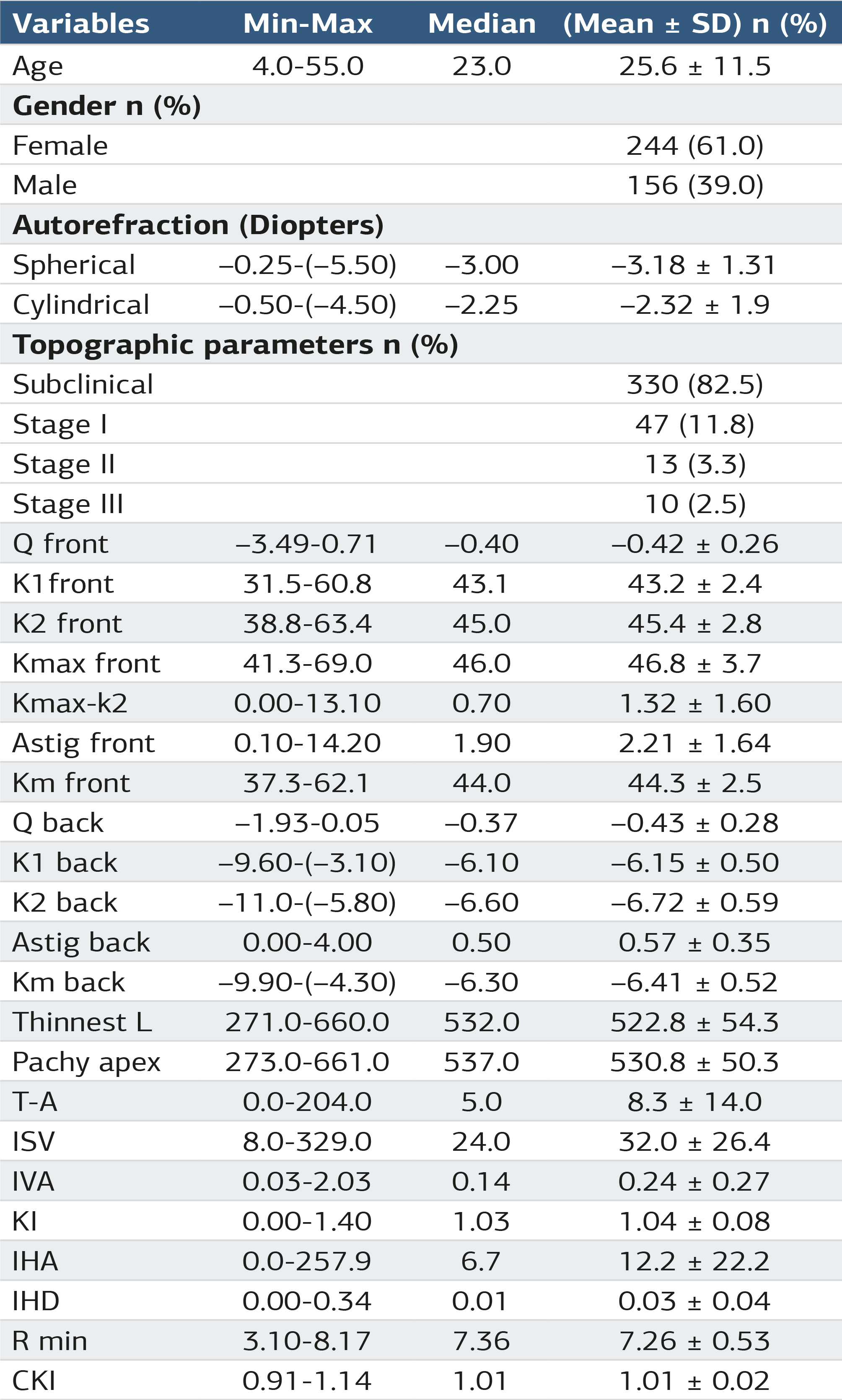
Q front: Asphericity front, K1front: Flat keratometry front , K2front: Steep keratometry front, Kmax front: Maximum keratometry front, Kmax-K2: Difference, Astig front: Corneal astigmatism front, Km front: Mean keratometry front, K1back: Flat keratometry back, K2back: Steep keratometry back, Km back: Mean keratometry back, Astig back: Corneal astigmatism back, Qback: Asphericity back ,Thinnest L: Thinnest location, Pachy apex: Corneal Apex Thickness, T-A: Thickness difference, ISV: Index of Surface Variance, IVA: Index of Vertical Asymmetry, KI: Keratoconus Index, IHA: Index of Height Asymmetry, IHD: Index of Height Decentration, Rmin: Minimum Corneal Curvature Radius, CKI: Central Keratoconus Index.
Table 2. Demographic characteristics, topographic parameter values, and statistical comparisons
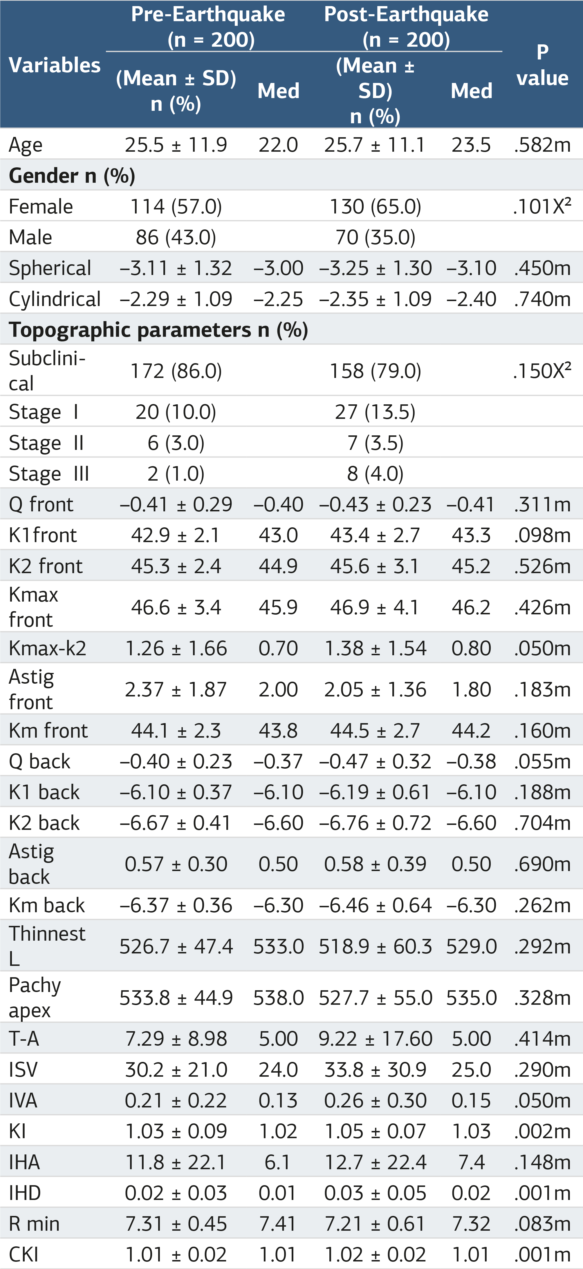
m: Mann-Whitney U test; χ²: chi-square test Q front: Asphericity front, K1 front: Flat keratometry front, K2 front: Steep keratometry front, Kmax front: Maximum keratometry front, Kmax-K2: Difference, Astig front: Corneal astigmatism front, Km front: Mean keratometry front, K 1back: Flat keratometry back, K2 back: Steep keratometry back, Km back: Mean keratometry back, Astig back: Corneal astigmatism back, Q back: Asphericity back, Thinnest L: Thinnest Location, Pachy apex: Corneal apex thickness, T-A: Thickness difference, ISV: Index of Surface Variance, IVA: Index of Vertical Asymmetry, KI: Keratoconus Index, IHA: Index of Height Asymmetry, IHD: Index of Height Decentration, Rmin: Minimum Corneal Curvature Radius, CKI: Central Keratoconus Index.
Additional Information
Publisher’s Note
Bayrakol MP remains neutral with regard to jurisdictional and institutional claims.
Rights and Permissions

This work is licensed under a Creative Commons Attribution-NonCommercial 4.0 International License (CC BY-NC 4.0). To view a copy of the license, visit https://creativecommons.org/licenses/by-nc/4.0/
About This Article
How to Cite This Article
Mübeccel Bulut, Ali Hakim Reyhan. An evaluation of the prevalence of keratoconus and corneal topographic alterations following the 2023 Türkiye earthquake. Ann Clin Anal Med 2025;16(9):658-662. doi:10.4328/ACAM.22822
Publication History
- Received:
- 21.07.2025
- Accepted:
- 25.08.2025
- Published Online:
- 31.08.2025
- Printed:
- 01.09.2025