
Prevalence of wrist ganglion cysts on magnetic resonance imaging in patients with wrist pain in the Turkish population
Wrist MRI ganglion prevalence
Authors
Abstract
Aim To investigate the prevalence of ganglion cysts on wrist magnetic resonance imaging (MRI) in the Turkish population and to evaluate their associations with age, sex, and laterality.
Methods A retrospective review was conducted of 2,099 patients who underwent wrist MRI at our institution between 2023 and 2024 due to wrist pain. Data on age, sex, side of involvement, and the presence of ganglion cysts based on MRI reports were collected. The relationship between ganglion cysts and sex or laterality was analyzed using the chi-square test, while age differences were assessed with the t-test. In addition, a multivariate logistic regression analysis was performed to evaluate the independent associations of age, sex, and side with the presence of ganglion cysts. A p-value < 0.05 was considered statistically significant.
Results Ganglion cysts were detected in 35.6% of the cases. The prevalence of cysts was significantly higher in females (37.7%) compared to males (31.5%), and in the left wrist (39.2%) compared to the right (32.6%) (p < 0.05 for both). In univariate analysis, age did not significantly differ between patients with and without cysts (p > 0.05). However, in multivariate logistic regression, younger age was found to be an independent predictor of ganglion cyst presence
(β = −0.0065, p = 0.040).
Conclusion Ganglion cysts are more frequently observed in female patients, on the left side, and in younger age groups in the Turkish population. These demographic patterns may aid clinicians in optimizing diagnostic strategies and imaging decisions.
Keywords
Introduction
Ganglion cysts are benign, fluid-filled synovial structures commonly found around the wrist.1 Patients may present with pain, limited range of motion, or cosmetic concerns.2 They are the most frequently encountered soft tissue masses in the wrist, typically located dorsally.3,4 The stalk of wrist ganglion cysts most frequently originates from the scapholunate ligament but may also arise from other capsular areas.5 Numerous studies have reported a higher prevalence in women.6,7 Although their exact etiology is unclear, some cysts appear to develop following trauma-related synovial fluid leakage and mucinous degeneration.8 Diagnosis is often made clinically through history and physical examination, but advanced imaging techniques such as ultrasonography and MRI can confirm the diagnosis.9 Treatment options include conservative methods such as aspiration and steroid injection; however, persistent or recurrent cases may require arthroscopic or open surgical excision.10,11
Although several studies have investigated the prevalence and demographic distribution of ganglion cysts, large-scale MRI-based data remain limited. This study aims to determine the prevalence of ganglion cysts in a large Turkish patient population undergoing wrist MRI and to assess their associations with age, sex, and laterality.
Materials and Methods
Patient SelectionThis retrospective study included patients who underwent wrist MRI between 2023 and 2024 due to various complaints. A total of 2,099 patients were included (Supplementary Figure S1). All examinations were performed using a 1.5-Tesla MRI scanner (Siemens Magnetom Avanto, Erlangen, Germany) with a dedicated wrist coil. Patients were positioned prone with the wrist in a neutral position. The standard institutional wrist MRI protocol included the following sequences: Axial, coronal, and sagittal T1-weighted spin-echo, Axial and coronal T2-weighted fat-suppressed (FS) or STIR, Sagittal and coronal proton-density (PD) FS. Ganglion cysts were identified by reviewing MRI reports. Ganglion cysts were defined as well-circumscribed, oval or round cystic lesions with low signal intensity on T1-weighted images and high signal intensity on T2-weighted images, homogenous internal content, and a clear connection to the adjacent joint capsule or tendon sheath. Cases with missing data or trauma-related MRIs were excluded. Variables analyzed included age, sex, side of involvement, and presence of ganglion cysts.
Ethical ApprovalThis study was approved by the Ethics Committee of Health Sciences University (Date: 2025-07-10, No: 2025/08-221).
Statistical AnalysisStatistical analysis was conducted using Python 3.10. Chi-square tests were used to evaluate categorical variables, while t-tests were used for continuous variables. In addition, multivariable logistic regression analysis was performed to identify independent predictors while adjusting for potential confounders. A p-value of < 0.05 was considered statistically significant.
Reporting GuidelinesThis study is reported in accordance with the STROBE guidelines.
Results
Of the 2,099 patients, 66.1% (n = 1,387) were female, and 33.9% (n = 711) were male. The mean age was 33.7 ± 14.6 years. Ganglion cysts were present in 747 patients (35.6%). Stratified by sex, the prevalence was 37.7% (523 / 1,387) in females and 31.5% (224 / 711) in males, with a statistically significant difference (p < 0.05). Regarding laterality, ganglion cysts were observed in 39.2% (379 / 968) of left wrists and 32.6% (368 / 1,130) of right wrists (p < 0.05). No significant difference in age was found between patients with and without ganglion cysts in univariate analysis (p > 0.05) (Table 1) (Supplementary Figure S2). However, multivariate logistic regression analysis demonstrated that younger age (β = −0.0065, p = 0.040), female sex (β = 0.289, p = 0.003), and left wrist involvement (β = −0.297, p = 0.001) were independently associated with the presence of ganglion cysts (Table 2).
Discussion
This study found a 35.6% prevalence of ganglion cysts on wrist MRI. Female patients were significantly more likely to have ganglion cysts, and cysts were more frequently located in the left wrist. Although age was not significantly associated with ganglion cyst presence in the univariate analysis, multivariate logistic regression revealed that younger age independently increased the likelihood of having a ganglion cyst. This suggests that the effect of age becomes apparent when other contributing factors are controlled. Ganglion cysts may therefore be more prevalent in younger individuals, potentially due to differences in tissue composition, joint activity, or degenerative changes that evolve with age. Previous studies have reported mixed results regarding the relationship between age and ganglion cysts, and further research is warranted to clarify the underlying pathophysiology.2,6,7
A study on asymptomatic chiropractic students demonstrated a high prevalence of wrist ganglion cysts even in the absence of symptoms. Bilateral wrist ultrasonography revealed cysts in approximately 50% of participants, with most being dorsally located and more common on the dominant side.12 These findings suggest that ganglion cysts may be widespread even in asymptomatic individuals and could influence diagnostic and therapeutic decisions.
The higher prevalence of wrist ganglion cysts in women has been consistently reported. Large-scale population-based studies have identified female sex as an independent risk factor, with a two- to three-fold higher likelihood compared to men.12,13,14 Although the exact mechanisms remain unclear, factors such as greater ligamentous laxity in women, potentially reducing joint stability and facilitating synovial fluid leakage, may play a role.7 Hormonal influences, particularly the effects of estrogen on connective tissue metabolism, and increased ligamentous elasticity during pregnancy or lactation, may also contribute, though these hypotheses have yet to be confirmed in clinical studies. Understanding the biological and mechanical factors underlying this sex-based difference is important for developing targeted preventive and therapeutic strategies.
A recent high-field MRI study involving 295 patients with symptomatic wrist ganglion cysts reported 49.3% of lesions on the left side. However, no statistically significant difference between sides was found, suggesting that random variation or factors such as hand dominance might influence laterality. Further research is needed to explore these individual-level variables.15
This study provides comprehensive MRI-based prevalence data on wrist ganglion cysts in the Turkish population.
Limitations
This study has several limitations. First, due to its retrospective design and reliance on archived MRI data, information regarding occupation, repetitive hand use, and hand dominance was not available, which limits the interpretation of laterality findings. Second, the study was based solely on imaging records, and therefore, symptomatic and asymptomatic ganglion cysts could not be distinguished. These factors may have clinical and functional implications and should be addressed in future prospective studies.
Conclusion
Ganglion cysts are common findings on wrist MRI and are significantly more prevalent in younger patients female patients and in the left wrist in the Turkish population. These demographic and anatomical patterns should be considered during diagnostic evaluation and follow-up planning. Further studies are warranted to elucidate the underlying causes and clinical implications of these differences.
Declarations
Ethics Declarations
Ethical approval for this study was obtained from the Health Sciences University Ethics Committee (Approval date: July 10, 2025; Decision No.: 2025/08-221).
Animal and Human Rights Statement
All procedures performed in this study were in accordance with the ethical standards of the institutional and/or national research committee and with the 1964 Helsinki Declaration and its later amendments or comparable ethical standards.
Informed Consent
Informed consent was waived by the ethics committee due to the retrospective design and the use of anonymized data.
Data Availability
The datasets used and/or analyzed during the current study are not publicly available due to patient privacy reasons but are available from the corresponding author on reasonable request.
Conflict of Interest
The authors declare that there is no conflict of interest.
Funding
None.
Author Contributions (CRediT Taxonomy)
Conceptualization: S.K., B.P.
Methodology: S.K., B.P.
Investigation: S.K., B.P.
Data curation: S.K., B.P.
Formal analysis: S.K.
Software: S.K.
Writing – original draft: S.K.
Writing – review & editing: S.K., B.P.
Supervision: S.K.
Scientific Responsibility Statement
The authors declare that they are responsible for the article’s scientific content, including study design, data collection, analysis and interpretation, writing, and some of the main line, or all of the preparation and scientific review of the contents, and approval of the final version of the article.
Abbreviations
β: Beta coefficient
CI: Confidence interval
FS: Fat-suppressed
MRI: Magnetic Resonance Imaging
N: Number of patients
PD: Proton density
SD: Standard deviation
Std: Standard
STIR: Short tau inversion recovery
T: Tesla
T1: T1-weighted
T2: T2-weighted
References
-
Gude W, Morelli V. Ganglion cysts of the wrist: pathophysiology, clinical picture, and management. Curr Rev Musculoskelet Med. 2008;1(3-4):205-211. doi:10.1007/s12178-008-9033-4
-
Cavit A, Civan O, Özcanlı H. Single-center, 14-year experience of ganglions of the hand and wrist. Sakarya Med J. Published online February 22, 2022. doi:10.31832/smj.1037576
-
Meena S, Gupta A. Dorsal wrist ganglion: current review of literature. J Clin Orthop Trauma. 2014;5(2):59-64. doi:10.1016/j.jcot.2014.01.006
-
Teefey SA, Dahiya N, Middleton WD, Gelberman RH, Boyer MI. Ganglia of the hand and wrist: a sonographic analysis. AJR Am J Roentgenol. 2008;191(3):716-720. doi:10.2214/AJR.07.3438
-
Angelides AC, Wallace PF. The dorsal ganglion of the wrist: its pathogenesis, gross and microscopic anatomy, and surgical treatment. J Hand Surg Am. 1976;1(3):228-235. doi:10.1016/S0363-5023(76)80042-1
-
Kuliński S, Gutkowska O, Mizia S, Gosk J. Ganglions of the hand and wrist: retrospective statistical analysis of 520 cases. Adv Clin Exp Med. 2017;26(1):95-100. doi:10.17219/acem/65070
-
Dworak TC, Balazs GC, Tropf J, Nanos GP, Tintle SM. Epidemiology of symptomatic dorsal wrist ganglia in active duty military and civilian populations. J Hand Surg Glob Online. 2020;2(6):349-353. doi:10.1016/j.jhsg.2020.08.001
-
Zhang A, Falkowski AL, Jacobson JA, Kim SM, Koh SH, Gaetke-Udager K. Sonography of Wrist Ganglion Cysts: Which Location Is Most Common? J Ultrasound Med. 2019;38(8):2155-2160. doi:10.1002/jum.14912
-
Neto N, Nunnes P. Spectrum of MRI features of ganglion and synovial cysts. Insights Imaging. 2016;7(2):179-86. doi:10.1007/s13244-016-0463-z
-
Head L, Gencarelli JR, Allen M, Boyd KU. Wrist ganglion treatment: systematic review and meta-analysis. J Hand Surg Am. 2015;40(3):546-553.e8. doi:10.1016/j.jhsa.2014.12.014
-
Shah AA, Raina AH, Ganie MA, Kumar IA. Comparison of aspiration followed by intra-lesional steroid injection and surgical excision in management of dorsal wrist ganglion. World J Plast Surg. 2019;8(2):181-184.
-
Haun D, Bradshaw K, St Aubin K, Alford C, Michener E, Perkins A. The prevalence of wrist ganglia in a chiropractic student population. Ultrasound Med Biol. 2015;41(4):S158-S159. doi:10.1016/j.ultrasmedbio.2014.12.610
-
Balazs GC, Dworak TC, Tropf J, Nanos GP, Tintle SM. Incidence and risk factors for volar wrist ganglia in the U.S. military and civilian populations. J Hand Surg Am. 2016;41(11):1064-1070. doi:10.1016/j.jhsa.2016.08.008
-
Coffey MJ, Fazlur Rahman M, Thirkannad SM. Pediatric ganglion cysts of the hand and wrist: an epidemiologic analysis. Hand. 2008;3(4):359-362. doi:10.1007/s11552-008-9122-2
-
Ferreira Branco D, Botti P, Bouvet C, et al. Dorsal wrist ganglion: clinical and imaging correlation in symptomatic population based on high-field MRI. Eur Radiol. 2024;34(12):7856. doi:10.1007/s00330-024-10831-3
Tables
Table 1. Distribution of ganglion cysts by gender, laterality, and age
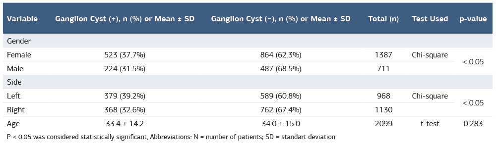
P < 0.05 was considered statistically significant, Abbreviations: N = number of patients; SD = standart deviation
Table 2. Logistic regression analysis for predictors of ganglion cyst presence
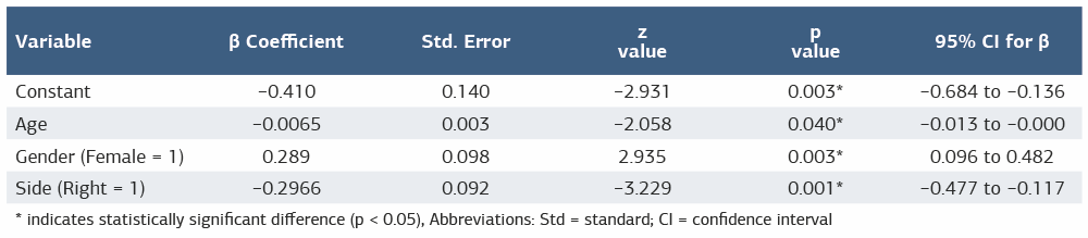
* indicates statistically significant difference (p < 0.05), Abbreviations: Std = standard; CI = confidence interval
Additional Information
Publisher’s Note
Bayrakol MP remains neutral with regard to jurisdictional and institutional claims.
Rights and Permissions

This work is licensed under a Creative Commons Attribution-NonCommercial 4.0 International License (CC BY-NC 4.0). To view a copy of the license, visit https://creativecommons.org/licenses/by-nc/4.0/
About This Article
How to Cite This Article
Salih Kaya, Basri Pür. Prevalence of wrist ganglion cysts on magnetic resonance imaging in patients with wrist pain in the Turkish population. Ann Clin Anal Med 2026;17(3):223-226
- Received:
- December 3, 2025
- Accepted:
- January 12, 2026
- Published Online:
- January 22, 2026
- Printed:
- March 1, 2026