
Abstract
AimDysplastic colorectal polyps, especially those with high-grade dysplasia, have a significant risk of malignant transformation. Immature granulocytes (IGs), markers of systemic inflammation, have shown prognostic value in various inflammatory and malignant diseases, but their role in colorectal polyp dysplasia is unclear. This study investigated the association between IG count and percentage and the histopathological features of colorectal polyps, and evaluated the predictive value of IG parameters for dysplasia.MethodsThis retrospective study included patients aged ≥18 years who underwent colonoscopy and were diagnosed with colorectal polyps. We analyzed demographic data, laboratory parameters, and colonoscopic and histopathological findings. We classified polyps by dysplasia grade, neoplastic status, and morphology (sessile or pedunculated). The immature granulocyte count and percentage were compared between groups using appropriate statistical tests.ResultsWe included 144 patients: 123 (85.4%) with low-grade dysplasia and 21 (14.6%) with high-grade dysplasia. Immature granulocyte count and percentage did not differ significantly between the low- and high-grade dysplasia groups. Similarly, IG parameters did not differ significantly according to neoplastic status or polyp morphology (sessile vs. pedunculated). We found no association between IG values and the histopathological characteristics of colorectal polyps.ConclusionThe immature granulocyte count and percentage do not appear to have predictive value in determining dysplasia grade, neoplastic status, or morphological features of colorectal polyps. These findings suggest that IG parameters may not be useful biomarkers for predicting dysplasia in colorectal polyps.
Keywords
Introduction
Dysplastic colon polyps, especially those with high-grade dysplasia, pose significant concerns regarding morbidity and mortality owing to their potential to harbor invasive malignancies or progress. The detection and management of these polyps have advanced with sophisticated screening programs aimed at limiting procedural morbidity while addressing the risk of occult lymph node metastasis.1 Various endoscopic techniques, such as mucosal resection and submucosal dissection, have been developed to manage low-risk lesions and reduce surgical morbidity. The incidence of colon polyps increases with age and is more common in Western countries. In the United States, the prevalence is 30% in people over 50 years of age and 6% in children.2,3 However, high-grade dysplasia and polyp location distant from splenic torsion are significant predictors of cancer risk. In such cases, with a 43% incidence of occult cancer, these polyps may not be suitable for less-invasive procedures during the learning curve of laparoscopic surgery.4 The management of serious dysplastic non-pedunculated lesions usually requires surgical intervention, as endoscopic sampling can be misleading, and these lesions must be treated with the same rigor as invasive cancers.5 In this study, we aimed to investigate the systemic effects of the inflammatory nature of dysplastic polyps and their relationship with immature granulocytes (IG), which are known to be early markers of inflammation in peripheral blood.
Materials and Methods
This study included patients who underwent colonoscopy at our center and were diagnosed with polyps. We conducted a comprehensive review of patients' medical records to obtain demographic information, medical history, laboratory results, and colonoscopy findings. Specifically, immature granulocyte percentage (IG%) levels and polyp findings were determined.Polyp EvaluationWe evaluated polyps based on several parameters, including size, number, histological classification, and the presence of dysplasia. The presence and degree of dysplasia were assessed by a pathologist.
We included patients over 18 years who underwent colonoscopic evaluation at our center, while we excluded patients in whom no polyps were detected during colonoscopy and whose pathological status could not be monitored.Ethical ApprovalThis study was approved by the Ethics Committee of Akdeniz University Faculty of Medicine Clinical Research (Date: 07.06.2023, Decision No: KAEK-459).Statistical AnalysisThe study results were evaluated using the Statistical Package for the Social Sciences (SPSS) for Windows, version 27.0. GraphPad Prism 9 software (Boston, MA) was used to graph the data collected in the study. Subsequently, the data were classified. Categorical data were defined as percentages and frequencies. Numerical data were defined, and a distribution analysis was performed. Data that followed a normal distribution were presented as mean ± standard deviation (SD). When data did not conform to a normal distribution, we reported the median and interquartile range (IQR). We used the chi-square test to assess relationships in categorical data. We used the t-test for normally distributed data and the Mann-Whitney U test for non-normally distributed data. P < .05 was considered statistically significant.
Reporting GuidelinesThis study was reported in accordance with the STROBE guidelines.
Results
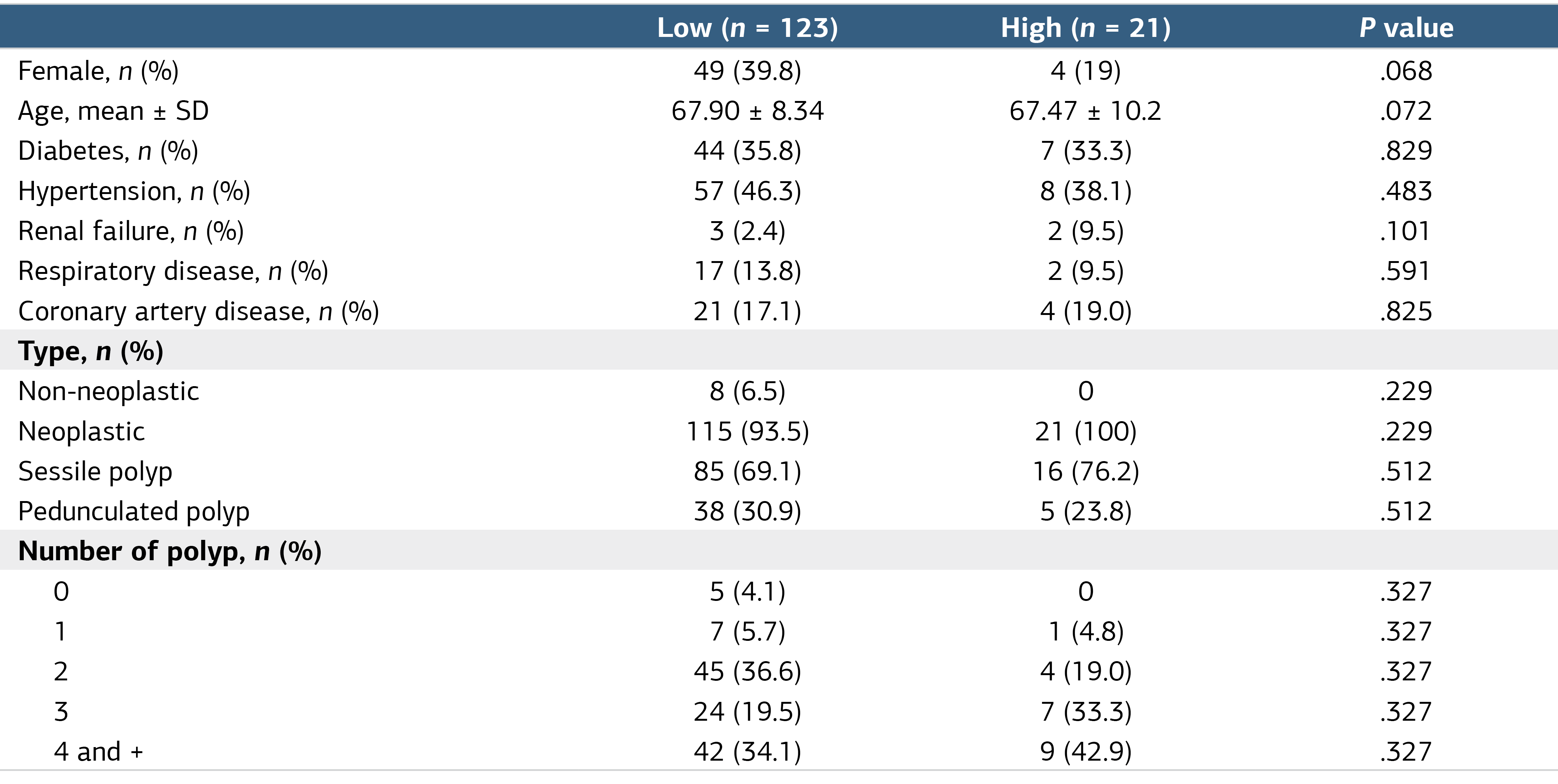
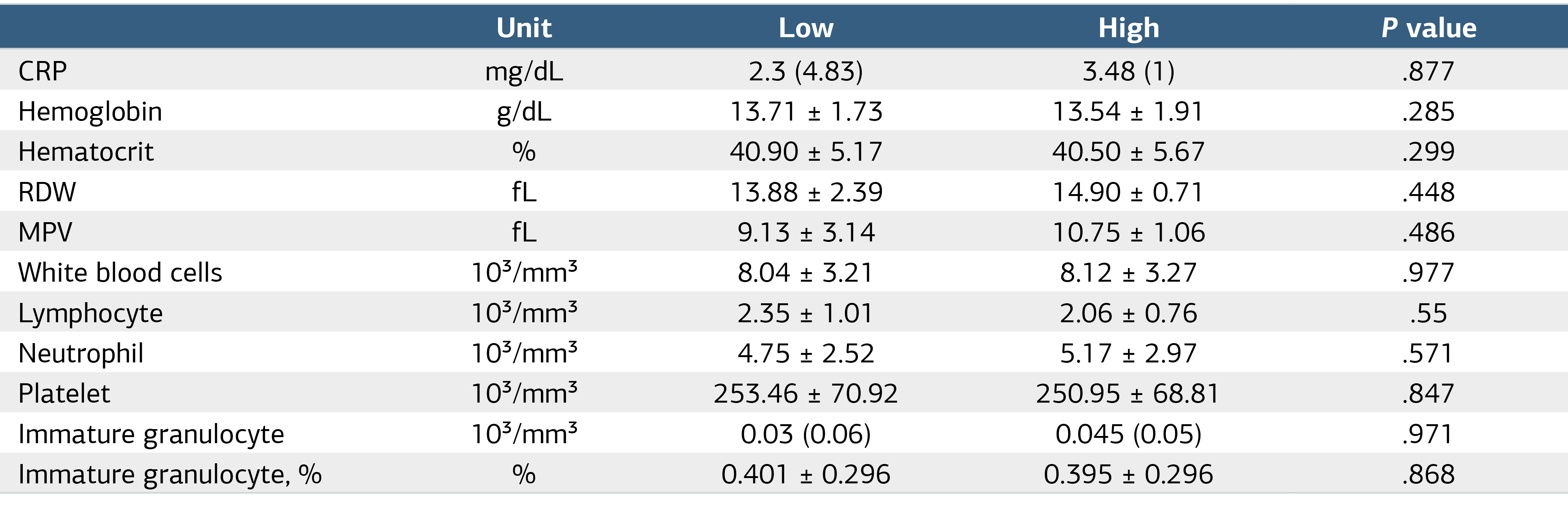
The study sample population comprised 144 patients. Of the patients in this study, 123 (85.4%) exhibited low-grade dysplasia and 21 (14.6%) demonstrated high-grade dysplasia. Table 1 presents the descriptive statistics. The mean hemogram value was 13.71 ± 1.73 in cases of low dysplasia and 13.54 ± 1.91 in cases of high dysplasia, with no statistically significant difference observed between the two groups (P = .285). Table 2 compares the biochemical values.
By neoplastic status, the median immature value was 0.02 (0.02-0.05) in non-neoplastic patients and 0.02 (0.01-0.04) in neoplastic patients. No significant difference was observed between the two groups (P = .925, Figure 1a). For the IG percentage, the median was 0.3 (0.23-0.58) for non-neoplastic patients and 0.3 (0.2-0.5) for neoplastic patients, with no significant difference between the groups (P = .110, Figure 1b).
The median IG count for sessile polyps was 0.02 (0.02–0.04), while the median IG count for pedunculated polyps was 0.02 (0.01–0.06), and no significant difference was observed between them (P = .406, Figure 1c). The median IG percentage for sessile polyps was 0.3 (0.2–0.4), and for pedunculated polyps it was 0.3 (0.2–0.5); no significant difference was observed (P = .441, Figure 1d).
Discussion
Predictive factors for dysplasia in colon polyps include patient demographics, polyp characteristics, and histological features, and are multifactorial. Age is a consistent predictor across studies; older patients have a higher prevalence of dysplasia in both pseudopolypoid lesions associated with inflammatory bowel disease (IBD) and sessile serrated lesions (SSLs) in the proximal colon. Polyp size is another critical factor; larger polyps, particularly pseudopolyps >5 mm and SSLs >10 mm, are more likely to exhibit dysplasia. Polyp location also matters, with right-sided colonic pseudopolyps and proximal SSLs more prone to dysplastic changes.6,7 Villous components and histological features such as high-grade dysplasia strongly indicate malignant transformation, emphasizing the need for comprehensive histopathological examination. Additionally, multiple and synchronous polyps, especially those with high-grade dysplasia or adenocarcinoma, increase the risk of dysplasia in other polyps.8,9 Sex differences have been noted, with male patients having a higher likelihood of developing adenomas, but high-grade dysplasia is more common in women. Hyperplastic polyps are associated with an increased risk of adenomas, suggesting they may be a marker of neoplastic evolution.10
IG, a type of myeloblast, is a young cell that has not yet matured into granulocytes. It appears to be a biomarker for various diseases in peripheral blood measurements. IG has emerged as an important predictive marker for various medical conditions and has demonstrated benefits in both diagnostic and prognostic contexts. In acute myocardial infarction (AMI), IG levels show high predictive value for short-term mortality (AUC 0.841), indicating effectiveness in predicting death shortly after the event, though they are less effective for long-term mortality prediction.11 Similarly, in acute pancreatitis, IG levels serve as an independent risk factor for persistent systemic inflammatory response syndrome (SIRS) with an AUC of 0.806 and perform better than other markers, such as APACHE II and CRP, in predicting persistent SIRS.12 In surgical settings such as cardiac surgery, the percentage of IG (IG%) has been useful in distinguishing between sepsis and non-infectious SIRS, with a predictive ability comparable to that of procalcitonin (PCT).13 Additionally, in diagnosing acute complicated appendicitis, IG count and percentage showed significant predictive value for early diagnosis, with AUC values of 0.796 and 0.693, respectively.14 In breast cancer, the IG count has demonstrated high sensitivity and specificity in detecting axillary metastasis, with an AUC of 0.976, suggesting its potential as a noninvasive diagnostic tool.15 Additionally, in pediatric respiratory syncytial virus bronchiolitis, IG% has been effective in predicting the need for intensive care (AUC 0.730), emphasizing its role in assessing disease severity.16 In acute cholecystitis, IG levels have been shown to predict the condition with reasonable sensitivity and specificity, further supporting its diagnostic value in emergency situations.17 Finally, in the context of upper gastrointestinal bleeding, IG% was a strong predictor of in-hospital mortality with an AUC of 0.98, highlighting its prognostic importance in critical care scenarios.18 It has also been observed as a mortality predictor due to inflammation after intrascerebellar hemorrhage, and it is used to differentiate peptic ulcer patients in the emergency department.19,20
In our study, we analyzed the characteristic structure of IG in colon polyps. It was observed that it did not play an effective role in determining the neoplastic structure of patients, identifying the polyp type, or distinguishing between pedunculated and sessile polyps.
The immature granulocyte structure causes a right shift in cases of inflammation, such as trauma, infection, and chronic inflammation. However, as observed in previous studies, it provides information on disease characteristics, mortality, and morbidity as a prognostic determinant. However, as seen in our study, systemic involvement did not differ among polyp types. Furthermore, we observed no descriptive differences in the numerical or percentage comparison of IG. This is thought to be due to the lack of effective changes in the inflammatory state.
Limitations
This study has several limitations. First, because of its retrospective, single-center design, causal relationships cannot be established, and the findings' generalizability is limited. Second, the relatively small sample size may have reduced the statistical power, particularly in subgroup analyses. Third, immature granulocyte (IG) levels were assessed at a single time point; therefore, dynamic changes in the inflammatory response could not be evaluated. In addition, we did not include other systemic inflammatory indices (such as SII, PIV, and SIRI), which may have limited a more comprehensive assessment of systemic inflammatory status. Future prospective multicenter studies with larger patient populations, serial IG measurements, and integrated inflammatory indices are needed to confirm and extend our findings.
Conclusion
Although immature granulocytes have no predictive value for identifying colon polyps, they do not differ significantly between sessile and pedunculated polyps.
Declarations
Ethics Declarations
This study was approved by the Ethics Committee of Akdeniz University Faculty of Medicine Clinical Research (Date: 2023-06-07; No: KAEK-459) and conducted in accordance with the Declaration of Helsinki.
Animal and Human Rights Statement
All procedures performed in this study were in accordance with the ethical standards of the institutional and/or national research committee and with the 1964 Helsinki Declaration and its later amendments or comparable ethical standards.
Informed Consent
Informed consent was waived due to the retrospective nature of the study.
Data Availability
The datasets used and/or analyzed during the current study are not publicly available due to patient privacy reasons but are available from the corresponding author on reasonable request.
Conflict of Interest
The authors declare that there is no conflict of interest.
Funding
None.
Author Contributions (CRediT Taxonomy)
Conceptualization: N.G.A., H.A.
Methodology: N.G.A., A.M.A., Ö.Z.
Software: N.G.A., P.M.
Validation: N.G.A., A.M.A.
Formal Analysis: Ö.Z.
Investigation: N.G.A.
Resources: A.M.A.
Data Curation: A.M.A.
Writing – Original Draft Preparation: N.G.A.
Writing – Review & Editing: N.G.A., P.M., A.M.A., Ö.Z., H.A.
Visualization: N.G.A., H.A.
Supervision: N.G.A., P.M., A.M.A., Ö.Z., H.A.
Project Administration: N.G.A., H.A.
Abbreviations
AMI: Acute myocardial infarction
AUC: Area under the curve
CRP: C-reactive protein
IBD: Inflammatory bowel disease
IG: Immature granulocyte
IQR: Interquartile range
PCT: Procalcitonin
SD: Standard deviation
SII: Systemic immune-inflammation index
SIRI: Systemic inflammation response index
SIRS: Systemic inflammatory response syndrome
SPSS: Statistical package for the social sciences
SSL: Sessile serrated lesion
References
- Rahman S, Yu J, Tsikitis VL, et al. Evaluation and management of malignant colorectal polyps. Surg Clin North Am. 2024;104(3):701-709. doi:10.1016/j.suc.2023.12.007
- Click B, Pinsky PF, Hickey T, et al. Association of colonoscopy adenoma findings with long-term colorectal cancer incidence. JAMA. 2018;319(19):2021-2031. doi:10.1001/jama.2018.5809
- Mouchli MA, Ouk L, Scheitel MR, et al. Colonoscopy surveillance for high-risk polyps does not always prevent colorectal cancer. World J Gastroenterol. 2018;24(8):905-916.
- Blumberg D. Laparoscopic colon resection of benign polyps: high grade dysplasia on endoscopic biopsy and polyp location predict risk of cancer. JSLS. 2009;19(3):255-257. doi:10.1097/sle.0b013e3181a6e494
- Arumugam P, Joseph A, Sweerts M, Haray PJ. Severe dysplastic lesions in the colon—how aggressive should we be? Colorectal Dis. 2002;4(5):345-347. doi:10.1046/j.1463-1318.2002.00397.x
- De Cristofaro E, Lolli E, Migliozzi S, et al. Frequency and predictors of dysplasia in pseudopolyp-like colorectal lesions in patients with long-standing inflammatory bowel disease. Cancers (Basel). 2023;15(13):3361. doi:10.3390/cancers15133361
- Tan YY, Tay GSK, Wong YJ, et al. Clinical features and predictors of dysplasia in proximal sessile serrated lesions. J Gastroenterol Hepatol. 2021;54(4):578-588. doi:10.5946/ce.2020.198
- Bretagne JF, Manfredi S, Piette C, Hamonic S, Durand G, Riou F. Yield of high-grade dysplasia based on polyp size detected at colonoscopy following a positive fecal occult blood test. Dis Colon Rectum. 2010;53(3):339-345. doi:10.1007/dcr.0b013e3181c37f9c
- Iordache S, Săftoiu A, Ciurea T, et al. Predictive parameters for advanced neoplastic adenomas and colorectal cancer in patients with colonic polyps. Rom J Morphol Embryol. 2006;47(3):239-243.
- Jass JR, Young PJ, Robinson EM. Predictors of presence, multiplicity, size and dysplasia of colorectal adenomas: a necropsy study in New Zealand. Gut. 1992;33(11):1508-1514. doi:10.1136/gut.33.11.1508
- Sedat G, Cihan HN, Serdar A, Güven FMK. Immature granulocyte in prediction of short- and long-term mortality of patients with acute myocardial infarction. Eur Res J. 2023;9(2):338-347.
- Tang C, Huang Y, Zhang L, et al. Predictive value of immature granulocytes for persistent systemic inflammatory response syndrome in acute pancreatitis. Chin Crit Care Med. 2018;30(12):1123-1127.
- Pavlik M, Vlasin L, Kolar P, et al. Immature granulocytes as a sepsis predictor in patients undergoing cardiac surgery. Interact Cardiovasc Thorac Surg. 2019;28(6):845-851.
- Gulsum Feyza T, Abdulkadir U, Hakan B. Predictive value of immature granulocyte in the diagnosis of acute complicated appendicitis. PLoS One. 2022;17(12):e0279316.
- Mehmet Buğra B, Fatih Y, Ilhami Taner K, et al. Immature granulocyte count and delta neutrophil index as new predictive factors for axillary metastasis of breast cancer. Clin Breast Cancer. 2022;32(2):220-225. doi:10.29271/jcpsp.2022.02.220
- Muhammet Furkan K, Ahmet K, Adalet M, et al. Immature granulocytes as an early clinical predictor in children with respiratory syncytial virus bronchiolitis. Pediatr Int. 2024;66(1):e15627.
- Mustafa K, Cihan B, Ramazan S, et al. Usefulness of immature granulocytes as a prognostic factor in ST-elevation myocardial infarction. Braz J Cardiovasc Surg. 2022;37(6):893-899.
- Hüseyin N, Mustafa B, Enver Ü, et al. Usefulness of the percentage of immature granulocytes in predicting in-hospital mortality in patients with upper gastrointestinal bleeding. Am J Emerg Med. 2021;46:646-650.
- Korkut M, Bedel C, Selvi F, Eyvaz K, Zortuk O. Is immature granulocyte a new predictor in the diagnosis of peptic ulcer perforation? Pol Przegl Chir. 2022;94(3):59-63. doi:10.5604/01.3001.0015.7360
- Bedel C, Korkut M, Selvi F, et al. Immature granulocyte count as a predictor of 30-day mortality in intracerebral hemorrhage patients. Int J Neurosci. 2021;10(2):114-120. doi:10.1055/s-0040-1721627
Tables
Figures
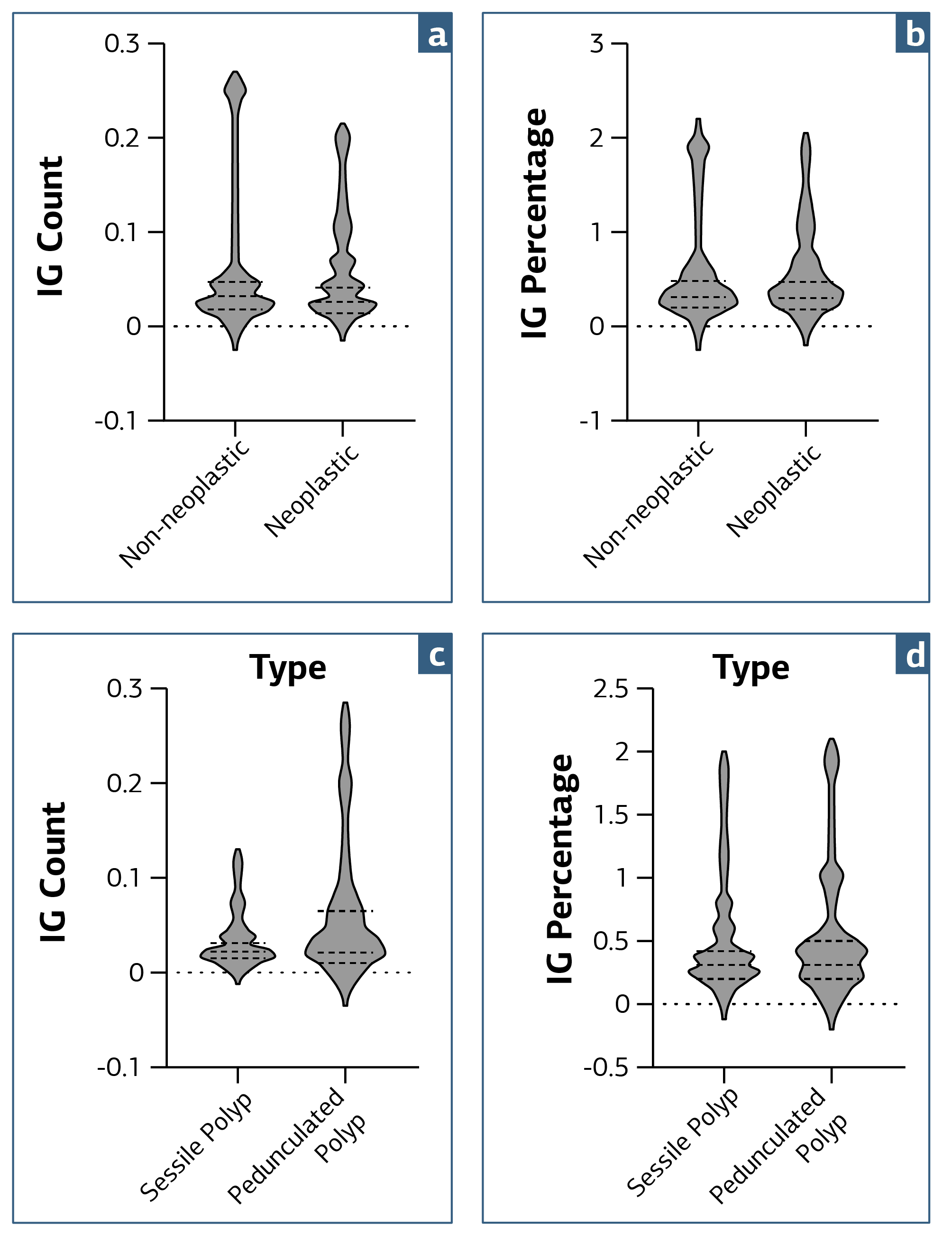
Figure 1. Comparison of Immature Granulocyte Counts and Percentages
a: Number of IG by type, b: Percentage of IG by type, c: Number of IG by stem condition, d: Percentage of IG by stem condition.
Additional Information
Publisher’s Note
Bayrakol MP remains neutral with regard to jurisdictional and institutional claims.
Rights and Permissions

This work is licensed under a Creative Commons Attribution-NonCommercial 4.0 International License (CC BY-NC 4.0). To view a copy of the license, visit https://creativecommons.org/licenses/by-nc/4.0/
About This Article
How to Cite This Article
Neslihan Güneş Aydemir, Püren Metin, Ahmet Mert Aydemir, Ökkeş Zortuk, Haydar Adanır. The role of immature granulocyte ratio in predicting dysplastic colon polyps. Ann Clin Anal Med. Articles in Press. doi:10.4328/ACAM.50024
Publication History
- Received:
- 13.01.2026
- Accepted:
- 16.04.2026
- Published Online:
- 16.04.2026
- Printed:
- 01.11.2026