
Abstract
IntroductionFibroepithelial polyps (FEPs) of the urethra are rare benign mesenchymal tumors, predominantly occurring in the posterior urethra of male children. Their occurrence in the distal urethra of female children is exceptionally rare.Case PresentationA 2-year-old female presented with a 2.5 cm pedunculated, fleshy mass protruding from the distal urethral meatus. The lesion was identified as an interlabial mass that became more prominent during straining. The patient underwent complete surgical excision using a vessel-sealing device at the Department of Pediatric Surgery. Histopathological analysis confirmed the diagnosis of a fibroepithelial polyp.ConclusionAlthough rare, FEP should be considered in the differential diagnosis of interlabial masses in prepubertal girls. Surgical excision is curative with an excellent prognosis.
Keywords
Introduction
Fibroepithelial polyps (FEPs) are benign, non-epithelial tumors of mesodermal origin that can occur throughout the urinary tract.1 While they are the most common benign tumors of the ureter, their occurrence in the urethra is rare, especially in the pediatric population.2 Historically, urethral FEPs have been described almost exclusively in male children, typically arising from the verumontanum in the posterior urethra.3 In female children, the presentation of a urethral polyp is considered exceptional.4 These lesions often present as an interlabial mass, which can lead to diagnostic confusion with more common conditions such as urethral prolapse, paraurethral cysts, or more serious malignancies like rhabdomyosarcoma.5
The aim of this case report is to present a rare case of distal urethral fibroepithelial polyp in a 2-year-old female and to highlight its diagnosis and surgical management.
Case Presentation
A 2-year-old female was brought to the pediatric surgery clinic with a complaint of a visible mass in the genital area. Physical examination in the frog-leg position revealed a pedunculated, reddish-pink, fleshy mass approximately 2.5 cm in length protruding from the distal urethral meatus (Figure 1). Under general anesthesia, the patient underwent surgical excision. A simple polypectomy was performed at the base of the stalk using a vessel-sealing device (LigaSure) to ensure precise hemostasis (Figure 2). A temporary urethral catheter was placed and removed after 24 hours. The patient was discharged on the first postoperative day without complications. Macroscopic examination revealed a 2.5 x 2.0 x 0.5 cm polypoid tissue fragment. Microscopic analysis showed a core of loose, vascularized fibroconnective stroma covered by benign squamous and transitional epithelium, confirming the diagnosis of a fibroepithelial polyp.Ethical ApprovalNot applicable.
This case report was prepared in accordance with the CARE guidelines.
Discussion
Urethral FEPs in children are rare, and their occurrence in females is even more so. A review of the literature suggests that fewer than 20 cases of female urethral polyps have been described in detail.6 Most pediatric urethral polyps are congenital and thought to arise from an error in the development of the urogenital sinus wall.7 The presentation of an interlabial mass in a young girl requires a broad differential diagnosis, including urethral prolapse, paraurethral cysts, and rhabdomyosarcoma.8
Limitations
The main limitations of this study are its single-case design and the lack of long-term follow-up data beyond the immediate postoperative period.
Conclusion
Distal urethral fibroepithelial polyps are a rare but important cause of interlabial masses in female children. Clinicians should maintain a high index of suspicion to differentiate these benign lesions from more aggressive pathologies. Complete surgical excision provides both a definitive diagnosis and a permanent cure.
Declarations
Animal and Human Rights Statement
All procedures performed in this case report were in accordance with the ethical standards of the institutional and/or national research committee and with the 1964 Declaration of Helsinki and its later amendments, or comparable ethical standards.
Informed Consent
Informed consent was obtained from the patient's legal guardians.
Data Availability
The datasets used and/or analyzed during the current study are not publicly available due to patient privacy reasons, but are available from the corresponding author on reasonable request.
Conflict of Interest
The authors declare no conflict of interest.
Funding
None.
Author Contributions (CRediT Taxonomy)
Conceptualization: S.D.
Methodology: S.D.
Software: S.D.
Formal Analysis: S.D.
Investigation: S.D.
Resources: S.D.
Data Curation: S.D.
Writing-Original Draft: S.D.
Visualization: S.D.
Supervision: S.D.
Project Administration: S.D.
Funding Acquisition: S.D.
Validation: I.A.
Writing-Review & Editing: I.A.
AI Usage Disclosure
The authors declare that no AI-assisted technologies were used.
Abbreviations
CARE: Case report guidelines
FEP: Fibroepithelial polyp
References
- Alhindi S, Alaradi H, Mubarak M. Fibroepithelial polyp in a child: a rare pathology of upper urinary tract obstruction. Cureus. 2020;12(6):e8748. doi:10.7759/cureus.8748
- Ksia A, Saad B M, Mekki M, et al. Fibroepithelial polyps of the urethra in infants: a report of three cases. Afr J Urol. 2014;20(1):31-34. doi:10.1016/j.afju.2013.12.001
- Williams TR, Wagner BJ, Corse WR, Vestevich JC. Fibroepithelial polyps of the urinary tract. Abdom Imaging. 2002;27(2):217-221. doi:10.1007/s00261-001-0066-z
- Demircan M, Ceran C, Karaman A, Uguralp S, Mizrak B. Urethral polyps in children: a review of the literature and report of two cases. Int J Urol. 2006;13(6):841-843. doi:10.1111/j.1442-2042.2006.01420.x
- Akbarzadeh A, Khorramirouz R, Saadat S, Hiradfar M, Kajbafzadeh A-M. Congenital urethral polyps in girls: as a differential diagnosis of interlabial masses. J Pediatr Adolesc Gynecol. 2014;27(6):330-334. doi:10.1016/j.jpag.2014.01.001
- Sancak EB, Ozaydin B, Bagla AG. A rare case of urethral fibroepithelial polyp in a young boy. Urol Case Rep. 2023;50:102525. doi:10.1016/j.eucr.2023.102525
- Gupta RK, Khobragade PM, Sanghvi BV, et al. Paediatric posterior urethral polyp: a case report. Pediatr Urol Case Rep. 2021;8(3):66-69.
- de Filippi G, Derchi LE, Coppi M, Biggi E. Sonographic diagnosis of urethral polyp in a child. Pediatr Radiol. 1983;13(6):351-352. doi:10.1007/BF01625967
Figures
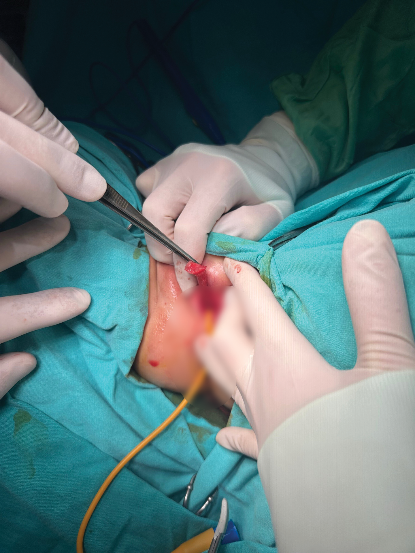
Figure 1. Intraoperative View of the Pedunculated Fibroepithelial Polyp Originating from the Distal Urethral Meatus
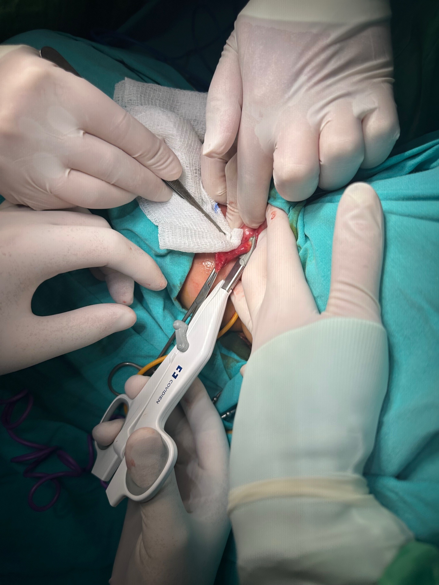
Figure 2. Intraoperative View Showing the Surgical Excision of the Polyp Base Using a Vessel-Sealing Device
Additional Information
Publisher’s Note
Bayrakol MP remains neutral with regard to jurisdictional and institutional claims.
Rights and Permissions

This work is licensed under a Creative Commons Attribution-NonCommercial 4.0 International License (CC BY-NC 4.0). To view a copy of the license, visit https://creativecommons.org/licenses/by-nc/4.0/
About This Article
How to Cite This Article
Sabit Dere, İbrahim Akkoyun. Distal urethral fibroepithelial polyp in a 2-year-old female: A Case Report. Ann Clin Anal Med. Articles in Press. doi:10.4328/ACAM.50043
Publication History
- Received:
- 20.01.2026
- Accepted:
- 31.03.2026
- Published Online:
- 04.04.2026
- Printed:
- 01.11.2026