
Abstract
AimTo investigate the association between first-trimester systemic inflammatory indices and the subsequent development of preeclampsia and to evaluate their predictive performance as early screening markers. MethodsThis retrospective case-control study included 123 women who subsequently developed preeclampsia and 121 healthy pregnant controls. Inflammatory indices, including neutrophil-to-lymphocyte ratio (NLR), platelet-to-lymphocyte ratio (PLR), systemic immune-inflammation index (SII), systemic inflammation response index (SIRI), aggregate index of systemic inflammation (AISI), and systemic inflammation marker index (SIMI), were calculated and compared between groups. Receiver operating characteristic (ROC) curve analysis was performed to assess their predictive value. ResultsMaternal age and gestational age at sampling were similar between groups, whereas parity was significantly higher in the preeclampsia group (P < .001). Women who subsequently developed preeclampsia had significantly lower neutrophil counts and higher lymphocyte counts than controls. Accordingly, NLR, SII, SIRI, and AISI were significantly lower in the preeclampsia group (P < .01), while PLR and SIMI did not differ significantly. ROC analysis demonstrated that NLR had the highest predictive performance (AUC: 0.677, 95% CI: 0.610–0.744), followed by SIRI (AUC: 0.648, 95% CI: 0.579–0.717). Although statistically significant, the overall discriminative abilities of these markers were limited. ConclusionFirst-trimester inflammatory indices are associated with the subsequent development of preeclampsia. Among the evaluated markers, NLR and SIRI demonstrated the best predictive performance; however, their clinical utility as standalone screening tools appears limited. These inexpensive biomarkers may contribute to multifactorial prediction models for identifying women at risk for preeclampsia.Keywords
Introduction
Preeclampsia remains one of the leading causes of maternal and perinatal morbidity and mortality worldwide, affecting approximately 2–8% of pregnancies. Characterized by new-onset hypertension and multisystem involvement after 20 weeks of gestation, preeclampsia is associated with severe maternal complications, including eclampsia, stroke, renal failure, and hepatic dysfunction, as well as adverse fetal outcomes such as fetal growth restriction, preterm birth, and perinatal death. Despite substantial advances in obstetric care, the ability to accurately predict preeclampsia before the onset of clinical manifestations remains limited, highlighting the need for reliable, accessible, and cost-effective early biomarkers 1,2
The pathophysiology of preeclampsia is complex and multifactorial. Although its exact etiology has not been fully elucidated, abnormal placentation during early pregnancy is considered a central event in disease development. Inadequate trophoblastic invasion and impaired remodeling of the spiral arteries result in placental hypoperfusion, oxidative stress, and the release of pro-inflammatory and antiangiogenic factors into the maternal circulation. These processes subsequently trigger widespread endothelial dysfunction and an exaggerated maternal inflammatory response, ultimately leading to the clinical manifestations of the disease.1,3 Increasing evidence suggests that inflammation is not merely a consequence of placental dysfunction but rather a critical component of the pathogenic cascade that begins during the first trimester.2,4
Routine complete blood count parameters have gained considerable attention as potential indicators of systemic inflammation. In recent years, several hemogram-derived inflammatory indices have been proposed as practical biomarkers reflecting the balance between innate and adaptive immune responses. Among these, the neutrophil-to-lymphocyte ratio (NLR), platelet-to-lymphocyte ratio (PLR), monocyte-to-lymphocyte ratio (MLR), systemic immune-inflammation index (SII), systemic inflammation response index (SIRI), aggregate index of systemic inflammation (AISI), and pan-immune-inflammation value (PIV) have demonstrated diagnostic and prognostic value in various diseases.3,5-7 These indices offer several advantages, including low cost, wide availability, and ease of calculation from routinely obtained laboratory data. Recent studies have explored the association between systemic inflammatory markers and adverse pregnancy outcomes, including preeclampsia. However, the majority of available investigations have focused on inflammatory parameters measured during the second or third trimester, often after the disease process has already become clinically apparent. Consequently, evidence regarding the predictive value of first-trimester inflammatory indices remains limited and inconsistent.3,8,9 Given that the pathological processes underlying preeclampsia begin well before the onset of symptoms, the identification of inflammatory alterations during early gestation may provide valuable opportunities for risk stratification and timely preventive interventions. Therefore, the present study aimed to evaluate the association between first-trimester systemic inflammatory indices and the subsequent development of preeclampsia. Additionally, we assessed the predictive performance of these biomarkers as screening tools in routine obstetric practice.
Materials and Methods
Study Design and Population This study was designed as a retrospective case-control study and included patients who presented to the Department of Obstetrics and Gynecology at Aksaray Training and Research Hospital during early pregnancy between December 2021 and July 2025. The study aimed to investigate the predictive value of first-trimester systemic inflammatory indices for the subsequent development of preeclampsia. Complete blood count (CBC) measurements were performed using an automated hematology analyzer (Mindray, BC-6000, China) according to the manufacturer's instructions. The analyzer was routinely calibrated and subjected to internal quality control procedures throughout the study period to ensure the accuracy and reliability of hematological measurements.This observational study was conducted and reported in accordance with the Strengthening the Reporting of Observational Studies in Epidemiology (STROBE) Statement. All applicable items of the STROBE checklist were considered during study design, data analysis, and manuscript preparation. A total of 244 pregnant women were included in the study. The preeclampsia group consisted of 123 women who subsequently developed preeclampsia during pregnancy, while the control group included 121 healthy pregnant women with uncomplicated pregnancies and no history of hypertensive disorders. Preeclampsia was diagnosed according to the criteria established by the American College of Obstetricians and Gynecologists. Briefly, preeclampsia was defined as new-onset hypertension (systolic blood pressure ≥140 mmHg and/or diastolic blood pressure ≥90 mmHg on at least two occasions four hours apart after 20 weeks of gestation) accompanied by proteinuria or evidence of maternal organ dysfunction. Maternal demographic characteristics and laboratory data were obtained from the hospital's electronic medical record system. Complete blood count parameters measured during routine first-trimester screening (11–14 weeks of gestation) were recorded. Based on these parameters, systemic inflammatory indices including the neutrophil-to-lymphocyte ratio (NLR), platelet-to-lymphocyte ratio (PLR), systemic immune-inflammation index (SII), systemic inflammation response index (SIRI), aggregate index of systemic inflammation (AISI), Systemic Inflammation Modulation Index (SIMI) were calculated. The control group was selected from pregnant women with singleton pregnancies who remained normotensive throughout gestation and delivered healthy neonates at term. To minimize potential confounding effects, women with chronic hypertension, pregestational diabetes mellitus, autoimmune diseases, acute or chronic inflammatory disorders, hematological diseases, active infections, multiple pregnancies, fetal congenital anomalies, or incomplete medical records were excluded from the study. All procedures were performed in accordance with the ethical standards of the institutional research committee and the principles of the Declaration of Helsinki. Since this was a retrospective study, no additional blood samples were collected from the participants. The inflammatory indices were calculated using the following formulas: • NLR = neutrophil / lymphocyte • PLR = platelet / lymphocyte • SII = (neutrophil × platelet) / lymphocyte • SIRI = (neutrophil × monocyte) / lymphocyte • AISI = (neutrophil × platelet × monocyte ) / lymphocyte • SIMI = (monocyte × platelet) / lymphocyte Inclusion and Exclusion Criteria This retrospective case–control study included pregnant women who underwent routine first-trimester screening at our institution between 11+0 and 13+6 weeks of gestation and had complete clinical and laboratory records available for review. Women who subsequently developed preeclampsia during pregnancy constituted the preeclampsia group, whereas healthy pregnant women who remained normotensive throughout gestation and experienced no major maternal or fetal complications were included in the control group. Only singleton pregnancies with available first-trimester complete blood count parameters were considered eligible for analysis. To ensure the reliability of systemic inflammatory indices and reduce potential confounding factors, several exclusion criteria were applied. Women with multiple pregnancies, pre-existing chronic hypertension, pregestational diabetes mellitus, chronic renal or hepatic disease, autoimmune disorders, hematological diseases, or active infections at the time of blood sampling were excluded. Pregnancies complicated by fetal congenital anomalies or chromosomal abnormalities were also excluded. In addition, patients receiving medications known to affect inflammatory or hematological parameters, as well as those with incomplete clinical, laboratory, or obstetric records, were not included in the study. Ethical Approval This study was approved by the Aksaray University Ethics Committee (Date: 25.12.2025; Decision No: SAGETİK 2025-165). Statistical Analysis Sample size calculation was performed using G-Power software (v3.1.2). Based on a medium effect size (Cohen’s d=0.50), an alpha level of 0.05, and the study sample sizes (123 Preeclampsi and 121 Controls), the statistical power of the study was calculated as 95%. Statistical analyses were performed using the MedCalc statistical software package (version 20.009; Ostend, Belgium). Arithmetic mean, standard deviation, median, 25th and 75th percentiles values were used to statistically describe the data. The Kolmogorov-Smirnov test was used to determine whether the groups conformed to a normal distribution. The Independent t-test was used for pairwise comparisons of groups. The Mann-Whitney U test was used for pairwise comparisons of groups that did not conform to a normal distribution. In the comparison tables, data conforming to a normal distribution were shown as mean and standard deviation (SD), while data that did not conform were shown as median (25th p – 75th p). ROC analysis was performed to evaluate and compare the diagnostic performance of the measured and calculated laboratory parameters. The Youden J index was used to obtain the optimal cutoff value, and the relevant sensitivity, specificity, positive predictive and negative predictive values, area under the curve (AUC), and 95% confidence intervals (CI) were presented. A significance level of P < .05 was used in interpreting the results. Reporting GuidelinesThis study was reported in accordance with the STROBE guideline.Results
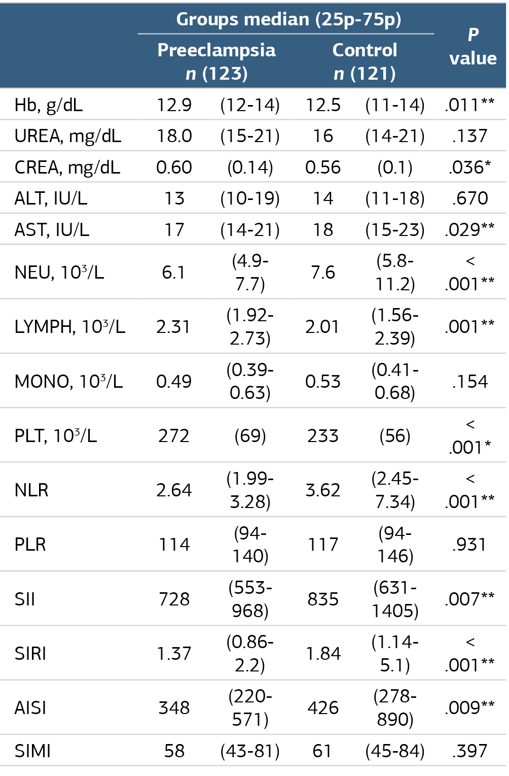
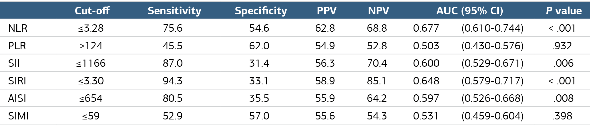
A total of 244 pregnant women were included in the study, comprising 123 patients who subsequently developed preeclampsia and 121 healthy controls. The demographic characteristics of the study population are presented in Table 1. Maternal age and gestational week at the time of first-trimester evaluation were comparable between the groups (P = .419 and P = .115, respectively). However, parity was significantly higher in the preeclampsia group than in the control group (P < .001). The comparison of hematological and biochemical parameters between the groups is shown in Table 2. Hemoglobin levels were significantly higher in women who later developed preeclampsia than in controls (P = .011). Similarly, serum creatinine levels were modestly but significantly elevated in the preeclampsia group (P = .036). In contrast, AST levels were slightly lower in the preeclampsia group compared with controls (P = .029), whereas no significant differences were observed for urea or ALT levels (P > .05). Analysis of complete blood count parameters revealed significantly lower neutrophil counts and higher lymphocyte counts in the preeclampsia group compared with controls (both p≤0.001). Platelet counts were also significantly elevated in women who developed preeclampsia (P < .001). No significant difference was detected in monocyte counts between the groups (P = .154). Regarding inflammatory indices, NLR, SII, SIRI, and AISI values were significantly lower in the preeclampsia group than in the control group (P < .01). In contrast, PLR and SIMI values did not differ significantly between the groups (P = .931 and P = .397, respectively). Receiver operating characteristic (ROC) curve analyses were performed to evaluate the predictive performance of inflammatory indices for preeclampsia (Table 3, Figure 2). NLR demonstrated limited discriminative ability, with an AUC of 0.677 (95% CI: 0.610–0.744, P < .001), yielding a sensitivity of 75.6% and a specificity of 54.6% at a cut-off value of ≤3.28. SIRI showed the highest sensitivity (94.3%) at a cut-off value of ≤3.30 and achieved an AUC of 0.648 (95% CI: 0.579–0.717, P < .001). SII and AISI also showed statistically significant but limited predictive performance, with AUC values of 0.600 and 0.597, respectively. PLR and SIMI did not demonstrate significant discriminative ability for predicting preeclampsia (AUC=0.503, P = .932 and AUC=0.531, P = .398, respectively).Discussion
In the present study, first-trimester inflammatory indices were evaluated in women who subsequently developed preeclampsia and compared with healthy controls. The main finding was that NLR, SII, SIRI, and AISI were significantly lower in the preeclampsia group, whereas PLR and SIMI did not differ significantly between groups. ROC analysis showed that NLR had the highest discriminative ability, followed by SIRI; however, the overall predictive performance of these markers was limited.
Preeclampsia is widely considered a disorder involving abnormal placentation, endothelial dysfunction, oxidative stress, and maternal immune dysregulation. Therefore, inflammatory indices derived from complete blood count parameters have attracted increasing attention as inexpensive and accessible biomarkers for early prediction. Several previous studies have reported higher first-trimester inflammatory markers in pregnancies that later developed preeclampsia. Gezer et al. found that elevated first-trimester NLR and PLR were independent predictors of subsequent preeclampsia, supporting the role of early systemic inflammation in disease development.3 Similarly, Oğlak et al. reported that first-trimester MPV, NLR, and PLR values may be clinically useful in predicting preeclampsia.8
More recently, studies evaluating composite inflammatory markers have also suggested a possible association between increased systemic inflammation and preeclampsia. Akdulum et al. reported that first-trimester SII was significantly higher in women who later developed preeclampsia, suggesting that SII may reflect early inflammatory activation.10 Özkan et al. similarly found that higher SII, SIRI, and PIV values were associated with an increased risk of future preeclampsia.11 These findings are partly inconsistent with our results, as we observed significantly lower NLR, SII, SIRI, and AISI values in the preeclampsia group.
This discrepancy may be explained by several factors. First, inflammatory indices in early pregnancy may not always reflect overt systemic inflammation; rather, they may represent the balance between maternal immune tolerance and inflammatory activation. In our cohort, lower neutrophil counts and higher lymphocyte counts in the preeclampsia group appear to be the main drivers of reduced NLR, SII, SIRI, and AISI values. This may indicate altered immune adaptation during early placentation rather than a simple increase in inflammation. Second, differences in study design, population characteristics, ethnicity, gestational age at blood sampling, disease severity, and exclusion criteria may influence the direction and magnitude of these associations.
Importantly, not all studies support a consistent increase in inflammatory indices before preeclampsia. Yu et al. reported no significant differences in NLR or PLR between preeclamptic and normotensive pregnancies across different trimesters.12 In addition, Seyhanli et al. found that first-trimester SIRI and PIV showed potential predictive value, whereas NLR, PLR, and SII were not uniformly strong predictors.5 These inconsistent findings suggest that hematological inflammatory indices may be affected by population-specific and methodological factors and should not be interpreted as universal standalone markers.
Our findings are particularly compatible with the recent study by Şahin et al., who also reported that first-trimester SII was significantly lower in women who later developed preeclampsia.13 This supports the possibility that, in some populations, preeclampsia may be associated with impaired or insufficient early inflammatory adaptation rather than exaggerated inflammatory activation. Therefore, the relationship between first-trimester inflammatory indices and preeclampsia may be more complex than previously assumed.
In the present study, NLR showed the highest AUC value, followed by SIRI, but both markers demonstrated only modest discriminative ability. This finding is consistent with the 2024 meta-analysis by Mészáros et al., which emphasized that although first-trimester NLR has been widely studied, its predictive value for preeclampsia remains limited and heterogeneous across studies.9 Therefore, our results suggest that NLR and SIRI may be useful as supportive markers, but they are unlikely to be sufficient for clinical prediction when used alone.
Overall, our study contributes to the growing body of evidence showing that first-trimester inflammatory indices are associated with later preeclampsia development. However, unlike many previous reports, our findings suggest that lower rather than higher inflammatory indices may be observed in women who subsequently develop preeclampsia. This highlights the need for larger prospective studies to clarify whether these differences reflect population characteristics, disease subtypes, or distinct immunological pathways in early pregnancy. Future prediction models may benefit from integrating CBC-derived inflammatory indices with maternal characteristics, uterine artery Doppler findings, and established biochemical markers rather than relying on inflammatory indices alone.
Limitations
The main limitations of this study include its retrospective single-center design and the lack of serial inflammatory marker measurements during pregnancy. Additionally, the moderate predictive performance observed in ROC analyses suggests that these indices alone may not be sufficient for clinical prediction of preeclampsia and require validation in larger prospective cohorts.
Conclusion
In conclusion, this study demonstrated that several first-trimester inflammatory indices derived from routine complete blood count parameters differ significantly between women who subsequently develop preeclampsia and healthy pregnancies. Specifically, NLR, SII, SIRI, and AISI were significantly lower in the preeclampsia group, whereas PLR and SIMI did not show meaningful differences. Among the evaluated markers, NLR exhibited the highest discriminative performance, followed by SIRI, although the overall predictive accuracies were limited. These findings suggest that alterations in the maternal inflammatory response may be detectable during the first trimester and contribute to the pathophysiological processes underlying preeclampsia. Given their low cost and ease of calculation, inflammatory indices such as NLR and SIRI may represent useful tools for early risk stratification. However, their predictive performance alone appears insufficient for clinical decision-making, and they should be interpreted in conjunction with established clinical and biochemical predictors. Further large-scale prospective studies are warranted to validate these findings and to determine whether the integration of inflammatory indices into multifactorial prediction models can improve the early identification of women at risk for preeclampsia.
Declarations
Animal and Human Rights Statement
All procedures performed in this study were in accordance with the ethical standards of the institutional and/or national research committee and with the 1964 Helsinki Declaration and its later amendments or comparable ethical standards.
Informed Consent
Informed consent was waived due to the retrospective design of the study.
Data Availability
The datasets used and/or analyzed during the current study are not publicly available due to patient privacy reasons but are available from the corresponding author on reasonable request.
Conflict of Interest
The authors declare that there is no conflict of interest.
Funding
None.
Author Contributions (CRediT Taxonomy)
Conceptualization: H.E, R.B
Methodology: H.E., R.B
Software: AB.G
Validation: H.E, R.B
Data curation: H. E, R.B, AB.G
Formal analysis: AB.G
Investigation: H.E., R.B, AB.G
Writing – original draft: H.E
Writing – review & editing: H.E., R.B, AB.G
Visualization: H.E., R.B, AB.G
Supervision: H.E
Project Administration: H.E
AI Usage Disclosure
Artificial intelligence tools were used only for language editing and improving readability. No AI tool was used for data analysis, interpretation of results, or generation of scientific content. The authors take full responsibility for the content of the manuscript.
Abbreviations
AISI: Aggregate index of systemic inflammation
AUC: Area under the curve
CI: Confidence interval
MLR: Monocyte-to-lymphocyte ratio
NLR: Neutrophil-to-lymphocyte ratio
NPV: Negative predictive value
PIV: Pan-immune-inflammation value
PLR: Platelet-to-lymphocyte ratio
PPV: Positive predictive value
ROC: Receiver operating characteristic curve
SII: Systemic immune–inflammation index
SIMI: Systemic inflammation marker index
SIRI: Systemic inflammation response index
References
- Redman CW, Staff AC. Preeclampsia, biomarkers, syncytiotrophoblast stress, and placental capacity. Am J Obstet Gynecol. 2015;213(4):9-11. doi:10.1016/j.ajog.2015.08.003
- Phipps E, Prasanna D, Brima W, Jim B. Preeclampsia: updates in pathogenesis, definitions, and guidelines. Clin J Am Soc Nephrol. 2016;11(6):1102-1113. doi:10.2215/cjn.12081115
- Gezer C, Ekin A, Ertas IE, et al. High first-trimester neutrophil-to-lymphocyte and platelet-to-lymphocyte ratios are indicators for early diagnosis of preeclampsia. Ginekol Pol. 2016;87(6):431-435. doi:10.5603/gp.2016.0021
- Burton GJ, Jauniaux E. Pathophysiology of placental-derived fetal growth restriction. Am J Obstet Gynecol. 2018;218(2 suppl). doi:10.1016/j.ajog.2017.11.577
- Seyhanli Z, Bayraktar B, Baysoz OB, et al. The role of first-trimester serum inflammatory indexes (NLR, PLR, MLR, SII, SIRI, and PIV) and the beta-hCG to PAPP-A ratio in predicting preeclampsia. J Reprod Immunol. 2024;162:104190. doi:10.1016/j.jri.2023.104190
- Genç Ş, Erdal H. Evaluating hematological inflammatory markers as predictors of preeclampsia. Clin Exp Obstet Gynecol. 2024;51(7).
- Erdal H, Kılıc AO, Yagcı BA, Yasar E. Can the pan-immune inflammation value, systemic inflammatory response index, and other hematological inflammatory indices be clinically used to predict pseudoexfoliation? J Clin Pract Res. 2024;46(1):19. doi:10.14744/cpr.2024.36036
- Oglak SC, Tunc S, Olmez F. First-trimester mean platelet volume, neutrophil-to-lymphocyte ratio, and platelet-to-lymphocyte ratio values are useful markers for predicting preeclampsia. Ochsner J. 2021;21(4):364-370.
- Meszaros B, Veres DS, Nagyistok L, Kovacs BG, Kukor Z, Valent S. A meta-analysis on first-trimester blood count parameters: is the neutrophil-to-lymphocyte ratio a potentially novel method for first-trimester preeclampsia screening? Front Med (Lausanne). 2024;11:1336764.
- Cevher Akdulum MF, Demirdag E, Arik SI, et al. Is the first-trimester systemic immune-inflammation index associated with preeclampsia? Cureus. 2023;15(8). doi:10.7759/cureus.44063
- Ozkan S, Dereli ML, Firatligil FB, et al. Role of systemic immune-inflammation index, systemic inflammation response index, and pan-immune inflammation value in the prediction of preeclampsia: a retrospective cohort study. Am J Reprod Immunol. 2024;92(6).
- Yu HB, Shi MD, Yao J, Jia J, Zhu CR, Zhou R. Value of neutrophil-to-lymphocyte ratio and platelet-to-lymphocyte ratio for predicting preeclampsia during pregnancy. Sichuan Da Xue Xue Bao Yi Xue Ban. 2022;53(6):1039-1044. Chinese.
- Sahin R, Inceoglu C, Tahiroglu V. The value of first-trimester inflammatory indices in predicting the development of preeclampsia in the third trimester. BMC Pregnancy Childbirth. 2025;25(1):713. doi:10.1186/s12884-025-07836-1
Tables

AUC, Area under curve; CI, Confidence interval; PPV, Positive predictive value; NPV, Negative predictive value
Figures
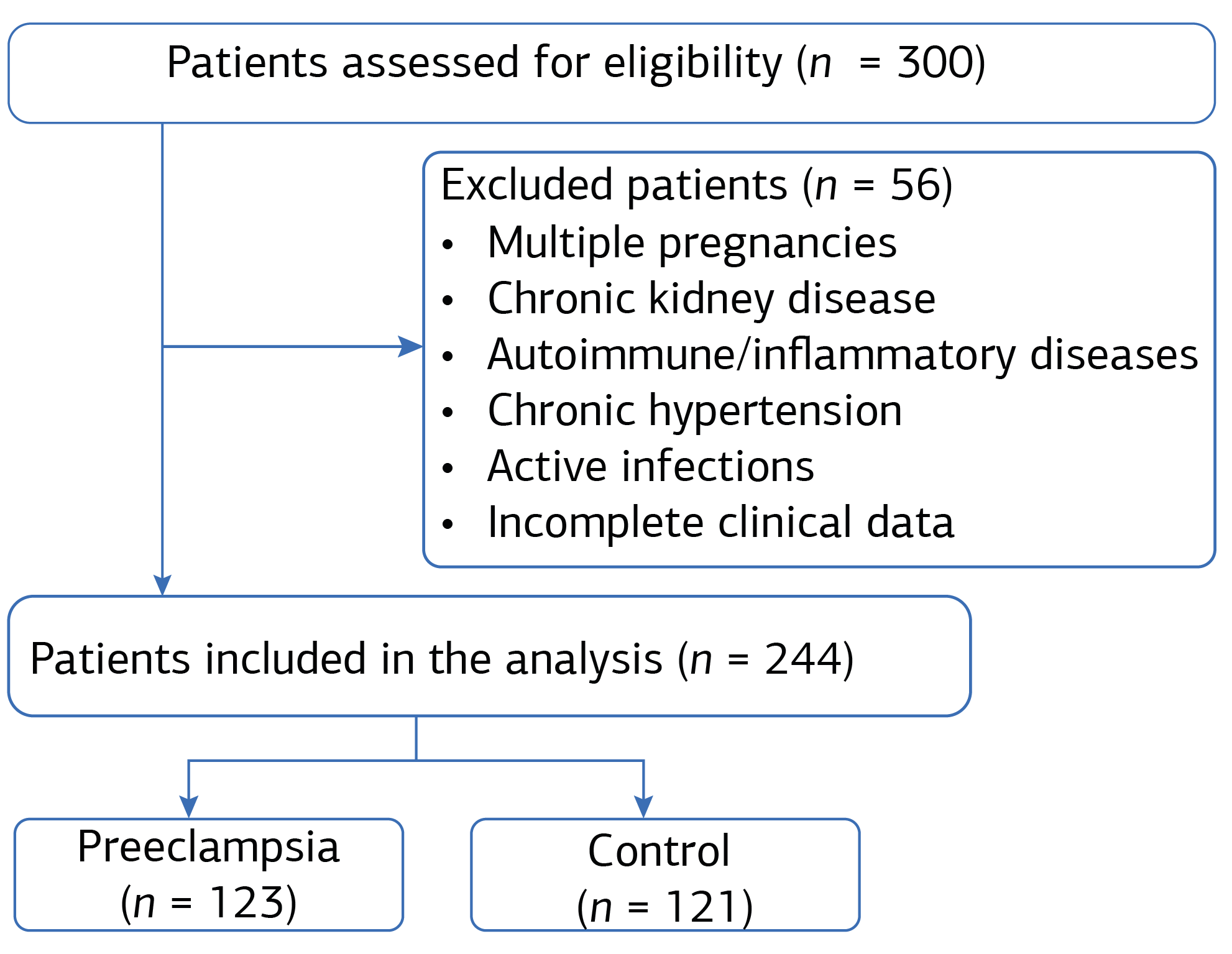
Figure 1. Flowchart of patient selection and group allocation in the study.
Additional Information
Publisher’s Note
Bayrakol MP remains neutral with regard to jurisdictional and institutional claims.
Rights and Permissions

This work is licensed under a Creative Commons Attribution-NonCommercial 4.0 International License (CC BY-NC 4.0). To view a copy of the license, visit https://creativecommons.org/licenses/by-nc/4.0/
About This Article
How to Cite This Article
Ramazan Bülbül, Hüseyin Erdal, Ahmet Burak Gürpınar. Systemic inflammatory indices in the first trimester: emerging tools for early prediction of preeclampsia. Ann Clin Anal Med 2026;17(9):958-963. doi:10.4328/ACAM.50241
Publication History
- Received:
- 13.06.2026
- Accepted:
- 05.08.2026
- Published Online:
- 13.08.2026
- Printed:
- 01.09.2026