
Abstract
AimBone fracture is a common injury necessitating surgical procedures for stabilization of the fracture or improvement of medical conditions. Tetracalcium phosphate (TTCP) is a component used in the production of some hydroxyapatite calcium phosphate cements to repair bone defects. Our study aimed to investigate the biomechanical effects of TTCP in an in vitro bone fracture model not previously studied.MethodsA total of 28 fresh chicken femurs (TFs) were obtained from 14 chickens of the same breed, age, and approximately the same weight for the study. Fresh TFs were randomly divided into 4 groups with 7 TFs per group: Control group, Plate group, Plate + TTCP group, and TTCP group. The maximum force (Newton) and maximum displacement (mm) values applied in the tests were recorded.ResultsThere was a significant difference in maximum force values between the groups (P < .001), but no significant difference was found in maximum displacement. In the post hoc analysis performed to determine the source of the significant difference between the groups, it was revealed that the Plate group (PG) and Plate + TTCP group (PTG) showed significantly higher values than the Control group (CG) (P = .007 for both), while the CG showed significantly higher values than the TTCP group (P = .024).ConclusionBased on the data obtained in our study, we conclude that TTCP did not have a positive biomechanical effect on the in vitro fracture model we created. According to our results, the use of plate and screws alone appears sufficient for small bone fractures.
Keywords
Introduction
Bone fracture is one of the most common injuries and is associated with treatment costs exceeding billions of dollars, societal productivity loss, and individual disability.1,2 Bone tissue is a dynamic structure where old or damaged tissue is replaced with new bone tissue through a process called remodeling. Fracture healing is a complex coordination of various cellular and mechanosensitive processes. Several influential mechanisms affect fracture healing, such as systemic biological factors, biochemical factors, hormonal factors, and biomechanical factors.3,4 A problem in this process can result in nonunion or delayed union.5 Approximately 5% to 10% of fractured bones result in nonunion and/or incomplete healing.6 A better understanding of factors affecting fracture healing has helped target earlier and more functional recovery processes. There are two types of bone healing: primary and secondary. Primary bone healing occurs when bone fragments are tightly secured under compression resulting from implantation. There is no callus formation, and the two bone pieces are bridged and healed directly through the activities of osteoclasts and osteoblasts.7,8 More commonly seen, secondary bone healing occurs when there is minimal movement at the fracture site. Interfragmentary movement leads to the formation of soft callus and results in secondary bone formation through both intramembranous and endochondral ossifications.7,9 Problems with bone healing are common, especially in fractures with bone defects. Excessive movement at the fracture site, large interfragmentary gap, and loss of vascularity negatively affect bone healing.10 Numerous surgical procedures are performed annually to stabilize fractured bones or to improve medical conditions that require bone fixation. Conventional fixation methods involve the use of metal plates, screws, etc., to stabilize the fracture.11 The primary issue in plate-screw fixation today is loosening and/or breakage due to the load applied to the plate and screws. Additionally, metallic fixation devices are associated with a high risk of infection and potential damage to surrounding tissues. Lastly, traditional methods are ineffective for treating small bone fragments or periarticular fractures.
The application of biodegradable bone adhesive molecules would be a simple and effective alternative solution for stabilizing bone fractures.12,13 Applying such a biologically degradable bone adhesive would eliminate the need for a second surgery to remove metallic fixation devices and reduce the risks of infection and other complications.
While there is no universally accepted definition for biodegradable bone adhesives, they must meet several requirements to function adequately in clinical practice. Primarily, they should exhibit strong adhesion to bone tissue in the presence of blood. For effective clinical application, a minimum bonding strength to bone of 200 kPa has been reported by Weber and Chapman 14, as lower bonding strengths have been shown to be inadequate for effective bone fixation. Additionally, bone adhesives should maintain bonding strength in the weeks and months following application and demonstrate sufficient mechanical stability under static and dynamic loads. Furthermore, the application of these adhesives to the surgical site should be straightforward within a clinically acceptable timeframe. They should not be toxic to the body during application or post-reaction. They should be easily manufacturable and sterilizable, as well as cost-effective.
TTCP is a component used in the production of some hydroxyapatite calcium phosphate cements used to repair bone defects.15 Injectable osteoconductive calcium phosphate cements are produced in various types and properties and are used in addition to intraosseous fixation. Unlike polymethylmethacrylate (PMMA) cements, which cause local cellular damage due to the exothermic reaction reaching temperatures as high as 60°C during hardening, these cements harden at normal physiological pH and body temperature, thereby not causing local cellular death or denaturing body proteins.16 They continue to crystallize and harden in vivo, reaching 50% of ultimate compressive strength within 1 hour and 80% within 4 hours. Therefore, when applied to areas with bone defects, they provide mechanical support.
An important advantage of TTCP cements is reducing the need for bone grafts, thereby eliminating significant donor site morbidity associated with bone graft procedures. Their general application supports the fixation of applied metal implants and requires implant support. They cannot provide fracture fixation alone and exhibit biomechanically weak resistance, especially against shear forces.17
In this experimental in vitro study, we aimed to investigate the biomechanical effects of TTCP, known for its mechanical support when used in early fracture treatment in small bone fractures.
Materials and Methods
Study DesignThis study was supported by the Scientific Research Projects Coordination Unit of Çanakkale Onsekiz Mart University Hospital (Project No: THD-2022-3797).
The study was conducted at the Department of Orthopedics and Traumatology of a tertiary university hospital. Our study was carried out experimentally through modeling.Specimen SelectionA total of 14 chickens of the same breed, age, and approximately the same weight were used in the study. A total of 28 fresh chicken femurs (TF) were used in the study. The TFs used were dissected without creating trauma to soft tissues. The dissected TFs did not undergo any chemical processing. After dissection, the TFs were kept at 4 °C until the time of the procedure (approximately 18 hours).Groups and Experimental ProceduresAfter dissection and storage at 4 °C, fresh TFs were randomized into four groups without distinction between right and left femurs, each consisting of 7 specimens. Except for the Control group (CG), the remaining groups (Group 2, 3, and 4) underwent transverse osteotomy at previously marked mid-diaphyseal regions using a motorized saw to create the fracture model. Subsequently, in the Plate group (PG), TFs with created fracture models were fixed using a 4-hole plate designed specifically for TF dimensions, secured with 3.5 mm diameter self-tapping cortical screws placed in each hole. In the Plate + TTCP group (PTG), TTCP was applied to the fracture lines of TFs followed by fixation using the same 4-hole plate and screws. The Tetracalcium Phosphate group (TKG) received fixation using TTCP application alone without additional plating.
Following these procedures, chicken femurs were stored at approximately 4 °C for 24 hours and subsequently subjected to biomechanical testing. Biomechanical tests were performed at the Biomechanics Laboratory, Department of Biomechanics, Çanakkale Onsekiz Mart University, using a Shimadzu AG-IS Autograph testing machine with a three-point bending test. The test speed was set at 5 mm/min, consistent with previous studies. Maximum force (Newton) and maximum displacement (mm) values applied during the test were recorded separately for each specimen on pre-established forms and archived.
To ensure standardization in biomechanical testing, the distance between the distal and proximal ends of chicken femurs in all experimental groups was measured, and the midpoint was marked (Figure 1). This marking designated the point where load application occurred and was marked for osteotomy (Figure 2).
All groups in the study, including the Control group (CG), underwent three-point bending tests to determine maximum resistance force at the bone's maximum resistance point and subsequent deformation, recorded on predefined forms. After completion of biomechanical testing, data from study forms were transferred to digital format for statistical analysis.Ethical ApprovalEthical approval was not required for this in vitro experimental study.Statistical AnalysisData obtained from our study and transferred to digital format were analyzed using SPSS version 26.0. Descriptive statistics including counts, percentages, median, interquartile range (IQR) (75th percentile - 25th percentile), minimum, and maximum values were used for data presentation. Before comparing two or more datasets, normality of data distribution was assessed using the Kolmogorov-Smirnov test. For analysis of categorical variables, Pearson's χ2 test was used when variables were five or more; Fisher's Exact Test was used when variables were fewer than five. Non-normally distributed bivariate data were analyzed using the Mann-Whitney U test, and for analysis of more than two variables that did not follow a normal distribution, the Kruskal-Wallis test was applied. Post hoc tests were conducted to identify the source of differences in parameters showing statistical significance from the Kruskal-Wallis test.
Statistical analysis was performed with a confidence interval of 95% and a significance level of P < .05.Reporting GuidelinesNo specific reporting guideline was applicable to this in vitro experimental study.
Results
Our study included a total of 28 TFs divided into 4 groups, each comprising 7 specimens. Maximum force and maximum displacement values observed in the three-point bending test were analyzed. The median maximum force values creating the fracture were notably high in PTG at 769.84 N and in PG at 674.38 N. In contrast, the median value in CG was measured at 394.53 N, while it was much lower at 36.88 N in TKG, where only TTCP was applied. Results showed that values in PG and PTG were higher than those in CG, whereas the level in bone models of TKG was lower than the average in CG. Additionally, the median maximum displacement values obtained in the three-point bending test were 2.19 mm in CG, 4.56 mm in PTG, 2.43 mm in TKG, and 2.04 mm in PG. These results indicated that the maximum displacement was highest in PTG and lowest in PG. Statistical analysis revealed significant differences in maximum force values among groups (P < .001), whereas no significant difference was found for maximum displacement (Tables 1 and 2). Following the identification of statistically significant differences among groups, post hoc analysis was conducted to determine the source of these differences. It was found that in terms of maximum force, PG and PTG were significantly higher than CG (P = .007 for both), and CG was significantly higher than TKG (P = .024). Furthermore, in the evaluation of maximum force results, PG was significantly higher than both CG and TKG (P = .007 and P < .001, respectively), and this difference was not observed between PG and PTG (P > .05). Median maximum force levels in PTG did not significantly differ from TKG (P > .05), but they were significantly higher compared to TKG (P < .001). There were no significant differences observed among the groups in terms of maximum displacement (Table 3).Discussion
In the current literature, while the effects of many bioactive cements on fracture healing have been studied extensively, there are limited studies specifically examining the impact of TTCP, a bioactive cement used in fracture healing. Our study investigated the early biomechanical effects of TTCP following fixation in a TF fracture model. At the outset of our study, we hypothesized that TTCP could have beneficial biomechanical effects in early small bone fracture fixation. Previous studies have shown that TTCP has beneficial biomechanical effects in fracture healing and fixation. However, our study had distinguishing features from others, such as the use of a small bone model with a small surface area, room temperature conditions, a transverse osteotomy line in the fracture model, and a pH different from living tissues. Based on the data obtained in the study, we conclude that TTCP did not have a positive biomechanical effect on our fracture model. According to our results, we believe that plate and screw combinations alone are sufficient for small bone fractures. The results we expected were that the implants used in the study had higher resistance than the group with the fractures. However, we believe that the addition of TTCP to plate and screw fixation was not an additional factor. We also believe that the use of TTCP alone provides insufficient stability.
Bone fracture is one of the most common injuries, and it is associated with high treatment costs, societal productivity loss, and individual disability. Plate-screw combinations have been widely used in orthopedic surgery for approximately a century to treat fractures. However, the primary problem in plate-screw fixation today is loosening and/or fractures due to the load on the plate and screw.18,19 TTCP is a component used in the formation of some hydroxyapatite calcium phosphate cements used to repair bone defects.20 Cadaver studies have shown that in some fractures of the distal radius, tibial plateau, proximal femur, and calcaneus, the use of Grandus® B-One in addition to traditional internal fixation can produce better stability, hardness, and strength than using implant fixation alone.17
Clinical studies have shown that TTCP reinforcement in tibial plateau and calcaneus fractures reduces the time required to achieve full postoperative loading, provides faster strength and range of motion recovery when used in distal radius fractures, and offers better stability in some hip fractures. In a study by Kaymaz et al. in a rat model, the use of TTCP was reported to be beneficial in early fracture healing.21 Furthermore, in a study by Neral et al. on distal radius fractures, the use of cement was reported not to cause any adverse effects and could facilitate reduction and fixation in comminuted fractures.22 Watson's study on eight patients with proximal or distal tibial metaphyseal fractures reported that calcium sulfate could be used safely.23 Su et al. mentioned that TTCP cement is biocompatible and has osteoconductive properties.24 In another study by Trenholm et al. on tibial plateau fractures, the use of calcium phosphate cement was reported to be safer than using bone autograft.25 Upon reviewing the literature, it is evident that minimally invasive interventions are becoming more prominent, and the importance of cement use is highlighted in enhancing reduction and bone healing success in these minimally invasive approaches.
Previously, TTCP has been shown to have positive outcomes in the treatment of large bone fractures and comminuted fractures. However, it is necessary to evaluate TTCP under appropriate conditions, especially in vivo, and in more comprehensive studies for small bone fractures.
Limitations
This study has several limitations. Previous research has demonstrated the influence of factors such as blood flow, body temperature, and pH on the efficacy of TTCP usage. In our study, the ineffectiveness of TTCP could be attributed to the use of non-living tissue, room temperature conditions, and the lack of desired pH levels. Furthermore, although there is insufficient data in the literature regarding dosage or applied surface area, we believe that the small applied surface area may have also impacted the results.
Conclusion
Based on the data obtained in the study, we conclude that TTCP did not have a positive biomechanical effect on the in vitro fracture model. According to our findings, TTCP is not yet an alternative treatment modality for plate fixation of fractures. However, it may be used as an augmentation in fractures with bone defects.
Declarations
Animal and Human Rights Statement
All procedures performed in this study were in accordance with the ethical standards of the institutional and/or national research committee and with the 1964 Helsinki Declaration and its later amendments or comparable ethical standards.
Informed Consent
Not applicable.
Data Availability
The datasets used and/or analyzed during the current study are available from the corresponding author on reasonable request.
Conflict of Interest
The authors declare that there is no conflict of interest.
Funding
This study titled 'Biomechanical Comparison of Plate and Tetracalcium Phosphate in an In Vitro Chicken Fracture Model' was supported by the Scientific Research Project Coordination Unit of Çanakkale 18 March University Hospital (Project No: THD-2022-3797).
Author Contributions (CRediT Taxonomy)
Conceptualization: TAO, CKU, MSK, KB
Methodology: TAO, CKU, MSK, KB Writing-Editing: TAO, CKU, MSK, KB
References
- Pivonka P, Dunstan CR. Role of mathematical modeling in bone fracture healing. Bonekey Rep. 2012;1:221. doi:10.1038/bonekey.2012.221
- Bonafede M, Espindle D, Bower AG. The direct and indirect costs of long bone fractures in a working-age US population. J Med Econ. 2013;16(1):169-178. doi:10.3111/13696998.2012.737391
- Bahney CS, Zondervan RL, Allison P, et al. Cellular biology of fracture healing. J Orthop Res. 2019;37(1):35-50. doi:10.1002/jor.24170
- Ghiasi MS, Chen J, Vaziri A, Rodriguez EK, Nazarian A. Bone fracture healing in mechanobiological modeling: a review of principles and methods. Bone Rep. 2017;6:87-100. doi:10.1016/j.bonr.2017.03.002
- Buckwalter JA, Einhorn TA, Marsh JL. Bone and joint healing. In: Bucholz RW, Heckman JD, eds. Rockwood and Green’s Fractures in Adults. Lippincott Williams & Wilkins; 2001:245-271.
- Praemer A, Furner SE, Rice DP. Selected musculoskeletal conditions. In: Praemer A, Furner S, Rice DP, eds. Musculoskeletal Conditions in the United States. American Academy of Orthopaedic Surgeons; 1999:168-192.
- Claes L, Recknagel S, Ignatius A. Fracture healing under healthy and inflammatory conditions. Nat Rev Rheumatol. 2012;8(3):133-143. doi:10.1038/nrrheum.2012.1
- Marsell R, Einhorn TA. The biology of fracture healing. Injury. 2011;42(6):551-555. doi:10.1016/j.injury.2011.03.031
- Gerstenfeld LC, Alkhiary YM, Krall EA, et al. Three-dimensional reconstruction of fracture callus morphogenesis. J Histochem Cytochem. 2006;54(11):1215-1228. doi:10.1369/jhc.6a6959.2006
- Rodriguez-Merchan EC, Forriol F. Nonunion: general principles and experimental data. Clin Orthop Relat Res. 2004;(419):4-12. doi:10.1097/00003086-200402000-00003
- Taljanovic MS, Jones MD, Ruth JT, et al. Fracture fixation. Radiographics. 2003;23(6):1569-1590. doi:10.1148/rg.236035159
- Sánchez-Fernández MJ, Hammoudeh H, Félix Lanao RP, et al. Bone-adhesive materials: clinical requirements, mechanisms of action, and future perspective. Adv Mater Interfaces. 2019;6:1802021.
- Tzagiollari A, McCarthy HO, Levingstone TJ, Dunne NJ. Biodegradable and biocompatible adhesives for the effective stabilisation, repair, and regeneration of bone. Bioengineering (Basel). 2022;9(6):250. doi:10.3390/bioengineering9060250
- Weber SC, Chapman MW. Adhesives in orthopaedic surgery: a review of the literature and in vitro bonding strengths of bone-bonding agents. Clin Orthop Relat Res. 1984;(191):249-261. doi:10.1097/00003086-198412000-00034
- Moseke C, Gbureck U. Tetracalcium phosphate: synthesis, properties, and biomedical applications. Acta Biomater. 2010;6(10):3815-3823. doi:10.1016/j.actbio.2010.04.020
- Cohen MS, Whitman K. Calcium phosphate bone cement—the Norian skeletal repair system in orthopedic surgery. AORN J. 1997;65:958-962. doi:10.1016/s0001-2092(06)62978-5
- Larsson S, Bauer TW. Use of injectable calcium phosphate cement for fracture fixation: a review. Clin Orthop Relat Res. 2002;(395):23-32. doi:10.1097/00003086-200202000-00004
- Blake CA, Boudrieau RJ, Torrance BS, et al. Single cycle to failure in bending of 3 standard and 5 locking plates and plate constructs. Vet Comp Orthop Traumatol. 2011;24(6):408-417. doi:10.3415/vcot-11-04-0061
- Miller EI, Acquaviva AE, Eisenmann DJ, Stone RT, Kraus KH. Perpendicular pull-out force of locking versus nonlocking plates in thin cortical bone using a canine mandibular ramus model. Vet Surg. 2011;40(7):870-874. doi:10.1111/j.1532-950x.2011.00893.x
- Bohner M, Gbureck U, Barralet JE. Technological issues for the development of more efficient calcium phosphate bone cements: a critical assessment. Biomaterials. 2005;26(33):6423-6429. doi:10.1016/j.biomaterials.2005.03.049
- Kaymaz B, Yılmaz O, Taşova AO, Anapa D. Tetracalcium phosphate treatment on an experimental fracture model in rats. Jt Dis Relat Surg. 2022;33(1):172-179. doi:10.52312/jdrs.2022.190
- Neral M, Solari M, Purnell C, Wollstein R. The use of bone cement in difficult distal radius fractures. Hand (N Y). 2013;8(4):387-391. doi:10.1007/s11552-013-9548-z
- Watson JT. The use of an injectable bone graft substitute in tibial metaphyseal fractures. Orthopedics. 2004;27(1 suppl). doi:10.3928/0147-7447-20040102-04
- Su Y, Cockerill I, Zheng Y, Tang L, Qin YX, Zhu D. Biofunctionalization of metallic implants by calcium phosphate coatings. Bioact Mater. 2019;4:196-206. doi:10.1016/j.bioactmat.2019.05.001
- Trenholm A, Landry S, McLaughlin K, et al. Comparative fixation of tibial plateau fractures using alpha-BSM, a calcium phosphate cement, versus cancellous bone graft. J Orthop Trauma. 2005;19(10):698-702. doi:10.1097/01.bot.0000183455.01491.bb
Tables
Table 1. Results of 3-Point Bending Test and Median Values
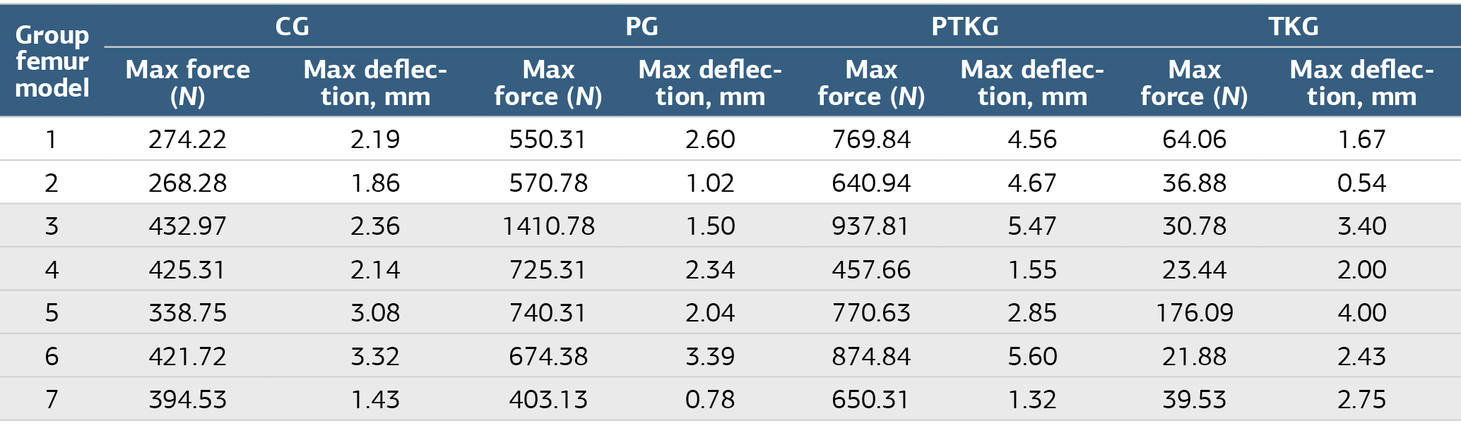
CG, Control Group; PG, Plate Group; PTKG, Plate+TTCP Group; TKG, TTCP Group IQR, Interquarter Range; N, Newton
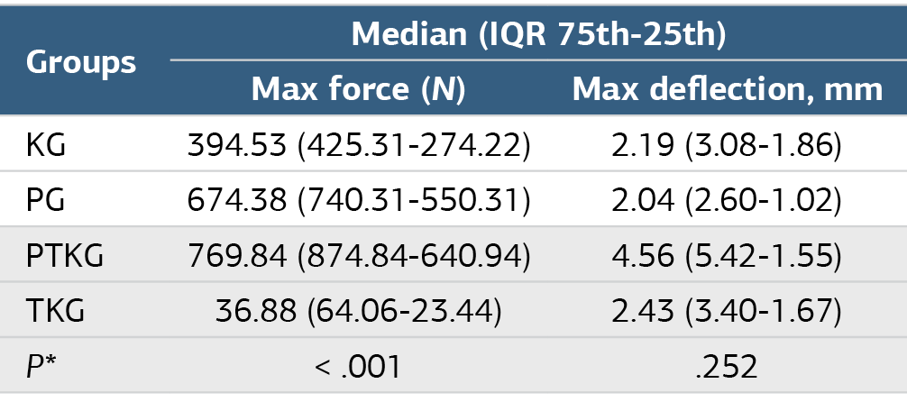
*: Kruskal-Wallis test was appliedCG, Control Group; PG: Plate Group; PTKG, Plate+TTCP Group; TKG, TTCP Group; IQR, Interquarter Range; N, Newton
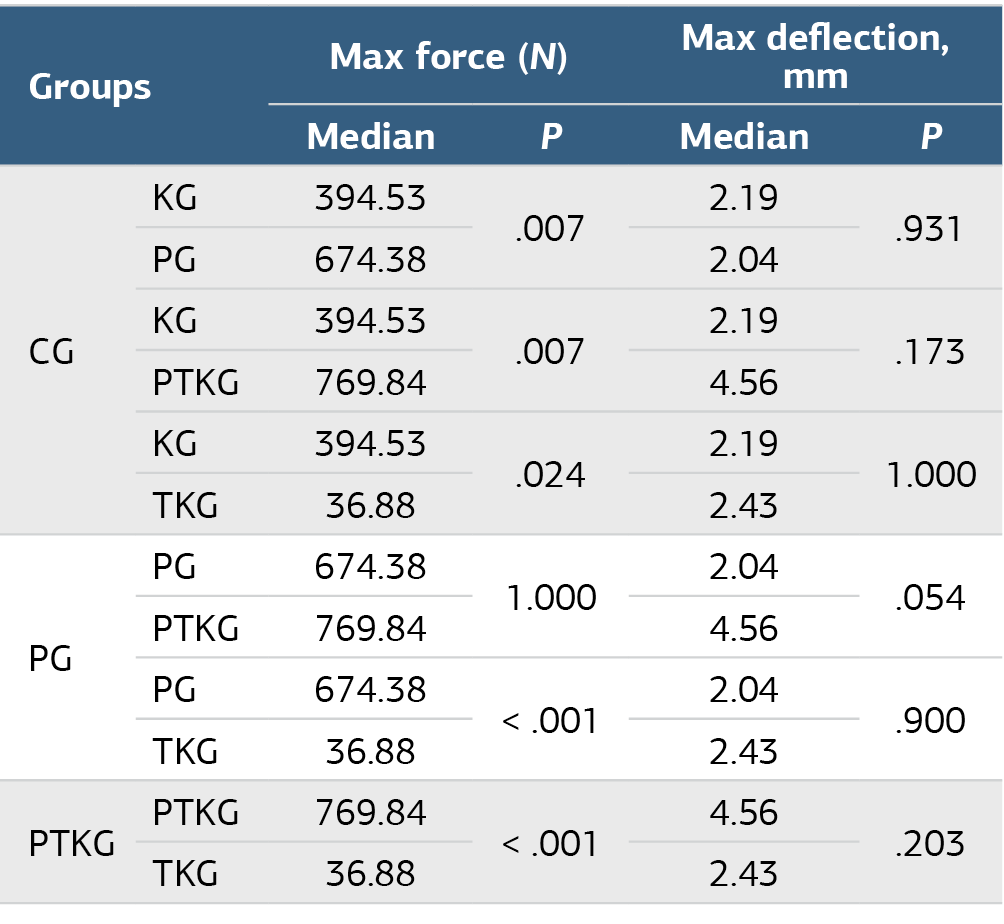
*, Post Hoc Testi; CG, Control Group; PG, Plate Group; PTKG, Plate+TTCP Group; TKG, TTCP Group; IQR, Interquarter Range; N, Newton
Figures
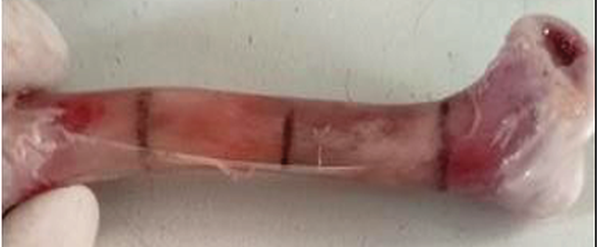
Figure 1. After the measurement, the midpoint was marked as the place where the force would be applied.
Additional Information
Publisher’s Note
Bayrakol MP remains neutral with regard to jurisdictional and institutional claims.
Rights and Permissions

This work is licensed under a Creative Commons Attribution-NonCommercial 4.0 International License (CC BY-NC 4.0). To view a copy of the license, visit https://creativecommons.org/licenses/by-nc/4.0/
About This Article
How to Cite This Article
Ali Osman Taşova, Ali Osman Taşova, Sedit Kıvanç Muratlı, Burak Kaymaz. Biomechanical comparison of plate and tetracalcium phosphate in an in vitro chicken fracture model. Ann Clin Anal Med 2026;17(9):969-973. doi:10.4328/ACAM.22386
Publication History
- Received:
- 26.08.2026
- Accepted:
- 28.08.2024
- Published Online:
- 02.08.2026
- Printed:
- 01.09.2026